. 2024 Nov;27(4):903-917.
doi: 10.1007/s10456-024-09948-2. Epub 2024 Sep 17.

Timed topical dexamethasone eye drops improve mitochondrial function to prevent severe retinopathy of prematurity

Affiliations

Timed topical dexamethasone eye drops improve mitochondrial function to prevent severe retinopathy of prematurity

Hitomi Yagi et al. Angiogenesis. 2024 Nov.

Abstract

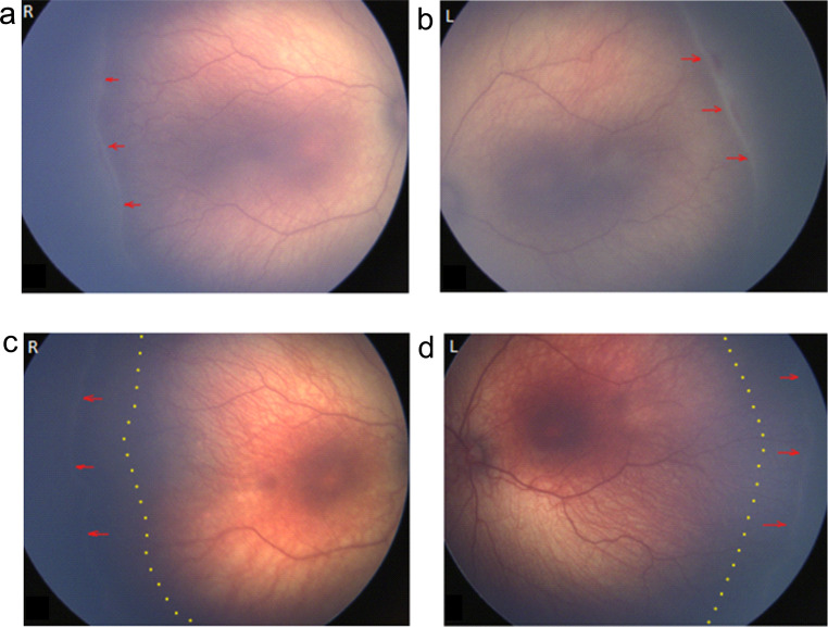
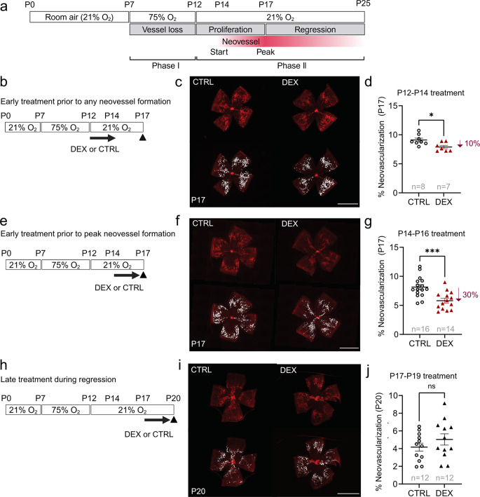
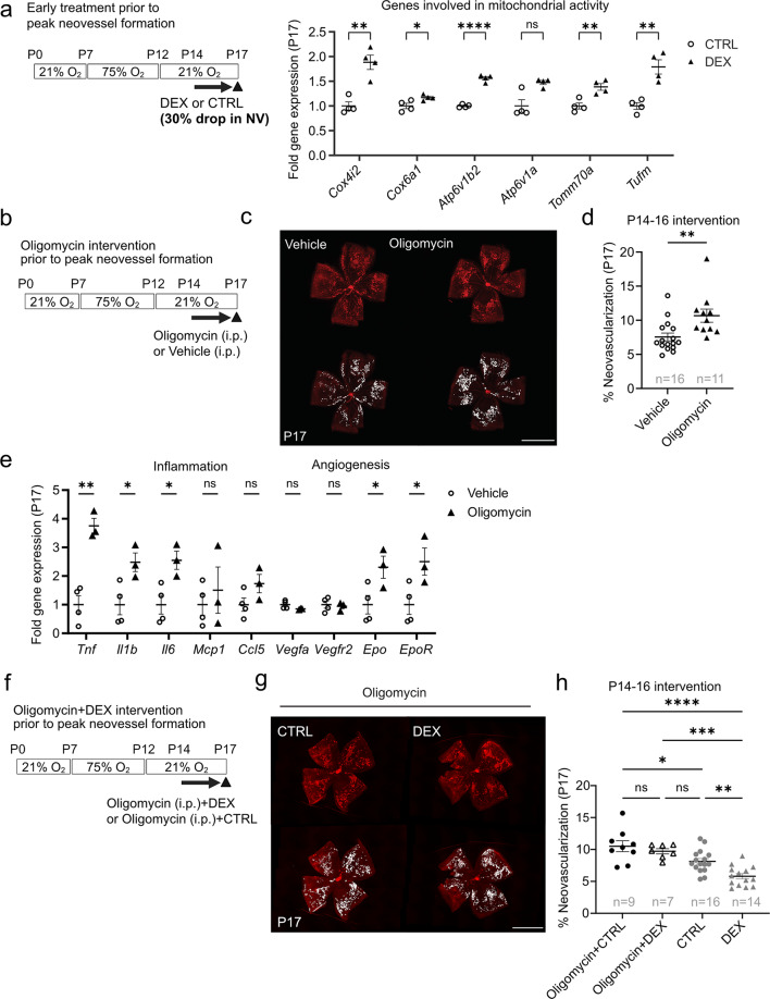
Pathological neovascularization in retinopathy of prematurity (ROP) can cause visual impairment in preterm infants. Current ROP treatments which are not preventative and only address late neovascular ROP, are costly and can lead to severe complications. We showed that topical 0.1% dexamethasone eye drops administered prior to peak neovessel formation prevented neovascularization in five extremely preterm infants at high risk for ROP and suppressed neovascularization by 30% in mouse oxygen-induced retinopathy (OIR) modeling ROP. In contrast, in OIR, topical dexamethasone treatment before any neovessel formation had limited efficacy in preventing later neovascularization, while treatment after peak neovessel formation had a non-statistically significant trend to exacerbating disease. Optimally timed topical dexamethasone suppression of neovascularization in OIR was associated with increased retinal mitochondrial gene expression and decreased inflammatory marker expression, predominantly found in immune cells. Blocking mitochondrial ATP synthetase reversed the inhibitory effect of dexamethasone on neovascularization in OIR. This study provides new insights into topical steroid effects in retinal neovascularization and into mitochondrial function in phase II ROP, and suggests a simple clinical approach to prevent severe ROP.

Keywords: Dexamethasone; Eye drops; Mitochondrial function; Neovascularization; Oxygen-induced retinopathy; Retinopathy of prematurity.


Conflict of interest statement

Declarations Competing interests The authors declare no competing interests.

Figures

Fig. 1
Fig. 1
A pilot clinical study of topical 0.1% dexamethasone eye drops Type 2 ROP prior to neovessel formation prevented progression to Type 1 ROP, and promoted normal vascular development. (a, b) Representative fundus images of Case 3 (Table 1). Born gestational age 25 weeks, birth weight 700 g, female. Start of dexamethasone treatment (a) in right eye (R) and (b) left eye (L) at postmenstrual age (PMA) 36 + 2 weeks. Red arrows indicate a ridge at the leading edge of developing retinal vasculature. (cd) Representative fundus images of Case 3 (Table 1), at the end of dexamethasone treatment (c) in R eye and (d) L eye at PMA 44 + 2 weeks. Red arrows indicate the extent of re-vascularization at the end of treatment. Dotted lines indicate the leading edge of vascular development at start of dexamethasone treatment (PMA 36 + 2)
Fig. 2
Fig. 2
Effect of topical 0.1% dexamethasone eye drops on neovascularization with treatment during different intervals during OIR development. (a) Schematic of mouse oxygen-induced retinopathy (OIR) and longitudinal development of neovascularization. Hyperoxia (75% O2) from postnatal day (P) 7 to P12 induces retinal vaso-obliteration. After returning to room air (21% O2) at P12 the avascular retina drives neovessel formation starting at P14, peaking at P17 with subsequent regression. (be, h) Schematics of topical 0.1% dexamethasone eye drops (DEX) or control eye drops (CTRL) treatment intervals in OIR. (b) P12 to P14 (prior to any neovessel formation) (e) P14 to P16 (prior to peak neovessel formation) (h) P17 to P19 (during peak neovessel formation and regression). Retinas were collected at P17 (b-g) or P20 (h-j). (cfi) Representative images of whole mounted retinas after OIR mice were treated with DEX or CTRL were shown. Retinal vessels (red, lectin), neovascular area highlighted in white. Scale bar, 2 mm. (d, g, j) Percentage of neovascular area over whole mounted retinas of OIR mice treated topically with DEX vs. CTRL from: (d) P12-P14, evaluated at P17: CTRL, n = 8; DEX, n = 7 retinas; Two-tailed unpaired t-test; *p < 0.05. Mean values ± SEM; (g) P14-P16 evaluated at P17: CTRL, n = 16; DEX, n = 14 retinas; Two-tailed unpaired t-test; ***p < 0.001. (j) P17-P19 evaluated at P20: CTRL, n = 12; DEX, n = 12 retinas; (d, g, j) Two-tailed unpaired ttest; ns, not significant
Fig. 3
Fig. 3
Label-free proteomic analyses of P17 OIR retinas with topical dexamethasone treatment prior to peak neovessel formation (P14-P16). (a) Overview of the experimental time course. Label-free LC-MS/MS-based proteomics of P17 OIR retinas treated topically with one drop per day of 0.1% DEX or CTRL from P14 to P16 (prior to peak neovessel formation). CTRL, n = 6; DEX, n = 6 mice (2 retinas from each mouse pooled for n = 1). (b) Principal component analysis plot of unfiltered proteome (p = 1 for n = 3,936 proteins; 2 or more unique peptides). (c) Number of identified and statistically significant (p < 0.05) proteins in the dataset. In DEX group versus CTRL group, 184 proteins with increased and 144 proteins with decreased in abundance. Volcano plot of differentially abundant proteins in P17 OIR DEX vs. CTRL retinas. Each data point in blue represents a unique protein considered significant (p < 0.05). Selected proteins (with low p-value or high fold change) involved in mitochondrial dysfunction, ion channel transport, and oxidative phosphorylation in Ingenuity Pathway Analysis (IPA) were labeled. (d) IPA activation Z-scores for selected canonical pathway in DEX group compared to CTRL group. Pathways were sorted by -log10 (p value). Significant pathways were defined as those with a z-score absolute value > 1 or an overlap p value < 0.05
Fig. 4
Fig. 4
Topical dexamethasone treatment prior to peak neovessel formation (P14-P16) suppresses P17 neovascularization via mitochondrial ATP production. (a) Quantification of mitochondrial mRNA gene expression levels in P17 retinas from OIR mice treated with one drop per eye per day of DEX or CTRL from P14 to P16 (prior to peak neovessel formation). CTRL, n = 4; DEX, n = 4 mice (2 retinas from each mouse pooled for n = 1). Two-tailed unpaired t-test for Cox4i2, Cox6a1, Atp6v1b2, Tomm70a, and Tufm; Mann-Whiteny U test for Atp6v1a; *p < 0.05; **p < 0.01; ***p < 0.001; ****p < 0.0001. Mean values ± SEM. (b) Schematic of oligomycin (mitochondrial ATP synthase inhibitor) intervention in OIR from P14 to P16 (prior to peak neovessel formation). (c) Representative images of P17 whole mounted retinas from OIR mice after oligomycin (0.25ug/g, i.p., daily) or vehicle P14-P16. Retinal vessels (red, lectin). NV was highlighted in white. Scale bar, 2 mm. (d) Percent neovascular area of whole retinal area in P17 OIR after oligomycin or control administration in (b). Vehicle control (Vehicle), n = 16; Oligomycin, n = 11 retinas; Two-tailed unpaired t-test; **p < 0.01; ns, not significant. (e) Quantification of inflammation- and angiogenesis-related mRNA expression levels in P17 retinas in OIR mice treated with oligomycin or vehicle control administration. Vehicle, n = 4; Oligomycin, n = 3 mice (2 retinas from each mouse pooled for n = 1). Two-tailed unpaired t-test; *p < 0.05; **p < 0.01; ns, not significant. Mean values ± SEM. (f) Schematic of intervention in OIR mice treated with oligomycin (0.25ug/g, i.p.) in addition to 0.1% dexamethasone eye drops (Oligomycin + DEX) or control eye drops (Oligomycin + CTRL) from P14 to P16 (prior to peak neovessel formation). (g) Representative images of whole mounted P17 retinas of OIR mice after treatment with Oligomycin + CTRL or Oligomycin + DEX from P14 to P16. Retinal vessels are visualized with lectin (red) and NV is highlighted in white. Scale bar, 2 mm. (h) Quantification of NV as percentage of total retinal area in P17 OIR retinas in mice treated with Oligomycin + CTRL or Oligomycin + DEX from P14 to P16. Oligomycin + CTRL, n = 9; Oligomycin + DEX, n = 7 retinas. DEX and CTRL groups from Fig. 2g are included for comparison. One-way ANOVA; *p < 0.05; **p < 0.01; ***p < 0.001; ****p < 0.0001; ns, not significant
Fig. 5
Fig. 5
Topical dexamethasone treatment at different intervals during OIR progression differentially suppressed pro-inflammatory and pro-angiogenic gene expression. (a-c) Quantification of inflammation- and angiogenesis-related mRNA expression levels in P17 (a, b) or P20 (c) retinas in OIR mice treated with DEX or CTRL (a) from P12 to P14 (prior to any neovessel formation). CTRL, n = 7; DEX, n = 7 mice (2 retinas from each mouse pooled for n = 1); Two-tailed unpaired t-test for Tnf, Il1b, Vegfa, Vegfr2, Epo, EpoR; Mann-Whitney U-test for Mcp1 and Ccl5; Welch’s test for Il6; *p < 0.05; **p < 0.01; ns, not significant. Mean values ± SEM. (b) from P14 to P16 (prior to peak neovessel formation). CTRL, n = 7; DEX, n = 8 mice (2 retinas from each mouse pooled for n = 1); Two-tailed unpaired t-test; *p < 0.05; **p < 0.01; ***p < 0.001; ns, not significant. Mean values ± SEM. (c) from P17 to P19 (during peak neovessels and neovessel regression). CTRL, n = 4; DEX, n = 4 mice (2 retinas from each mouse pooled for n = 1); Two-tailed unpaired t-test; *p < 0.05; ns, not significant. Mean values ± SEM

Update of

References

    1. Hellstrom A, Smith LE, Dammann O (2013) Retinopathy of prematurity. Lancet 382(9902):1445–1457. 10.1016/S0140-6736(13)60178-6 - PMC - PubMed
    1. Fu Z, Nilsson AK, Hellstrom A, Smith LEH (2022) Retinopathy of prematurity: metabolic risk factors. Elife 11. 10.7554/eLife.80550 - PMC - PubMed
    1. Sato T, Wada K, Arahori H, Kuno N, Imoto K, Iwahashi-Shima C, Kusaka S (2012) Serum concentrations of bevacizumab (avastin) and vascular endothelial growth factor in infants with retinopathy of prematurity. Am J Ophthalmol 153(2):327–333e321. 10.1016/j.ajo.2011.07.005 - PubMed
    1. Zehden JA, Mortensen XM, Reddy A, Zhang AY (2022) Systemic and ocular adverse events with Intravitreal Anti-VEGF therapy used in the treatment of Diabetic Retinopathy: a review. Curr Diab Rep 22(10):525–536. 10.1007/s11892-022-01491-y - PubMed
    1. Yossuck P, Yan Y, Tadesse M, Higgins RD (2000) Dexamethasone and critical effect of timing on retinopathy. Invest Ophthalmol Vis Sci 41(10):3095–3099 - PubMed

MeSH terms

LinkOut - more resources