Case Reports
. 2024 Dec;23(6):2679-2683.
doi: 10.1007/s12311-024-01734-6. Epub 2024 Sep 17.

CACNA1G Causes Dominantly Inherited Myoclonus-Ataxia with Intellectual Disability: A Case Report

Affiliations
Case Reports

CACNA1G Causes Dominantly Inherited Myoclonus-Ataxia with Intellectual Disability: A Case Report

Martina De Riggi et al. Cerebellum. 2024 Dec.

Abstract

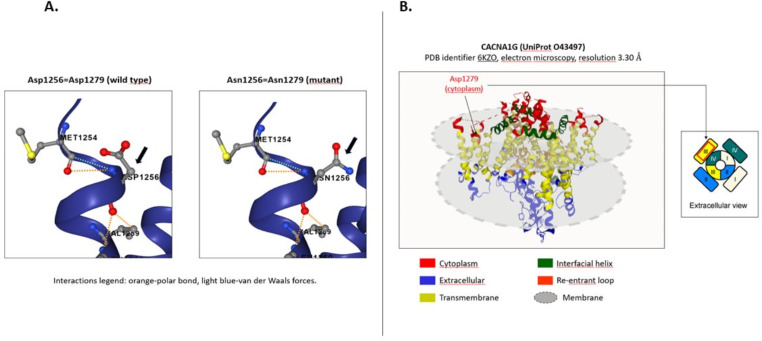
Spinocerebellar ataxias (SCAs) are characterized by substantial phenotypic variability. Among them, SCA42 is a rare non-expansion entity presenting with slowly progressive cerebellar syndrome but whose clinical spectrum may be also wider. A 53-year-old male presented with progressive myoclonus-ataxia and intellectual disability. Genetic screening revealed a novel c.3835G > A (p. Asp1279Asn) variant in the CACNA1G gene. SCA42 is a rare non-expansion SCA caused by mutations in CACNA1G on chromosome 17q21, encoding the Ca(V)3.1, a low-threshold voltage-gated T-type calcium channel. The novel variant we identified is potentially involved in channel activity. This case expands the knowledge regarding CACNA1G-associated phenotype and highlights the importance of genetic screening in myoclonus-ataxia disorders.

Keywords: CACNA1G; Ataxia; Genetic; Intellectual disability; Myoclonus.


Conflict of interest statement

Declarations. Ethical Approval: The authors confirm that the approval of an institutional review board was not required for this work. A written informed consent of the patient was obtained for the publication of his data. The authors have read the Journal’s position on issues involved in the ethical publication and affirm that this work is consistent with the guidelines. Informed Consent: Patient signed informed consent regarding publishing his data and photographs. Competing Interests: The authors declare no competing interests.

Figures

Fig. 1
Fig. 1
Brain magnetic resonance imaging (MRI): (A) T1-weighted axial image and (B) T1-weighted sagittal image show a slight widening of the subarachnoid spaces of the cranial vault (grey arrows) without evidence of cerebellar or brainstem atrophy
Fig. 2
Fig. 2
Neurophysiological assessment: (A) Electromyographic (EMG) trace: EMG1 + = Right wrist extensor, EMG2 + = Right wrist flexor recorded with the arms outstretched in front of the chest. The EMG trace shows frequent bursts of very short duration (30 ms) on EMG1+, which are often observed synchronously on the antagonist muscle EMG2+, compatible with myoclonus. The number of bursts per second is irregular and varies during the recording; (B) Jerk-Locked Back Averaging (JLBA): the JLBA performed on 71 EMG discharges at about 30 milliseconds shows a positive-negative wave on the contralateral central area (C3), with maximum positivity at -15 milliseconds from the onset of the myoclonus (time 0). No recognizable waves are seen on C4; (C) the averaging of the 71 myoclonic jerks, visible synchronously on EMG1 + and, with lower amplitude, also on EMG2+; (D) a voltage map shows the temporal trend of EEG voltages at -40, -26, -14, and 0 ms from the myoclonus. Red, white, and blue colors on the voltage map indicate respectively positive, isoelectric, and negative voltage. The highest positive voltage can be observed at -16 milliseconds over C3 area, that turns into a negative voltage at 0 milliseconds. (E) Cortico-muscular coherence analysis shows high peaks of coherence within the beta frequencies band (from 16 to 29 Hz) with the highest peak on 22–23 Hz. The horizontal black line indicates the 95% significant coherence level, set at 0.055. EEG–EMG data analysis was performed with Brainstorm which is documented and freely available for download online under the GNU general public license (http://neuroimage.usc.edu/brainstorm)
Fig. 3
Fig. 3
In silico analysis of mutated residue. (A) Assessment of amino acid localization was performed with Protein Data Bank of Transmembrane Proteins (PDBTM) using an experimentally determined protein structure (PDB entry: 6KZO) and revealed that aspartate in position 1279 is placed in the cytoplasmic side of S1III domain. (B) Representation of wild-type and mutant residues within protein structure. Dotted orange line = polar bond, dotted light blue line = van der Waals forces, grey spheres = carbon atoms, red spheres = oxygen atoms, blue spheres = nitrogen atoms, yellow spheres = sulfur atoms

References

    1. Sullivan R, Yau WY, O’Connor E, Houlden H. Spinocerebellar ataxia: an update. J Neurol Febbraio. 2019;266(2):533–44. - PMC - PubMed
    1. Morino H, Matsuda Y, Muguruma K, Miyamoto R, Ohsawa R, Ohtake T, et al. A mutation in the low voltage-gated calcium channel CACNA1G alters the physiological properties of the channel, causing spinocerebellar ataxia. Mol Brain Dicembre. 2015;8(1):89. - PMC - PubMed
    1. Kimura M, Yabe I, Hama Y, Eguchi K, Ura S, Tsuzaka K, et al. SCA42 mutation analysis in a case series of Japanese patients with spinocerebellar ataxia. J Hum Genet Settembre. 2017;62(9):857–9. - PubMed
    1. Li X, Zhou C, Cui L, Zhu L, Du H, Liu J, et al. A case of a novel CACNA1G mutation from a Chinese family with SCA42: a case report and literature review. Med (Baltimore) Settembre. 2018;97(36):e12148. - PMC - PubMed
    1. Tadel F, Baillet S, Mosher JC, Pantazis D, Leahy RM. Brainstorm: a user-friendly application for MEG/EEG analysis. Comput Intell Neurosci. 2011;2011:1–13. - PMC - PubMed

Publication types

Substances

LinkOut - more resources