. 2024 Dec 2;84(23):4049-4065.
doi: 10.1158/0008-5472.CAN-24-0984.

mLumiOpto Is a Mitochondrial-Targeted Gene Therapy for Treating Cancer

Affiliations

mLumiOpto Is a Mitochondrial-Targeted Gene Therapy for Treating Cancer

Kai Chen et al. Cancer Res. .

Abstract

Mitochondria are important in various aspects of cancer development and progression. Targeting mitochondria in cancer cells holds great therapeutic promise, yet current strategies to specifically and effectively destroy cancer mitochondria in vivo are limited. Here, we developed mitochondrial luminoptogenetics (mLumiOpto), an innovative mitochondrial-targeted luminoptogenetics gene therapy designed to directly disrupt the inner mitochondrial membrane potential and induce cancer cell death. The therapeutic approach included synthesis of a blue light-gated cationic channelrhodopsin in the inner mitochondrial membrane and coexpression of a blue bioluminescence-emitting nanoluciferase in the cytosol of the same cells. The mLumiOpto genes were selectively delivered to cancer cells in vivo by an adeno-associated virus carrying a cancer-specific promoter or cancer-targeted mAB-tagged exosome-associated adeno-associated virus. Induction with nanoluciferase luciferin elicited robust endogenous bioluminescence, which activated cationic channelrhodopsin, triggering cancer cell mitochondrial depolarization and subsequent cell death. Importantly, mLumiOpto demonstrated remarkable efficacy in reducing tumor burden and killing tumor cells in glioblastoma and triple-negative breast cancer xenograft mouse models. Furthermore, the approach induced an antitumor immune response, increasing infiltration of dendritic cells and CD8+ T cells in the tumor microenvironment. These findings establish mLumiOpto as a promising therapeutic strategy by targeting cancer cell mitochondria in vivo. Significance: mLumiOpto is a next generation optogenetic approach that employs selective delivery of genes to cancer cells to trigger mitochondrial depolarization, effectively inducing cell death and reducing tumor burden.


Conflict of interest statement

No disclosures were reported.

Figures

None
Graphical abstract
Figure 1.
Figure 1.
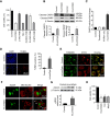
Development and in vitro characterization of mLumiOpto. A, Map of mLumiOpto expression vector. The luciferase (i.e., NLuc) and mitochondrial rhodopsin (i.e., ABCB-CoChR) genes are linked via a cleavable 2A linker. B, Photostimulation with LED light (0.5 mW/mm2, 24 hours) induced mitochondrial depolarization (measured by MitoView dye) in ABCB-ChR2– and ABCB-CoChR–expressing cells, with a more significant effect in ABCB-CoChR cells. C, NLuc emitted much stronger luminescence than RLuc when coupled with ViviRen. D, Confocal images showing high-level mitochondrial CoChR expression in HeLa and MDA-MB-231 cells, indicated by strong overlap of eYFP (fused to CoChR) and MitoTracker dye. E, Confocal images showing NLuc-GFP and CoChR-mCherry coexpression in mLumiOpto-transfected MDA-MB-231 cells. F, IVIS images showing ViviRen-induced dose-dependent luminescent responses in mLumiOpto-transfected cancer cells. G, Confocal images showing ViviRen-induced dose-dependent mitochondrial depolarization in mLumiOpto-transfected MDA-MB-231 cells. H, ViviRen induced dose-dependent cell death in mLumiOpto-transfected MDA-MB-231 cells, with no cytotoxic effect in mock-transfected cells. I, ViviRen (40 µmol/L) caused substantial cytotoxicity in mLumiOpto-transfected cancer cell lines, including GBM cells (U251 and U87) and TNBC cells (BT-20 and MDA-MB-468). *, P < 0.05 vs. control; #, P < 0.05 vs. ChR2. n = 4–6/group.
Figure 2.
Figure 2.
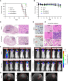
Mechanistic pathways underlying mLumiOpto-mediated cytotoxicity. A, Z-VAD-FMK (pan-caspase inhibitor), Z-DEVD-FMK (Casp-3 inhibitor) and Z-LEHD-FMK (caspase-9 inhibitor) significantly alleviated mLumiOpto-mediated cytotoxicity in MDA-MB-231 cells, whereas Nec-1 (necrosis inhibitor) and Z-IETD-FMK (caspase-8 inhibitor) had no effect. B, Expression of apoptotic markers cleaved CASP3 and PARP significantly increased mLumiOpto-treated cells compared with controls. C, Casp-3 activity significantly increased in mLumiOpto-treated cells compared with controls. D, The number of TUNEL-positive cells was significantly higher in mLumiOpto-treated cancer cells than that in controls. E, Immunofluorescence imaging showed CYCS release in mLumiOpto-treated cancer cells but not in controls. F, Syto24 staining revealed mLumiOpto-induced substantial DNA damage. G, Western blotting confirmed DNA damage by increased γH2AX expression in mLumiOpto-treated cells. H, CsA and MitoQ had no significant effect on mLumiOpto-mediated cancer cell death. *, P < 0.05 vs. mLumiOpto (A) or control (else). n = 4/group.
Figure 3.
Figure 3.
Characterization of AAV-mediated mLumiOpto gene delivery in the GBM xenograft mouse model. A, TEM imaging showed AAV DJ/8 particles with correct morphology and size (∼20 nm). B, Western blotting confirmed AAV viral capsid proteins VP1, VP2, and VP3. C, ViviRen (30 µmol/L) induced strong NLuc luminescence in mLumiOpto AAV-transduced GBM U87 cells. D, mLumiOpto (AAV+ViviRen) induced dramatic mitochondrial depolarization in U87 cells, as measured by MitoView. E, mLumiOpto killed 90% to 99% of GBM U251, U251-TMZ, LN229, and GL261 cells within 72 hours, whereas AAV or ViviRen alone had no significant cytotoxic effect. F, AAV administered through i.c.v. injection led to remarkably higher mLumiOpto gene (NLuc) expression in GBM tumors than through i.v. injection. G, ViviRen elicited strong luminescence in mLumiOpto AAV-transduced GBM xenografts. H,Ex vivo IVIS imaging confirmed mLumiOpto expression in GBM xenografts but not in normal organs. n = 4–6/group.
Figure 4.
Figure 4.
Evaluations of anticancer efficacy of mLumiOpto in the GBM U87 xenograft mouse model. A, mLumiOpto treatment significantly extended the survival of GBM xenograft mice. B, Body weight trends were similar among the control and treatment groups. C, H&E staining of the paraffin section slides of GBM xenograft demonstrated tumor burden reduction by mLumiOpto. D, IHC staining of tumor slides with antibodies of cleaved CASP3 and Ki67 indicated apoptosis-induced cell death and inhibition of proliferation, respectively, post-treatment. E, H&E staining did not detect obvious injury or toxicity in normal organs of the treatment group. F, IVIS imaging revealed >10-fold reduction in GBM tumor size in mLumiOpto-treated groups compared with controls (saline and AAV only). Vertical dividing lines (red) indicate mice from difference source images. G, MRI images taken in the late stage of the survival study (i.e., 23 days after the last ViviRen injection) confirmed a significant reduction of GBM tumor burden in mLumiOpto-treated groups. *, P < 0.0001 vs. saline. n = 8–10/group.
Figure 5.
Figure 5.
Evaluation of anticancer efficacy of mLumiOpto in the GBM PDX mouse model. A, MRI at the endpoint (14 weeks post PDX) showed reduced tumor burden in mLumiOpto-treated mice compared with controls. B, H&E staining confirmed reduced tumor cells in GBM PDXs. C, mLumiOpto treatment significantly extended survival of GBM PDX mice. D, Body weight profiles were comparable between groups. E, IHC staining with cleaved CASP3 and Ki67 antibodies indicated apoptosis activation and proliferation inhibition. F, H&E staining showed no injury or toxicity in normal organs of the treated group as compared with the control group. *, P < 0.0001 vs. saline. n = 10–15/group.
Figure 6.
Figure 6.
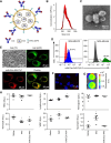
Construction and in vitro characterization of mAb-Exo-AAV. A, Schematic description of mAb-Exo-AAV. The anti-EGFR mAb is tagged to the surface of Exo-AAV via DMPE-PEG-NHS linker. B, mAb-Exo-AAV size distribution determined by NanoSight. C, TEM imaging showed expected mAb-Exo-AAV morphology and size. D, Flow cytometry showed that mAb-Exo-AAV bound strongly to TNBC cells (MDA-MB-468 and MDA-MB-231). E, Confocal imaging confirmed that mAb-Exo-AAV-Cy5.5 (red) is bound to the surface and internalized by MDA-MB-468 cells. F, Confocal imaging showed that AAV-Cy3 (red) transduced more than 95% of TNBC cells and started accumulating around the nucleus (blue) 30 minutes after incubation. G, IVIS imaging confirmed ViviRen-induced bright NLuc luminescence in mAb-Exo-AAV–transduced MDA-MB-231 cells. H, Complete blood cell count demonstrated low peripheral toxicity of mAb-Exo-AAV in BALB/cJ mice. *, P < 0.05 vs. saline. n = 5/group. RBC, red blood cell; WBC, white blood cell.
Figure 7.
Figure 7.
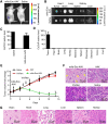
Evaluation of anticancer efficacy of mAb-Exo-AAV–delivered mLumiOpto in the human TNBC MDA-MB-231 xenograft mouse model. A, Live animal IVIS imaging revealed NLuc luminescence overlay with TNBC xenograft. B,Ex vivo IVIS imaging showed mAb-Exo-AAV accumulation in TNBC xenografts, not in normal organs. C,CoChR gene expression was detected in tumor of mLumiOpto mAb-Exo-AAV mice but not in saline controls. D, High CoChR expression in TNBC tumors, undetectable in normal tissues of mAb-Exo-AAV mice. E, Tumor volume reduced in mLumiOpto-treated TNBC xenograft mice compared with control groups (saline, AAV, and ViviRen alone). F, H&E staining revealed significant cell death in tumors of mLumiOpto-treated mice. G, H&E staining showed no injury in normal organs of mLumiOpto-treated mice. *, P < 0.05 vs. controls. n = 6/group.
Figure 8.
Figure 8.
Evaluation of anticancer efficacy of mAb-Exo-AAV–delivered mLumiOpto in the mouse 4T1 immunocompetent xenograft mouse model. A, ViviRen triggered strong NLuc bioluminescence in mAb-Exo-AAV mice. B, Prolonged ViviRen administration effectively inhibited tumor growth in mAb-Exo-AAV mice compared with controls (saline). C, Terminal tumor wet weight in mAb-Exo-AAV mice was significantly lower than controls. D, Flow cytometry showed enriched CD11C+ DC (left) and CD8+ T (right) cells in the TME of mLumiOpto-treated mice compared with controls (saline). E, Luminex assay of tumor tissues showed significant upregulation of cytokines TFNγ, IL1β, IL2, IL4, IL13, and IL12p70 and minimal changes in immune suppressors IL17A and IL23. *, P < 0.05 vs. saline. n = 4–6/group.

Update of

Similar articles

Cited by

References

    1. Stanley WC, Recchia FA, Lopaschuk GD. Myocardial substrate metabolism in the normal and failing heart. Physiol Rev 2005;85:1093–129. - PubMed
    1. Scheid AD, Beadnell TC, Welch DR. Roles of mitochondria in the hallmarks of metastasis. Br J Cancer 2021;124:124–35. - PMC - PubMed
    1. Armstrong JS. Mitochondria: a target for cancer therapy. Br J Pharmacol 2006;147:239–48. - PMC - PubMed
    1. Neuzil J, Wang X-F, Dong L-F, Low P, Ralph SJ. Molecular mechanism of ‘mitocan’-induced apoptosis in cancer cells epitomizes the multiple roles of reactive oxygen species and Bcl-2 family proteins. FEBS Lett 2006;580:5125–9. - PubMed
    1. Horobin RW, Trapp S, Weissig V. Mitochondriotropics: a review of their mode of action, and their applications for drug and DNA delivery to mammalian mitochondria. J Control Release 2007;121:125–36. - PubMed

Substances