. 2024 Sep 17;15(9):680.
doi: 10.1038/s41419-024-07063-0.

Iron chelation as a new therapeutic approach to prevent senescence and liver fibrosis progression

Affiliations

Iron chelation as a new therapeutic approach to prevent senescence and liver fibrosis progression

Josep Amengual et al. Cell Death Dis. .

Abstract

Iron overload and cellular senescence have been implicated in liver fibrosis, but their possible mechanistic connection has not been explored. To address this, we have delved into the role of iron and senescence in an experimental model of chronic liver injury, analyzing whether an iron chelator would prevent liver fibrosis by decreasing hepatocyte senescence. The model of carbon tetrachloride (CCl4) in mice was used as an experimental model of liver fibrosis. Results demonstrated that during the progression of liver fibrosis, accumulation of iron occurs, concomitant with the appearance of fibrotic areas and cells undergoing senescence. Isolated parenchymal hepatocytes from CCl4-treated mice present a gene transcriptomic signature compatible with iron accumulation and senescence, which correlates with induction of Reactive Oxygen Species (ROS)-related genes, activation of the Transforming Growth Factor-beta (TGF-β) pathway and inhibition of oxidative metabolism. Analysis of the iron-related gene signature in a published single-cell RNA-seq dataset from CCl4-treated livers showed iron accumulation correlating with senescence in other non-parenchymal liver cells. Treatment with deferiprone, an iron chelator, attenuated iron accumulation, fibrosis and senescence, concomitant with relevant changes in the senescent-associated secretome (SASP), which switched toward a more anti-inflammatory profile of cytokines. In vitro experiments in human hepatocyte HH4 cells demonstrated that iron accumulates in response to a senescence-inducing reagent, doxorubicin, being deferiprone able to prevent senescence and SASP, attenuating growth arrest and cell death. However, deferiprone did not significantly affect senescence induced by two different agents (doxorubicin and deoxycholic acid) or activation markers in human hepatic stellate LX-2 cells. Transcriptomic data from patients with different etiologies demonstrated the relevance of iron accumulation in the progression of liver chronic damage and fibrosis, correlating with a SASP-related gene signature and pivotal hallmarks of fibrotic changes. Altogether, our study establishes iron accumulation as a clinically exploitable driver to attenuate pathological senescence in hepatocytes.


Conflict of interest statement

MS is shareholder of Senolytic Therapeutics, Inc., Life Biosciences, Inc., Rejuveron Senescence Therapeutics, AG, and Altos Labs, Inc.

Figures

Fig. 1
Fig. 1. Characterization of fibrosis, iron accumulation and senescence in a CCl4-induced liver fibrosis murine model at 4 and 8 weeks.
A Picro-Sirius Red staining was conducted to reveal collagen deposition. B Immunostaining of α-SMA as a marker of myofibroblasts. C To simultaneously detect fibrotic areas and iron accumulation, Picro-Sirius Red/FastGreen staining was combined with enhanced Perl’s Prussian Blue (EPPB). D Immunohistochemistry for the senescent marker p21. Images of representative areas are shown. In (A and B), the percentage of positive stained area was calculated using ImageJ software. Each dot represents the arithmetic mean of the percentage of positive stained area from three different regions for each animal. In (D), the percentage of positive nuclei was calculated using QuPath software. Each dot represents the percentage of positive nuclei quantified in a whole liver lobe for each animal. Data are presented as mean (SD) for each group. Number of animals per group is as follows: M. Oil 4 weeks (5), CCl4 4 weeks (7), M. Oil 8 weeks (4), CCl4 8 weeks (6). Statistical analysis was done using GraphPad Prism software (two-tailed Mann–Whitney U-test). *p < 0.05; **p < 0.01; ***p < 0.001; ****p < 0.0001.
Fig. 2
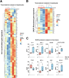
Fig. 2. Analysis of the iron accumulation and SASP gene signatures in hepatocytes and proteomic analysis of SASP in liver tissues in the CCl4-induced liver fibrosis murine model.
RNA-seq analysis was performed in hepatocytes isolated from mice treated with CCl4 or mineral oil (M. Oil) for 4 weeks (n = 3 mice/group). A Heatmap showing changes in gene expression for an iron accumulation gene signature (designed by Maus et al. [19]) in hepatocytes from CCl4-treated mice compared to mineral oil. B Heatmap showing changes in gene expression of the SASP gene signature from the Reactome collection in the same samples. C Liver tissue was collected from mice treated with CCl4 or mineral oil for 4 weeks, and a panel of 45 SASP-related factors was quantified at the protein level (n = 5 mice/group). A selection of some cytokines relevant to fibrosis is shown. Statistical analysis was done with two-tailed Mann–Whitney U-test. *p < 0.05.
Fig. 3
Fig. 3. Effects of deferiprone on iron accumulation in the model of CCl4-induced liver fibrosis.
A Schematic representation of the experimental design to induce liver fibrosis by intraperitoneal injections of CCl4 (480 mg/kg) or mineral oil (M. Oil) twice a week for 4 weeks and treatment without or with deferiprone 1 mg/mL in the drinking water. B Representative images of the livers from each group at sacrifice. C Variation in body weight along the experiment. D Iron accumulation analyzed by enhanced Perl’s Prussian Blue (EPPB) staining in paraffin-embedded liver sections. Representative areas are shown for each group. Percentage of positive stained area was quantified for each animal using ImageJ software. Each dot represents the arithmetic mean of the percentage of positive stained area from three different regions for each animal. Data are presented as mean (SD) (n = 10 mice/group). Statistical analysis was done with one-way ANOVA with Sidak’s correction. *p < 0.05; **p < 0.01; ***p < 0.001; ****p < 0.0001.
Fig. 4
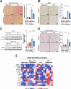
Fig. 4. Effects of deferiprone on fibrosis and senescence hallmarks in the mouse CCl4-induced liver fibrosis model.
Fibrosis was analyzed in paraffin-embedded tissue sections after 4 weeks of CCl4 treatment: A Picro-Sirius Red staining and B α-SMA immunohistochemistry as a marker of myofibroblasts. C Western blot analysis of α-SMA protein levels in the liver tissue. D Fresh liver tissues embedded in OCT were cut in sections and stained for detection of β-Galactosidase activity. For (AC), images of representative areas are shown for each group. Percentage of positive stained area was quantified for each animal using ImageJ software. Each dot represents the arithmetic mean of the percentage of positive stained area from three different regions for each animal. Results are shown as mean (SD) (n = 10 mice/group). Two-tailed Mann–Whitney U-test was used for statistical analysis. *p < 0,05; **p < 0.01; ***p < 0.001; ****p < 0.0001. E A panel of 45 cytokines and chemokines characteristic of the SASP were analyzed at the protein level in tissue samples, n = 5 mice/group. Data were analyzed carefully, and those factors with statistically significant changes (one-way ANOVA with Sidak’s post-hoc test, p-value < 0.05) are summarized in a heatmap format.
Fig. 5
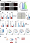
Fig. 5. Effects of deferiprone on iron accumulation, senescence and cell viability in human hepatocytes.
The human hepatocyte cell line HH4 was cultured as described in Supplementary Fig. 8A. A FerroOrange dye was used to analyze iron accumulation. Treatment for 24 h with iron (660 µM) was used as positive control. Representative images were taken with a fluorescence microscope (left) and quantification was performed by flow cytometry (right). B Gene expression of HMOX1 and HAMP as relevant genes related to iron metabolism and CDKN1A as a marker of senescence were analyzed by RT-qPCR. C β-Galactosidase staining assay was performed to analyze senescence. At least 10 images were taken from random fields by phase contrast microscopy (left) and percentage of positive cells was quantified (right). D RT-qPCR of a panel of SASP-related genes (CX3CL1, CCL22, IL6, CCL20 and LIF). E Cell viability was assessed by crystal violet and expressed as loss of viability versus the respective control (vehicle or deferiprone). F Flow cytometry with DAPI staining was used to determine the percentage of dead cells upon 6 days of treatments. G Diagram summarizing the message of the manuscript in a visual way. Briefly, iron accumulation during chronic liver damage induces senescence and SASP in hepatocytes, which would contribute to liver inflammation and fibrosis, and these hallmarks can be attenuated by deferiprone. All analyses in panels (AF) were done on day 6 post-treatment. Statistical analysis was done with one-way ANOVA with Sidak’s correction (B) or two-tailed Mann–Whitney U-test (CE) (n = 3 independent experiments). In (A, C and F), a representative experiment is shown. *p < 0.05; **p < 0.01; ***p < 0.001; ****p < 0.0001.
Fig. 6
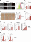
Fig. 6. Effects of deferiprone on iron accumulation, senescence and activation markers in human HSC.
The human HSC cell line LX-2 was cultured, as detailed in Supplementary Fig. 8B, with doxorubicin as a senescent inducer. A FerroOrange dye was used to analyze iron accumulation. Treatment for 24 h with iron (660 µM) was used as positive control. Representative images were taken with a fluorescence microscope (left) and quantification was performed by flow cytometry (right). B TFR1 mRNA levels were analyzed by RT-qPCR. C β-Galactosidase staining assay was performed to analyze senescence. Six images were taken from random fields by phase contrast microscopy (left) and percentage of positive cells was quantified (right). DF Analysis of mRNA levels by RT-qPCR of CDKN1A, a panel of SASP-related genes (IL1A, IL1B, CCL20 and LIF) and HSC activation-related genes (COL1A1, FN1). All analyses in panels (AF) were done on day 6 post-treatment. Statistical analysis was done with one-way ANOVA with Sidak’s correction (n = 3 independent experiments). In (A and C), a representative experiment is shown *p < 0.05; **p < 0.01; ***p < 0.001; ****p < 0.0001.
Fig. 7
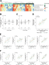
Fig. 7. Analysis of iron accumulation and SASP expression across liver fibrosis stages in human patients.
A Heatmap showing changes in gene expression for the following gene signatures: iron accumulation (designed by Maus et al. [19]), SASP (SenMayo and Reactome), collagen formation (Reactome) and ECM organization (Reactome) in the Fujiwara et al. cohort of liver fibrosis patients [34], classified by fibrosis stage. B Boxplot of relative enrichment (GSVA score) for iron accumulation gene signature across fibrosis stages. C Boxplot of SASP (SenMayo) gene signature relative enrichment (GSVA score) across fibrosis stages. D Pearson correlation analysis of the relative enrichment of iron accumulation gene signature with SASP (SenMayo) gene signature. E, F Pearson correlation analyses of the relative enrichment in the gene signatures related to iron accumulation (D) and relative enrichment of SASP (SenMayo) (E) with ROS pathway (Hallmark), TGF-β signaling (Hallmark), collagen formation (Reactome) and ECM organization (Reactome) gene signatures. Each dot is a sample (color indicates the fibrosis stage). Kendall’s τ was used to assess the association between the gene signatures and fibrosis stage. All analyses were adjusted for multiple testing with Bonferroni test correction.
Fig. 8
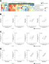
Fig. 8. Analysis of iron accumulation and SASP expression in HCC-naïve ASH and alcoholic cirrhosis patients.
A Heatmap showing changes in gene expression for the following gene signatures: iron accumulation (designed by Maus et al. [19]), SASP (SenMayo and Reactome), collagen formation (Reactome) and ECM organization (Reactome) in the Trepo et al. cohort of HCC-naïve ASH and alcoholic cirrhosis patients [35]. B Pearson correlation analysis of the relative enrichment of iron accumulation gene signature with SASP (SenMayo and Reactome) gene signatures in ASH (left) and alcoholic cirrhosis (right) patients. C, D Pearson correlation analyses of the relative enrichment in the gene signatures related to iron accumulation (C) and relative enrichment of SASP (SenMayo) (D) with collagen formation (Reactome) and ECM organization (Reactome) gene signatures in ASH (left) and alcoholic cirrhosis (right) patients. Each dot is a sample. All analyses were adjusted for multiple testing with Bonferroni test correction.

References

    1. Kisseleva T, Brenner D. Molecular and cellular mechanisms of liver fibrosis and its regression. Nat Rev Gastroenterol Hepatol. 2021;18:151–66. - PubMed
    1. Canbay A, Taimr P, Torok N, Higuchi H, Friedman S, Gores GJ. Apoptotic body engulfment by a human stellate cell line is profibrogenic. Lab Invest. 2003;83:655–63. - PubMed
    1. Hammerich L, Tacke F. Hepatic inflammatory responses in liver fibrosis. Nat Rev Gastroenterol Hepatol. 2023;20:633–46. - PubMed
    1. Tsuchida T, Friedman SL. Mechanisms of hepatic stellate cell activation. Nat Rev Gastroenterol Hepatol. 2017;14:397–411. - PubMed
    1. Kim HY, Sakane S, Eguileor A, Carvalho Gontijo Weber R, Lee W, Liu X, et al. The origin and fate of liver myofibroblasts. Cell Mol Gastroenterol Hepatol. 2024;17:93–106. - PMC - PubMed