. 2024 Sep 17;15(1):8139.
doi: 10.1038/s41467-024-51652-8.

Distinct medial amygdala oxytocin receptor neurons projections respectively control consolation or aggression in male mandarin voles

Affiliations

Distinct medial amygdala oxytocin receptor neurons projections respectively control consolation or aggression in male mandarin voles

Yishan Qu et al. Nat Commun. .

Abstract

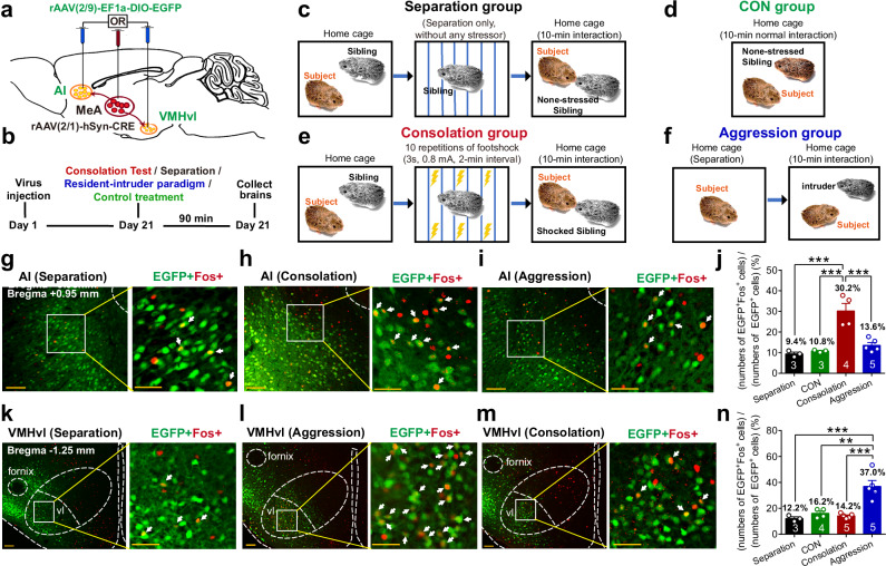
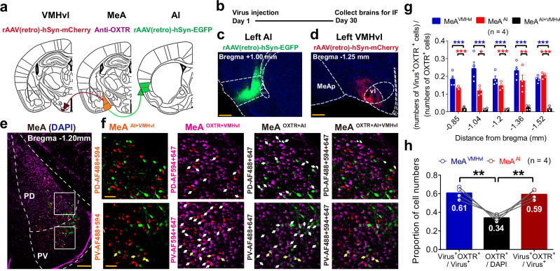
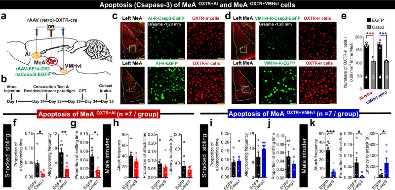
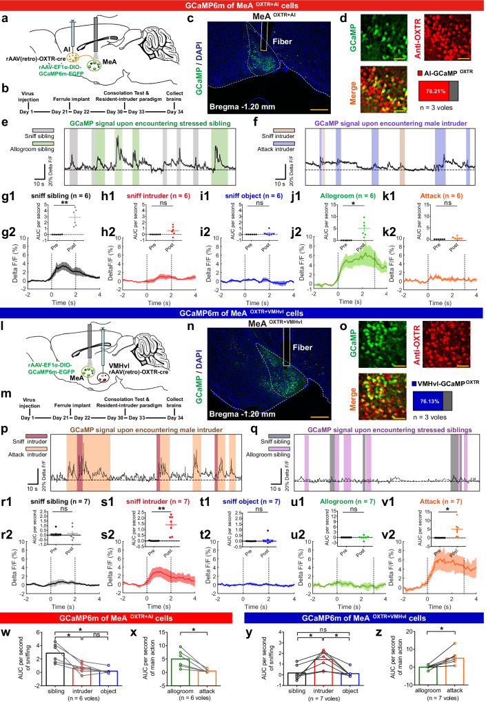
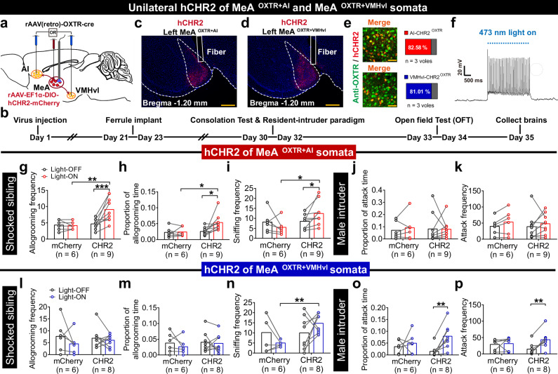
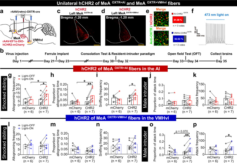
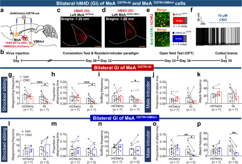
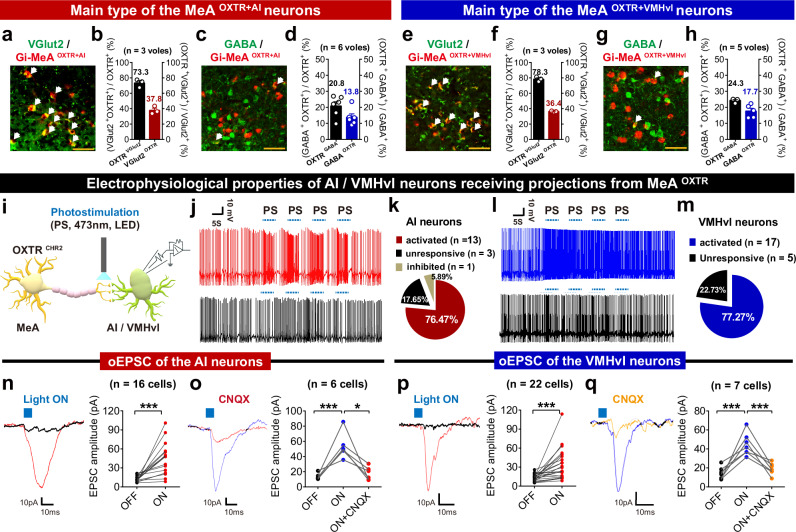
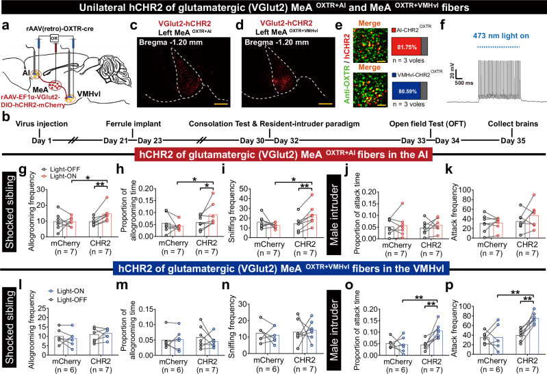
The individuals often show consolation to distressed companions or show aggression to the intruders. The circuit mechanisms underlying switching between consolation and aggression remain unclear. In the present study, using male mandarin voles, we identified that two distinct subtypes of oxytocin receptor (OXTR) neurons in the medial amygdala (MeA) projecting to the anterior insula (AI) and ventrolateral aspect of ventromedial hypothalamus (VMHvl) response differently to stressed siblings or unfamiliar intruders using c-Fos or calcium recording. Oxytocin release and activities of PVN neurons projecting to MeA increased upon consoling and attacking. OXTR antagonist injection to the MeA reduced consoling and attacking. Apoptosis, optogenetic or pharmacogenetic manipulation of these two populations of neurons altered behavioral responses to these two social stimuli respectively. Here, we show that two subtypes of OXTR neurons in the MeA projecting to the AI or VMHvl causally control consolation or aggression that may underlie switch between consolation and aggression.


Conflict of interest statement

The authors declare no competing interests.

Figures

Fig. 1
Fig. 1. The morphological distribution of the MeA neurons projecting to the AI and VMHvl.
a, b Diagram showing virus injection regimen (adapted from The Mouse Brain in Stereotaxic Coordinates by Paxinos and Franklin, a) and schedule (b). c, d Representative images of rAAV (retro)-mCherry (red) and rAAV (retro)-EGFP (green) injection sites at the AI (c) and VMHvl (d). Scale bars, 500 μm. 5 independent repetitions with similar results in (c) and (d). e, f Representative images with retro-virus labeling at both the posterior dorsal (PD) and posterior ventral (PV) subregions of MeA. The selected boxed areas were magnified (300 μm × 300 μm). The MeAAI and MeAVMHvl were characterized by white arrows. The MeAAI+VMHvl were characterized by yellow arrows. Scale bars, 200 μm in e and 50 μm in (f). g Proportions of MeAAI, MeAVMHvl, MeAAI+VMHvl at the MeA along the anteroposterior axis. * and # implies a discrepancy between the Overlap and MeAAI / MeAVMHvl, respectively. Overlap versus MeAAI: p = 0.0099, 0.0390, 0.0246, 0.0042, and 0.0010 at bregma −0.85, –1.04, −1.20, −1.36, and −1.50 mm, respectively; Overlap versus MeAVMHvl: p = 0.0021, 0.0028, 0.0031, 0.0010 and 0.0035 at bregma −0.85, −1.04, −1.20, −1.36, and −1.50 mm, respectively. h Proportions of MeAAI, MeAVMHvl, MeA AI+VMHvl at the PV and PD. At the PD subregion, MeAAI versus Overlap, p = 0.006; MeAVMHvl versus Overlap, p = 0.003. At the PV subregion, MeAAI versus Overlap, p = 0.021; MeAVMHvl versus Overlap, p = 0.006. n = 5 voles in (g) and (h). ***p < 0.001, ###p < 0.001, **p < 0.01, ##p < 0.01, *p < 0.05. Data was analyzed by repeated measure two-way (g) and one-way (h) ANOVA with Sidak’s multiple comparison test. Data are presented as the means +/− SEM. Statistical details are presented in Supplementary Data. 1 file. Source data are provided as a Source Data file.
Fig. 2
Fig. 2. The positive involvement of MeA–AI in consolation and MeA–VMHvl in aggression, respectively.
a, b Diagram showing injection protocol (adapted from The Mouse Brain in Stereotaxic Coordinates by Paxinos and Franklin, a) and schedule (b) of anterograde monosynaptic labeling virus. cf Experimental process diagrams of different treatments in Separation, CON, Consolation, and Aggression groups. gi Representative overlapped images of anterograde monosynaptic virus (EGFP, green) and c-Fos (Cy3, red) at the AI after separation treatment (Separation group, g), consolation test (Consolation group, h) and resident-intruder paradigm (Aggression group, i). km Representative overlapped images of anterograde monosynaptic virus and c-Fos at the VMHvl in Separation, Consolation, and Aggression groups. Scale bars, 100 μm in gi and km. The selected boxed areas were magnified (200 μm × 200 μm) (scale bars, 50 μm). Co-labeled neurons were characterized by white arrows. j, n Comparison of percentage of EGFP and c-Fos co-labeling neurons in whole anterograde virus-marked neurons at the AI (n = 3, 3, 4 and 5 voles) or VMHvl (n = 3, 4, 5, and 5 voles) in Separation, CON, Consolation and Aggression groups, respectively. At the AI (j), Separation versus Consolation, p = 0.0004; CON versus Consolation, p = 0.0007; Aggression versus Consolation, p = 0.0008. At the VMHvl (n), Separation versus Aggression, p = 0.0006; CON versus Aggression, p = 0.0013; Consolation versus Aggression, p = 0.0003. ***p < 0.001, **p < 0.01. Data was analyzed by one-way ANOVA with Sidak’s multiple comparison test in j and n. Data are presented as the means +/− SEM. Statistical details are presented in Supplementary Data. 1 file. Source data are provided as a Source Data file.
Fig. 3
Fig. 3. The morphological distribution of the MeAOXTR+AI and MeAOXTR+VMHvl populations.
a, b Diagram showing virus injection regimen (adapted from The Mouse Brain in Stereotaxic Coordinates by Paxinos and Franklin, a) and schedule (b). c, d Representative images of rAAV-retro-mCherry (red) and rAAV-retro-EGFP (green) injection sites at the AI (c) and VMHvl (d). Scale bars, 500 μm. 4 independent repetitions with similar results in (c) and (d). e, f Representative overlapped images of dual-retrograde rAAVs tracing and OXTR antibody (AF647, magenta) at both PD and PV subregions (e). The enlarged views of the selected boxed areas (300 μm × 300 μm). f White arrows indicate MeAAI+OXTR and MeAVMHvl+OXTR. Yellow arrows indicate merged neurons (MeAAI+OXTR+VMHvl). Scale bars, 200 μm (e) and 50 μm (f). g Proportion of different retrograde virus-positive and overlapped neurons expressing OXTR along the anteroposterior axis of the MeA. MeAAI versus MeAAI+VMHvl, p < 0.001, =0.036, <0.001, =0.004 and <0.001 at bregma −0.85, −1.04, −1.20, −1.36 and −1.50 mm, respectively; MeAVMHvl versus MeAAI+VMHvl, p < 0.001, <0.001, <0.001, <0.001, and <0.001 at bregma −0.85, −1.04, −1.20, −1.36, and −1.50 mm, respectively. h Proportion of different virus-positive cells expressing OXTR and of the anti-OXTR cells in the total MeA cells. MeAAI versus MeAOverall, p = 0.0017; MeAVMHvl versus MeAOverall, p = 0.0013; n = 4 voles in (g) and (h). ***p < 0.001, **p < 0.01, *p < 0.05. Data was analyzed by repeated measure two-way (g) and one-way (h) ANOVA with Sidak’s multiple comparison test. Data are presented as the means +/− SEM. Statistical details are presented in Supplementary Data. 1 file. Source data are provided as a Source Data file. IF  immunofluorescence experiment.
Fig. 4
Fig. 4. The effects of apoptosis of the MeAOXTR+AI and MeAOXTR+VMHvl on consolation and aggression.
a, b Diagram showing protocol (adapted from The Mouse Brain in Stereotaxic Coordinates by Paxinos and Franklin, a) and schedule (b) of virus injection for apoptosis of bilateral MeAOXTR+AI or MeAOXTR+VMHvl. c, d Representative co-labeling images of AI-retro (c)/VMHvl-retro (d) Caspase3 or EGFP (green) and anti-OXTR (OXTR-ir, Cy3, red) cells in the MeA (scale bars, 200 μm). The enlarged views of the selected boxed areas (300 μm × 300 μm) (scale bars, 50 μm). e Numbers of OXTR-ir cells in the MeA between AI-retro EGFP and Casp3 groups, and between VMHvl-retro EGFP and Caspase3 groups. AI-retro EGFP versus Casp3, p = 0.0007; VMHvl-retro EGFP versus Casp3, p = 0.0004. n = 4, 3, 4, and 3 voles in AI-retro EGFP, AI-retro Casp3, VMHvl-retro EGFP and VMHvl-retro Casp3 groups, respectively. f, g, i, j Duration proportion and frequency of allogrooming, and frequency of sniffing siblings between AI-retro EGFP and Csap3 groups (f, g), and between VMHvl-retro EGFP and Csap3 groups (i, j). h, k Frequency, duration proportion of attack, and latency to attack between the AI-retro EGFP and Csap3 groups (h), and between the VMHvl-retro EGFP and Csap3 groups (k). In fh, AI-retro EGFP versus AI-retro Casp3, allogrooming time proportion: p = 0.014; allogrooming frequency: p = 0.006; sniffing time proportion: p = 0.02; attack frequency: p = 0.196; attack time proportion: p = 0.533; latency to attack: p = 0.532. In ik, VMHvl-retro EGFP versus VMHvl-retro Casp3, allogrooming time proportion: p = 0.278; allogrooming frequency: p = 0.181; sniffing time proportion: p = 0.278; attack frequency: p < 0.001; attack time proportion: p = 0.042; latency to attack: p = 0.033. n = 7 voles/group in fk. ***p < 0.001, **p < 0.01, *p < 0.05. Data was analyzed by two-tailed unpaired t-test (e) and paired t-test (fk). Data are presented as the means +/− SEM. Statistical details are presented in Supplementary Data. 1 file. Source data are provided as a Source Data file.
Fig. 5
Fig. 5. Dynamics of GCaMP6m-fluorescence signal in the MeAOXTR+AI and MeAOXTR+VMHvl during social behaviors.
a, b, l, m Virus regimen (adapted from The Mouse Brain in Stereotaxic Coordinates by Paxinos and Franklin, a, l) and schedule (b, m). c, n Images of GCaMP6m of MeAOXTR+AI(VMHvl) and actual fiber tracts (scale bars, 500 μm). 6 and 7 independent repetitions with similar results in (c) and (n). d, o Overlapped images of GCaMP6m (EGFP, green) and OXTR (Cy3, red) in the MeAOXTR+AI(VMHvl) (scale bars, 50 μm). n = 3 voles/group. e, f, p, q Representative traces of calcium signal in the MeAOXTR+AI(VMHvl) when facing stressed siblings and male intruders. g1k1, r1v1 Changes of calcium signals in the MeAOXTR+AI(VMHvl) before and after sniffing siblings (g1, p < 0.05; r1), intruder (h1; s1, p < 0.01) and object (i1; t1), allogroom (j1, p = 0.013; u1) and attack (k1; v1, p = 0.022). g2k2, r2v2 Peri-event plot of calcium signal (Delta F/F, %) in the MeAOXTR+AI(VMHvl) aligned to onsets of social behaviors. Colored lines indicate group averages of 6-s calcium signal and shaded areas indicate S.E.M. Dotted lines define time windows for analyzing AUC per second of a single event. wz AUC per second distinctions of calcium signal traces in the MeAOXTR+AI(VMHvl). GCaMP6m of MeAOXTR+AI, sniffing: sibling versus intruder, p = 0.029; sibling versus object, p = 0.015. Allogrooming versus attack, p = 0.015. GCaMP6m of MeAOXTR+VMHvl, sniffing: sibling versus intruder, p = 0.041; intruder versus object, p = 0.014. Attack versus allogrooming, p = 0.018. n = 6 and 7 voles in (w, x) and (y, z). Data was analyzed by a two-tail paired t-test (g1k1, r1v1, x, z) and Repeated measure one-way ANOVA with Sidak’s multiple comparison test (w, y). **p < 0.01, *p < 0.05. Data are presented as the means +/− SEM. Statistical details are presented in Supplementary Data. 1 file. Source data are provided as a Source Data file. AUC area under the curve, Delta F/F (%) change in fluorescence as a function of baseline fluorescence, ns no significance.
Fig. 6
Fig. 6. Effects of optogenetic activation of MeAOXTR+AI and MeAOXTR+VMHvl somata on consolation and aggression.
a, b Virus regimen (adapted from The Mouse Brain in Stereotaxic Coordinates by Paxinos and Franklin, a) and schedule (b) for optogenetic activation of the somata. c, d Images of OXTR-hCHR2 expression (mCherry, red) in the MeAOXTR+AI and MeAOXTR+VMHvl (scale bars, 500 μm). 9 and 8 independent repetitions with similar results in (c) and (d). e Overlapped images of hCHR2 (mCherry) and anti-OXTR (AF488, green) cells in the MeAOXTR+AI and MeAOXTR+VMHvl (scale bars, 50 μm). n = 3 voles/group. f Representative trace from electrophysiological recordings showing photoactivation of the MeAOXTR+AI or MeAOXTR+VMHvl soma. gk Comparison of allogrooming and sniffing siblings, and of attacking intruders between the AI-retro CHR2 and mCherry groups. n (mCherry) = 6 voles; n (CHR2) = 9 voles. Allogrooming frequency: CHR2-Light OFF versus CHR2-Light ON, p < 0.001; mCherry-Light ON versus CHR2-Light ON, p = 0.008; Allogrooming time proportion: CHR2-Light OFF versus CHR2-Light ON, p = 0.026; mCherry-Light ON versus CHR2-Light ON, p = 0.026; sniffing frequency: CHR2-Light OFF versus CHR2-Light ON, p = 0.040; mCherry-Light ON versus CHR2-Light ON, p = 0.027. lp Comparison of allogrooming and sniffing siblings, and of attacking intruders between the VMHvl-retro CHR2 and mCherry groups. n (mCherry) = 6 voles, n (CHR2) = 8 voles. Sniffing frequency: mCherry-Light ON versus CHR2-Light ON, p < 0.001; attack time proportion: CHR2-Light OFF versus CHR2-Light ON, p = 0.002; attack frequency: CHR2-Light OFF versus CHR2-Light ON, p = 0.002. Data was analyzed by repeated measure two-way ANOVA with Sidak’s multiple comparison test (gp). ***p < 0.001, **p < 0.01, *p < 0.05. Data are presented as the means +/− SEM. Statistical details are presented in Supplementary Data. 1 file. Source data are provided as a Source Data file.
Fig. 7
Fig. 7. Effects of optogenetic activation of MeAOXTR+AI and MeAOXTR+VMHvl fibers on consolation and aggression.
a, b Virus regimen (adapted from The Mouse Brain in Stereotaxic Coordinates by Paxinos and Franklin, a) and schedule (b) for optogenetic activation of the fibers. c, d Images of OXTR-hCHR2 expression (mCherry, red) in MeAOXTR+AI and MeAOXTR+VMHvl (scale bars, 500 μm). 7 independent repetitions with similar results in (c) and (d). e Overlapped images of hCHR2 (mCherry) and anti-OXTR (AF488, green) cells in the MeAOXTR+AI and MeAOXTR+VMHvl (scale bars, 50 μm). n = 3 voles/group. f Representative traces from electrophysiological recordings showing photoactivation of the MeAOXTR+AI or MeAOXTR+VMHvl somata. gk Comparison of allogrooming and sniffing siblings, and of attacking intruders between the AI-retro CHR2 and mCherry groups. n (mCherry) = 6 voles; n (CHR2) = 7 voles. In gk, allogrooming frequency: CHR2-Light OFF versus CHR2-Light ON, p = 0.0195; mCherry-Light ON versus CHR2-Light ON, p = 0.0158; allogrooming time proportion: CHR2-Light OFF versus CHR2-Light ON, p = 0.004; mCherry-Light ON versus CHR2-Light ON, p = 0.031; sniffing frequency: CHR2-Light OFF versus CHR2-Light ON, p = 0.0375. lp Comparison of allogrooming and sniffing siblings, and of attacking intruders between the VMHvl-retro CHR2 and mCherry groups. n (mCherry) = 6 voles, n (CHR2) = 7 voles. In lp, attack time proportion: CHR2-Light OFF versus CHR2-Light ON, p = 0.006; attack frequency: CHR2-Light OFF versus CHR2-Light ON, p = 0.0321. Data was analyzed by Repeated measure two-way ANOVA with Sidak’s multiple comparison test (gp). ***p < 0.001, **p < 0.01, *p < 0.05. Data are presented as the means +/− SEM. Statistical details are presented in Supplementary Data. 1 file. Source data are provided as a Source Data file.
Fig. 8
Fig. 8. Effects of pharmacogenetic inhibition of the MeAOXTR+AI and MeAOXTR+VMHvl on consolation and aggression.
a, b Virus regimen (adapted from The Mouse Brain in Stereotaxic Coordinates by Paxinos and Franklin, a) and schedule (b) for pharmacogenetic inhibition of the MeAOXTR+AI and MeAOXTR+VMHvl. c, d Images of OXTR-hM4D(Gi)-mCherry (red) expression in the MeAOXTR+AI (c) and MeAOXTR+VMHvl (d). Scale bars, 500 μm. 7 independent repetitions with similar results in (c) and (d). e Overlapped images of OXTR-Gi-mCherry and anti-OXTR (AF488, green) in the MeAOXTR+AI and MeAOXTR+VMHvl (scale bars, 50 μm). n = 3 voles/group. f Representative traces from pharmacogenetic inhibition of the MeAOXTR+AI or MeAOXTR+VMHvl. gk, lp Comparison of allogrooming and sniffing siblings, and of attacking intruders between the Gi and mCherry groups in the MeAOXTR+AI (gk) and MeAOXTR+VMHvl (lp). n (mCherry) = 7 voles, n (Gi) = 7 voles in (gk). n (mCherry) = 7 voles, n (Gi) = 5 voles in lp. In gk, allogrooming frequency: Gi-saline versus Gi-CNO, p = 0.0243; mCherry-CNO versus Gi-CNO, p < 0.001; allogrooming time proportion: Gi-saline versus Gi-CNO, p = 0.020; mCherry-CNO versus Gi-CNO, p < 0.001; sniffing frequency: Gi-saline versus Gi-CNO, p = 0.015. In l–p, attack time proportion: Gi-saline versus Gi-CNO, p = 0.0327; mCherry-CNO versus Gi-CNO, p = 0.008; attack frequency: Gi-saline versus Gi-CNO, p = 0.003; mCherry-CNO versus Gi-CNO, p = 0.098. Data was analyzed by Repeated measure two-way ANOVA with Sidak’s multiple comparison test (gp). ***p < 0.001, **p < 0.01, *p < 0.05. Data are presented as the means +/− SEM. Statistical details are presented in Supplementary Data. 1 file. Source data are provided as a Source Data file. Veh Vehicle (saline), CNO clozapine N-oxide.
Fig. 9
Fig. 9. Electrophysiological characteristics of the AI and VMHvl neurons receiving monosynaptic projections from the MeA OXTR neurons.
a, c, e, g Overlapped images of hM4D (Gi) (mCherry, red) and VGlut2 positive (AF488, green) neurons, and Gi and GABA positive (AF488, green) neurons in the MeAOXTR+AI (a, c) and MeAOXTR+VMHvl (e, g). Scale bars, 50 μm. b, d, f, h Quantification of the percentage of VGlut2 (GABA)-expressing Gi neurons and Gi-expressing VGlut2 (GABA) neurons in the MeAOXTR+AI (b, d) and MeAOXTR+VMHvl (f, h). n = 3 voles in (b) and (f), 6 voles in (d) and 5 voles in (h). i Experimental regimen showing photostimulation (PS) site and electrophysiological recording. jm Representative activated and unresponsive spontaneous discharge rate traces under PS (j, l). Pie charts showing the percentage of activated neurons in the AI (n = 17 cells, k) and VMHvl (n = 22 cells, m). n, p EPSC traces under PS, and quantified activated effect in the AI (n = 16 cells, n) and VMHvl (n = 22 cells, p). In (n), OFF versus ON, p = 0.0002; In (p), OFF versus ON, p = 0.0007. o, q EPSC traces under PS with CNQX, and quantified inhibited effect in the AI (n = 6 cells, o) and VMHvl (n = 7 cells, q). In (o), OFF versus ON, p = 0.009; ON versus ON + CNQX, p = 0.029; In (q), OFF versus ON, p = 0.009; ON versus ON + CNQX, p = 0.006. Data was analyzed by two-tail paired t-test (n, p) and Repeated measure one-way ANOVA with Sidak’s multiple comparison test (o, q). ***p < 0.001, *p < 0.05. Data are presented as the means +/− SEM. Statistical details are presented in Supplementary Data. 1 file. Source data are provided as a Source Data file. PS photostimulation, oEPSC optically evoked excitatory postsynaptic current, CNQX 6-cyano-7-nitroquinoxaline-2,3-dione (AMPA receptor antagonist).
Fig. 10
Fig. 10. Effects of optogenetic activation of glutamatergic MeAOXTR+AI and MeAOXTR+VMHvl fibers on consolation and aggression.
a, b Virus regimen (adapted from The Mouse Brain in Stereotaxic Coordinates by Paxinos and Franklin, a) and schedule (b) for optogenetic activation of the glutamatergic (VGlut2) fibers of the MeAOXTR+AI and MeAOXTR+VMHvl. c, d Images of VGlut2-hCHR2 expression (mCherry, red) in MeAOXTR+AI and MeAOXTR+VMHvl. Scale bars, 500 μm. 7 independent repetitions with similar results in (c) and (d). e Representative overlapped images of VGlut2-hCHR2 somata (mCherry) and anti-OXTR (AF488, green) cells. Scale bars, 50 μm. n = 3 voles/group. f Representative traces from electrophysiological recordings showing photoactivation of the glutamatergic MeAOXTR+AI or MeAOXTR+VMHvl somata. gk Comparison of allogrooming and sniffing siblings, and of attacking intruders between the AI-retro VGlut2-CHR2 and VGlut2-mCherry groups. n (mCherry) = 7 voles; n (CHR2) = 7 voles. Allogrooming frequency: CHR2-Light OFF versus CHR2-Light ON, p = 0.0041; mCherry-Light ON versus CHR2-Light ON, p = 0.0406; Allogrooming time proportion: CHR2-Light OFF versus CHR2-Light ON, p = 0.038; mCherry-Light ON versus CHR2-Light ON, p = 0.032; Sniffing frequency: CHR2-Light OFF versus CHR2-Light ON, p = 0.006; mCherry-Light ON versus CHR2-Light ON, p = 0.025. lp Comparison of allogrooming and sniffing siblings, and of attacking intruders between the VMHvl-retro VGlut2-CHR2 and VGlut2-mCherry groups. n (mCherry) = 6 voles, n (CHR2) = 7 voles. Attack time proportion: CHR2-Light OFF versus CHR2-Light ON, p = 0.003; mCherry-Light ON versus CHR2-Light ON, p = 0.008; Attack frequency: CHR2-Light OFF versus CHR2-Light ON, p = 0.002; mCherry-Light ON versus CHR2-Light ON, p = 0.004. Data was analyzed by repeated measure two-way ANOVA with Sidak’s multiple comparison test (gp). ***p < 0.001, **p < 0.01, *p < 0.05. Data are presented as the means +/− SEM. Statistical details are presented in the Supplementary Data 1 file. Source data are provided as a Source Data file. VGlut2 vesicular glutamate transporter 2.

References

    1. Lischinsky, J. E. & Lin, D. Neural mechanisms of aggression across species. Nat. Neurosci.23, 1317–1328 (2020). - PubMed
    1. Wu, Y. E. & Hong, W. Neural basis of prosocial behavior. Trends Neurosci.45, 749–762 (2022). - PMC - PubMed
    1. Baron-Cohen, S. & Wheelwright, S. The empathy quotient: an investigation of adults with Asperger syndrome or high functioning autism, and normal sex differences. J. Autism Dev. Disord.34, 163–175 (2004). - PubMed
    1. Bonfils, K. A., Lysaker, P. H., Minor, K. S. & Salyers, M. P. Affective empathy in schizophrenia: a meta-analysis. Schizophrenia Res.175, 109–117 (2016). - PubMed
    1. Perroud, N., Baud, P., Mouthon, D., Courtet, P. & Malafosse, A. Impulsivity, aggression and suicidal behavior in unipolar and bipolar disorders. J. Affect. Disord.134, 112–118 (2011). - PubMed

Publication types

LinkOut - more resources