. 2024 Sep 17;8(1):206.
doi: 10.1038/s41698-024-00707-6.

Mechanistic insights into lethal hyper progressive disease induced by PD-L1 inhibitor in metastatic urothelial carcinoma

Affiliations

Mechanistic insights into lethal hyper progressive disease induced by PD-L1 inhibitor in metastatic urothelial carcinoma

Kazuki Nishimura et al. NPJ Precis Oncol. .

Abstract

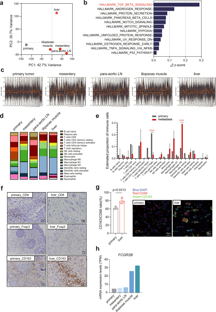
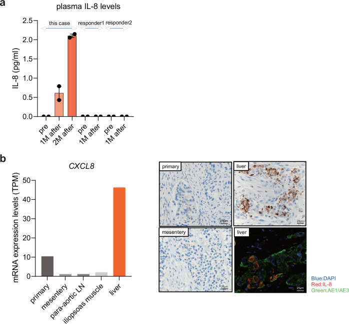
Hyper progressive disease (HPD) is a paradoxical phenomenon characterized by accelerated tumor growth following treatment with immune checkpoint inhibitors. However, the pathogenic causality and its predictor remain unknown. We herein report a fatal case of HPD in a 50-year-old man with metastatic bladder cancer. He had achieved a complete response (CR) through chemoradiation therapy followed by twelve cycles of chemotherapy, maintaining CR for 24 months. Three weeks after initiating maintenance use of a PD-L1 inhibitor, avelumab, a massive amount of metastases developed, leading to the patient's demise. Omics analysis, utilizing metastatic tissues obtained from an immediate autopsy, implied the contribution of M2 macrophages, TGF-β signaling, and interleukin-8 to HPD pathogenesis.


Conflict of interest statement

The authors declare no competing interests.

Figures

Fig. 1
Fig. 1. Clinical timeline of the patient.
a Clinical course and radiographic images of this case along with representative pathogenetic information. b Changes in levels of C-reactive protein and corrected calcium concentration in peripheral blood after avelumab treatment. Day 0 corresponds to the day of the first avelumab administration.
Fig. 2
Fig. 2. The analysis of next generation sequencing (NGS) results and pathological validation.
a Principal component analysis for whole transcriptome data in primary and metastatic samples. b Activated gene sets in metastatic tumors compared to primary tumor in hallmark gene sets. ⊿z-score represents the difference in z-score between the average of metastatic tumors and primary tumor. c Genome-wide frequency plot of copy number alterations in primary and metastatic tumors. The log2 ratio for each of the genomic clones is plotted according to chromosome positions. d Estimated proportion of immune infiltration in primary and metastatic tumors using the CIBERSORTx. e The difference of immune infiltration between primary and metastatic tumors. Gray and red bar represent estimated proportion of immune cells in primary tumor and metastatic tumor, respectively. Error bars show the 95% confidence interval. p values were calculated by two-way ANOVA. p < 0.0001****. f Representative images of CD8, FOXP3 and CD163 immunohistochemistry in primary and liver metastatic tumors. g The ratio of CD163/CD68 positive cells detected by immunohistochemistry in primary and liver metastatic tumors (left) and representative multiple immunofluorescent staining (right). Nuclei, pan-macrophages, and M2-macrophages were stained blue, red, and green, respectively, using specific antibodies (shown in Methods). P value was calculated by Welch’s test. h FCGR2B mRNA expression levels in primary and metastatic tumors were shown in transcript per million (TPM).
Fig. 3
Fig. 3. Plasma IL-8 level chronologically increased in the HPD case.
a ELISA analysis of 19 cytokines was conducted on plasma samples from three patients treated with avelumab (including this case and two avelumab responders). The plasma samples were collected before avelumab treatment, and at one and two months after the first avelumab treatment. b Left: mRNA expression level (transcripts per million: TPM) analysis of CXCL8 (IL-8). Right: Representative IL-8 immunohistochemistry and multiple immunofluorescent staining of primary and metastatic (mesentery and liver) tumors. Nuclei, tumor cells, and IL-8 were stained blue, green, and red, respectively, using specific antibodies (shown in Methods).

References

    1. Bray, F. et al. Global cancer statistics 2018: GLOBOCAN estimates of incidence and mortality worldwide for 36 cancers in 185 countries. CA Cancer J. Clin.68, 394–424 (2018). - PubMed
    1. Siegel, R. L., Miller, K. D. & Jemal, A. Cancer statistics, 2019. CA Cancer J. Clin.69, 7–34 (2019). - PubMed
    1. von der Maase, H. et al. Long-term survival results of a randomized trial comparing gemcitabine plus cisplatin, with methotrexate, vinblastine, doxorubicin, plus cisplatin in patients with bladder cancer. J. Clin. Oncol.23, 4602–4608 (2005). - PubMed
    1. von der Maase, H. et al. Gemcitabine and Cisplatin versus methotrexate, vinblastine, doxorubicin, and cisplatin in advanced or metastatic bladder cancer: results of a large, randomized, multinational, multicenter, phase III study. J. Clin. Oncol.41, 3881–3890 (2023). - PubMed
    1. Bellmunt, J. et al. Pembrolizumab as second-line therapy for advanced urothelial carcinoma. N. Engl. J. Med.376, 1015–1026 (2017). - PMC - PubMed

LinkOut - more resources