. 2024 Aug 26;32(3):200864.
doi: 10.1016/j.omton.2024.200864. eCollection 2024 Sep 19.

Lemur tail kinase 3 serves as a predictor of patient outcomes and a target for the treatment of ovarian cancer

Affiliations

Lemur tail kinase 3 serves as a predictor of patient outcomes and a target for the treatment of ovarian cancer

Ghassan M Saed et al. Mol Ther Oncol. .

Abstract

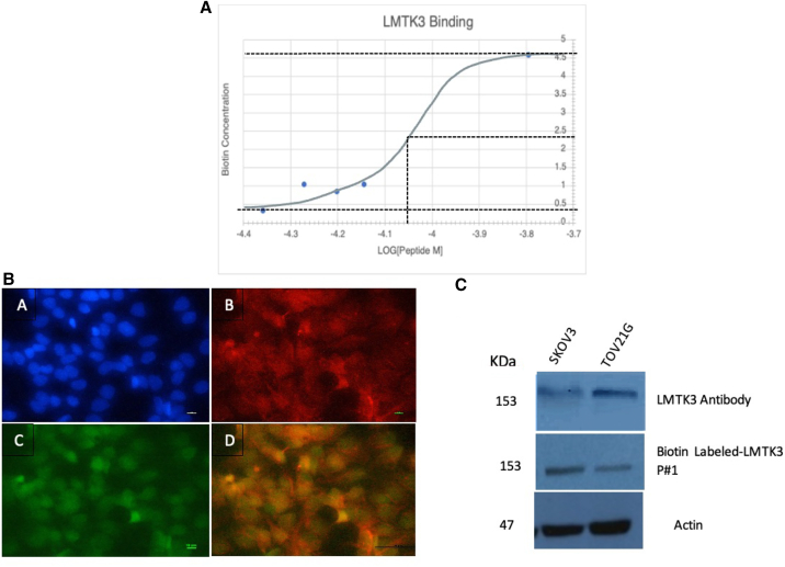
Lemur tail kinase 3 (LMTK3) belongs to a family of tyrosine kinases that are known to correlate with tumor grade and patient survival in some cancers. Here, we validated LMTK3 as a specific target and a prognostic biomarker in ovarian cancer (OC). In samples from 204 stage I-II OC patients, immunohistochemical studies revealed a higher cytoplasmic-to-nuclear staining intensity of LMTK3, which correlated with worse overall survival (p < 0.001). Efficacy studies utilizing novel LMTK3 binding peptides (LMTK3BPs) showed that all chemosensitive and chemoresistant OC cells were killed without affecting normal cells (p < 0.005), with synergistic effects shown following cisplatin and docetaxel treatment. In an orthotopic xenograft mouse model of OC, we saw a 35% tumor reduction in response to intravenous injections of 2 mg/kg LMTK3BP given three times a week for 3 weeks. Furthermore, in vivo safety studies showed no signs of toxicity after LMTK3BP treatment, even at doses as high as 40 mg/kg. This study highlights LMTK3 as a predictor of patient clinical outcomes. More importantly, novel LMTK3BPs represent potential safe treatment options, either alone or in combination with therapies, for OC.

Keywords: binding peptides; chemoresistance; lemur tail kinase 3; ovarian cancer; targeted therapy.


Conflict of interest statement

The authors declare no competing interests.

Figures

None
Graphical abstract
Figure 1
Figure 1
LMTK3 expression as a predictor of overall survival Nuclear, cytoplasmic, and cytoplasmic:nuclear LMTK3 ratios were established through IHC staining of LMTK3 in 197 human ovarian carcinomas from various stages and histotypes. High cytoplasmic LMTK3 staining significantly correlated with poor prognosis. Significance was determined by one-way ANOVA testing (p < 0.05).
Figure 2
Figure 2
Targeting LMTK3 specifically induced killing of ovarian cancer cells (A) Cytotoxicity was determined in human normal ovarian epithelial cells as well as in chemosensitive and chemoresistant epithelial ovarian cancer cell lines treated with 5 μg/mL and 10 μg/mL of the isotype control, LMTK3 antibody, and LMTK3BP #1 and #2 by the MTT assay. (B) Synergistic effect of LMTK3 peptide and chemotherapy in ovarian cancer. Ovarian cancer cell lines and their chemoresistant counterparts were treated with a combination of LMTK3BP #1 and cisplatin or docetaxel. The MTT assay was used to determine cytotoxicity after treatment. The Compusyn software was utilized to generate Chou-Talalay plots (x axis: fractional activity [Fa], reflects the fraction of cellular viability affected by treatment relative to controls; y axis: combination index with <1, =1, and >1 indicating synergistic, additive, and antagonistic effects, respectively. Each point represents a different combination of LMTK3BP concentration tested.) Experiments were performed in triplicate.
Figure 3
Figure 3
Silencing LMTK3 expression by LMTK3-specific siRNA (A) Normal and ovarian cancer cells were treated with various doses of siRNA. Viability of cells was determined by the TACS MTT proliferation assay kit. (B) LMTK3 levels in the lysate of ovarian cancer cells. LMTK3 protein levels were measured by commercially available LMTK3 ELISA kits. LMTK3 levels were below the sensitivity range in the lysate of normal epithelial cell lines (normal epithelial ovarian cells and HOSEpiC); thus, they are not depicted in the graph. (C) LMTK3BPs induced apoptosis in ovarian cancer cells. Caspase-3 activity was determined by the Caspase-3 Colorimetric Activity Assay Kit in ovarian cancer cell lines treated with 10 μg/mL of LMTK3BP #1 (P1) and #2 (P2) for 24 h.
Figure 4
Figure 4
Binding affinity of LMTK3BP (A) A colorimetric biotin assay was performed with increasing concentrations (3.6, 4.5, 5.4, 6.2, and 18 μg) of N-terminal biotin-labeled LMTK3 peptide #1 and 50 μg of protein isolated from A2780 cells. Using Microsoft Excel, a plot of the relationship between the increasing concentration of the labeled peptide with a fixed amount of protein was constructed, and the Kd was extrapolated from the tangents. (B) Binding of LMTK3 peptide to its target in situ. Cells grown in 8-well chamber slides were fixed and stained with DAPI (A), LMTK3 antibody (Invitrogen 1:200) (B), and LMTK3 biotin-labeled peptide 1 (100 μg/mL) (C); (A) and (B) were merged to show LMTK3 cytoplasmic localization (D). Orange/yellow color indicates the co-localization of the LMTK3 antibody and peptide. Images were taken at 40× magnification. (C) LMTK3 antibody and LMTK3BPs bind the same target. Western blot membranes were hybridized with LMTK3 biotin-labeled peptide #1 and LMTK3 antibody, followed by detection with the avidin-biotin complex.
Figure 5
Figure 5
In vivo safety and efficacy (A) The total body weight of mice was determined over 20 days during 2, 10, and 40 mg/kg LMTKBP1 treatment 3 times/week for 3 weeks. (B) Liver and spleen weight of mice treated with 2, 10, and 40 mg/kg LMTKBP1 3 times/week for 3 weeks were determined. (C) Histological appearance of various organs. Representative H&E staining of brain, original magnification 18×; thoracic structures (heart), 8×; lung parenchyma, 200×; esophagus, 100×; kidney, 6×; small intestine and duodenum, 200×; adrenal gland, 50×; and liver, 5×. (D) Tumor weight at sacrifice after 8 doses of vehicle, peptide #1, peptide #2, peptide #3 (negative control), or cisplatin. Bonferroni post hoc test found statistically significant differences between vehicle and peptide #1, vehicle and peptide #2, and vehicle and cisplatin (one-way ANOVA test, ∗∗p = 0.006).
Figure 6
Figure 6
Proposed mechanism of survival in epithelial ovarian cancer cells (A) Integrin αV and integrin β1 subunit levels were determined with commercially available ELISA kits on the 5 and 10 μg/mL recombinant LMTK3 protein-treated TOV112D cell line. (B) LMTK3-activated αV/β1 integrin, in turn, activates MPO, which produces the one-electron nitrosonium cation from NO. Nitrosylation of caspase-3 leads to its inactivation. NO, nitric oxide; NO+, nitrosonium cation; iNOS, inducible nitric oxide synthase; MPO, myeloperoxidase.

References

    1. Reid B.M., Permuth J.B., Sellers T.A. Epidemiology of ovarian cancer: a review. Cancer Biol. Med. 2017;14:9–32. - PMC - PubMed
    1. Allemani C., Weir H.K., Carreira H., Harewood R., Spika D., Wang X.S., Bannon F., Ahn J.V., Johnson C.J., Bonaventure A., et al. Global Surveillance of Cancer Survival 1995-2009: Analysis of Individual Data for 25,676,887 Patients from 279 Population-Based Registries in 67 Countries (CONCORD-2) Lancet. 2015;385:977–1010. - PMC - PubMed
    1. Cortez A.J., Tudrej P., Kujawa K.A., Lisowska K.M. Advances in ovarian cancer therapy. Cancer Chemother. Pharmacol. 2018;81:17–38. - PMC - PubMed
    1. Anbarasu K., Jayanthi S. Identification of curcumin derivatives as human LMTK3 inhibitors for breast cancer: a docking, dynamics, and MM/PBSA approach. 3 Biotech. 2018;8:228. - PMC - PubMed
    1. Jiang T., Lu X., Yang F., Wang M., Yang H., Xing N. LMTK3 promotes tumorigenesis in bladder cancer via the ERK/MAPK pathway. FEBS Open Bio. 2020;10:2107–2121. - PMC - PubMed

LinkOut - more resources