. 2024 Sep 4:42:299-315.
doi: 10.1016/j.bioactmat.2024.08.045. eCollection 2024 Dec.

Low-molecular-weight estrogenic phytoprotein suppresses osteoporosis development through positive modulation of skeletal estrogen receptors

Affiliations

Low-molecular-weight estrogenic phytoprotein suppresses osteoporosis development through positive modulation of skeletal estrogen receptors

John Akrofi Kubi et al. Bioact Mater. .

Abstract

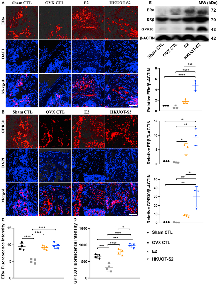
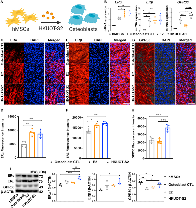
Age-related osteoporosis is a metabolic skeletal disorder caused by estrogen deficiency in postmenopausal women. Prolonged use of anti-osteoporotic drugs such as bisphosphonates and FDA-approved anti-resorptive selective estrogen receptor modulators (SERMs) has been associated with various clinical drawbacks. We recently discovered a low-molecular-weight biocompatible and osteoanabolic phytoprotein, called HKUOT-S2 protein (32 kDa), from Dioscorea opposita Thunb that can accelerate bone defect healing. Here, we demonstrated that the HKUOT-S2 protein treatment can enhance osteoblasts-induced ossification and suppress osteoporosis development by upregulating skeletal estrogen receptors (ERs) ERα, ERβ, and GPR30 expressions in vivo. Also, HKUOT-S2 protein estrogenic activities promoted hMSCs-osteoblasts differentiation and functions by increasing osteogenic markers, ALP, and RUNX2 expressions, ALP activity, and osteoblast biomineralization in vitro. Fulvestrant treatment impaired the HKUOT-S2 protein-induced ERs expressions, osteoblasts differentiation, and functions. Finally, we demonstrated that the HKUOT-S2 protein could bind to ERs to exert osteogenic and osteoanabolic properties. Our results showed that the biocompatible HKUOT-S2 protein can exert estrogenic and osteoanabolic properties by positively modulating skeletal estrogen receptor signaling to promote ossification and suppress osteoporosis. Currently, there is no or limited data if any, on osteoanabolic SERMs. The HKUOT-S2 protein can be applied as a new osteoanabolic SERM for osteoporosis treatment.

Keywords: Estrogen receptors (ERs); HKUOT-S2 protein; Osteoblast functions; Osteoporosis; Ovariectomy (OVX).


Conflict of interest statement

Kelvin Wai Kwok Yeung is an associate editor and Wei Qiao an editorial board member for Bioactive Materials and were not involved in the editorial review or the decision to publish this article. The authors declare that they have no conflict of interest.

Figures

Image 1
HKUOT-S2 protein treatment suppressed osteoporosis via positive modulation of skeletal estrogen signaling.
Scheme 1
Scheme 1
Proposed biological mechanisms of HKUOT-S2 protein. Schematic diagram illustrating that HKUOT-S2 protein suppressed osteoporosis development through estrogen receptor signaling.
Fig. 1
Fig. 1
HKUOT-S2 protein suppressed osteoporosis development of the femur. A) Schematic diagram showing that HKUOT-S2 protein treatment prevented osteoporosis progression in the OVX mouse model. B) Representative μCT scans of the femoral bone. C) μCT analysis of BV/TV, BS/TV, Tb.th, Tb. N of the femoral bone. D) Representative images of H&E staining of bone tissues. E) Representative image of Masson Trichrome staining of the femur. F) Representative image of Calcein green fluorescence intensity of the femur. G) Analysis of Calcein green fluorescence intensity of the femur. The values were shown as mean ± SEM, n = 8 for μCT scan analysis, and n = 4 for calcein green labeling. black scale bars = 100 μm *p < 0.05, **p < 0.01, ***p < 0.001 and ****p < 0.0001.
Fig. 2
Fig. 2
HKUOT-S2 protein promoted osteoblasts differentiation and activities to prevent bone loss in OVX mice. A) HKUOT-S2 protein treatment increased osteogenic markers ALP, COL1A1, and RUNX2 expressions. B, C) Representative immunofluorescence staining images and quantified fluorescence intensities of ALP protein expression in the experimental mice. D) Quantified bone tissue ALP activity in experimental mice. E, F) Representative immunofluorescence staining images and quantified fluorescence intensities of RUNX2 protein expression in experimental mice G) Representative image of TRACP/ALP staining of the femur. H) Quantification of TRAP + cells and ALP + cells. The values were shown as mean ± SEM. n = 3 for qPCR data and TRACP/ALP staining analyses, n = 4 for immunofluorescence staining, white scale bars = 50 μm, black scale bars = 100 μm, *p < 0.05, **p < 0.01, ***p < 0.001 and ****p < 0.0001.
Fig. 3
Fig. 3
HKUOT-S2 protein upregulated estrogen receptors (ERs) in the experimental mice to prevent osteoporosis. A-D) Representative immunofluorescence staining images and quantified fluorescence intensities of estrogen receptors ERα and GPR30 in the experimental mice. E) Western blot analyses of ERα, ERβ, and GPR30 in experimental mice. The values were shown as mean ± SEM, n = 4 for immunofluorescence staining, n = 3 for Western blot, white scale bars = 50 μm, *p < 0.05, **p < 0.01, ***p < 0.001 and ****p < 0.0001.
Fig. 4
Fig. 4
HKUOT-S2 protein modulated ERs during hMSCs-osteoblasts differentiation. A) hMSCs-osteoblasts differentiation w/o HKUOT-S2 protein. B) Relative mRNA expressions of ERα, ERβ, and GPR30 during hMSCs-osteoblasts differentiation. C-H) Representative immunofluorescence staining images and quantification of ERα, ERβ, and GPR30 protein expressions in osteoblasts. I) Western blot analyses of ERα, ERβ, and GPR30 expressions relative to β-ACTIN during hMSCs-osteoblasts differentiation. n = 4 for qPCR and immunofluorescence staining, n = 3 for Western blot, white scale bars = 50 μm. The values were shown as mean ± SEM. *p < 0.05, **p < 0.01, ***p < 0.001 and ****p < 0.0001.
Fig. 5
Fig. 5
HKUOT-S2 protein promoted osteoblasts differentiation and functions. A) HKUOT-S2 protein treatment increased osteogenic markers ALP, COL1A1, and RUNX2 mRNA expressions in hMSCs-derived osteoblasts. B-E) Representative immunofluorescence staining images and quantification of ALP and RUNX2 protein expressions in osteoblasts. F) Representative Western blot images and quantification of ALP and RUNX2 protein expressions in osteoblasts. G) HKUOT-S2 protein treatment significantly enhanced osteoblastic ALP activity. H) HKUOT-S2 protein treatment significantly increased osteoblast biomineralization. n = 4, white scale bars = 50 μm, black and yellow scale bars = 100 μm. The values were shown as mean ± SEM, n = 4. *p < 0.05, **p < 0.01, ***p < 0.001 and ****p < 0.0001.
Fig. 6
Fig. 6
Fulvestrant treatment suppressed estrogen receptor expressions in osteoblasts. A) Schematic diagram showing that fulvestrant treatment suppressed estrogen receptors ERα, ERβ, and GPR30 during hMSCs-osteoblasts differentiation. B) Effects of fulvestrant treatment on ERα, ERβ, and GPR30 expressions during hMSCs-osteoblasts differentiation. (C–H) Representative immunofluorescence staining images and quantified fluorescence intensities of estrogen receptor ERα, ERβ, and GPR30 in fulvestrant treated osteoblasts. I) Western blot analysis of ERα, ERβ, and GPR30 protein expressions in fulvestrant-treated osteoblasts. The values were shown as mean ± SEM, n = 4, white scale bars = 50 μm, *p < 0.05, **p < 0.01, ***p < 0.001 and ****p < 0.0001.
Fig. 7
Fig. 7
Fulvestrant treatment suppressed HKUOT-S2 protein-induced osteoblasts differentiation. A) qPCR analyses of osteogenic gene expressions in fulvestrant treated osteoblasts. B-E) Representative immunofluorescence staining images and quantified fluorescence intensities of ALP and RUNX2 expressions in fulvestrant-treated osteoblasts. F) Representative Western blot images and quantifications of ALP and RUNX2 expressions relative to the housekeeping protein β-ACTIN during fulvestrant-treated hMSCs-osteoblasts differentiation. n = 4 for qPCR and immunofluorescence staining, n = 3 for Western blot, white scale bars = 50 μm. The values were shown as mean ± SEM. *p < 0.05, **p < 0.01, ***p < 0.001 and ****p < 0.0001.
Fig. 8
Fig. 8
HKUOT-S2 protein interacts with estrogen receptors (ERs. A) Immunoprecipitation (IP) showing the interaction between HKUOT-S2 protein and ERα, ERβ, and GPR30 antibodies. B) Western blot and silver staining analyses of HKUOT-S2 protein in SDS-PAGE. C) Western blot and silver staining analyses of the IP elute proteins separated in SDS-PAGE. ERα U= IP elute with HKUOT-S2 protein unbound to ERα antibody, ERα = IP elute with HKUOT-S2 protein bound to ERα, ERβ U= IP elute with HKUOT-S2 protein unbound to ERβ antibody, ERβ = IP elute with HKUOT-S2 protein bound to ERβ, GPR30 U= IP elute with HKUOT-S2 protein unbound to GPR30 antibody, GPR30= IP elute with HKUOT-S2 protein bound to GPR30, IgG U= IP elute with HKUOT-S2 protein unbound to IgG U antibody, IgG = IP elute with HKUOT-S2 protein bound to IgG.

References

    1. Foger-Samwald U., Dovjak P., Azizi-Semrad U., Kerschan-Schindl K., Pietschmann P. Osteoporosis: pathophysiology and therapeutic options. EXCLI J. 2020;19:1017–1037. - PMC - PubMed
    1. Tang C.H. Osteoporosis: from molecular mechanisms to therapies 3.0. Int. J. Mol. Sci. 2021;22(23) - PMC - PubMed
    1. Ensrud K.E., Crandall C.J. Osteoporosis. Ann. Intern. Med. 2017;167(3):ITC17–ITC32. - PubMed
    1. Foessl I., Dimai H.P., Obermayer-Pietsch B. Long-term and sequential treatment for osteoporosis. Nat. Rev. Endocrinol. 2023;19(9):520–533. - PubMed
    1. Akkawi I., Zmerly H. Osteoporosis: current concepts. Joints. 2018;6(2):122–127. - PMC - PubMed

LinkOut - more resources