

Enhancing long-segmental tracheal restoration: A self-repairing hydrogel loaded with chondrocytokines for sutureless anastomosis and cartilage regeneration
- PMID: 39290468
- PMCID: PMC11405917
- DOI: 10.1016/j.mtbio.2024.101208
Enhancing long-segmental tracheal restoration: A self-repairing hydrogel loaded with chondrocytokines for sutureless anastomosis and cartilage regeneration
Abstract
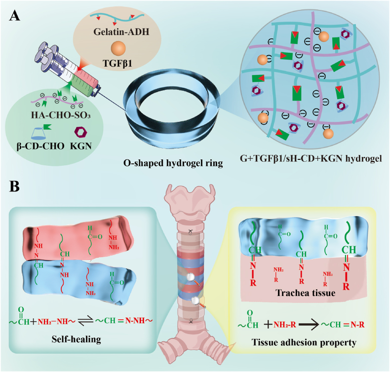
Artificial tracheal substitutes encounter significant challenges during long-segmental tracheal defects (LSTD) reconstruction, notably early postoperative anastomotic stenosis and tracheal chondromalacia. Mitigating early anastomotic stenosis by creating a compliant sutureless substitute is pivotal. Enhancing its chondrogenic capacity is equally critical for sustained healthy tracheal cartilage regeneration. This study proposes a self-healing hydrogel for sutureless tracheal anastomosis to mitigate anastomotic stenosis, enriched with kartogenin (KGN) and transforming growth factor-β1 (TGFβ1) to bolster chondrogenic properties. Initially, two precursor solutions were prepared: 1) aldehyde-modified hyaluronic acid with sulfonation and β-cyclodextrin-CHO loaded with KGN; 2) hydrazide-grafted gelatin loaded with TGFβ1. Coextrusion of these solutions resulted in a gelated G + TGFβ1/sH-CD + KGN hydrogel, characterized by a robust covalent bonding network of acylhydrazones between hydrazide and aldehyde groups, imparting excellent self-healing properties. The G + TGFβ1/sH-CD + KGN hydrogels, showcasing favorable cytocompatibility, excellent injectability, and rapid gelation, were loaded with bone marrow stem cells. These were customized into O-shaped rings and assembled into a malleable tracheal substitute using our established ring-to-tube method. This resultant compliant substitute facilitated sutureless anastomosis of LSTD in a rabbit model, attributed to the Schiff base reaction between the hydrogel's carbonyl group and the tissue's amino group. Notably, the tracheal substitute reduced early postoperative anastomotic stenosis, maintained tracheal patency, alleviated sputum blockage, promoted reepithelization, and increased the survival rate of the experimental rabbits. The sustained release of chondrocytokines resulted in excellent tracheal cartilage regeneration. Employing chondrocytokines-loaded hydrogels with self-healing properties represents a significant advancement in sutureless tracheal anastomosis and tracheal cartilage regeneration, holding promising potential in inhibiting early postoperative anastomotic stenosis and tracheal chondromalacia when treating LSTD.
Keywords: Dual-drug encapsulation; Hydrogel; Self-healing; Suture-free; Tracheal reconstruction.
© 2024 The Authors.
Conflict of interest statement
The authors declare no conflict of interest.
Figures


Similar articles
-
Injectable and self-healing sulfated hyaluronic acid/gelatin hydrogel as dual drug delivery system for circumferential tracheal repair.Int J Biol Macromol. 2024 Nov;279(Pt 2):134978. doi: 10.1016/j.ijbiomac.2024.134978. Epub 2024 Aug 23. Int J Biol Macromol. 2024. PMID: 39182860
-
Effect of kartogenin-loaded gelatin methacryloyl hydrogel scaffold with bone marrow stimulation for enthesis healing in rotator cuff repair.J Shoulder Elbow Surg. 2021 Mar;30(3):544-553. doi: 10.1016/j.jse.2020.06.013. Epub 2020 Jul 7. J Shoulder Elbow Surg. 2021. PMID: 32650072
-
Temporal control in shell-core structured nanofilm for tracheal cartilage regeneration: synergistic optimization of anti-inflammation and chondrogenesis.Regen Biomater. 2024 Apr 11;11:rbae040. doi: 10.1093/rb/rbae040. eCollection 2024. Regen Biomater. 2024. PMID: 38769993 Free PMC article.
-
Exosomes loaded with chondrogenic stimuli agents combined with 3D bioprinting hydrogel in the treatment of osteoarthritis and cartilage degeneration.Biomed Pharmacother. 2023 Dec;168:115715. doi: 10.1016/j.biopha.2023.115715. Epub 2023 Oct 17. Biomed Pharmacother. 2023. PMID: 37857246 Review.
-
Sutureless and reduced suture anastomosis of hollow vessels with fibrin glue: a review.J Invest Surg. 1999 Sep-Oct;12(5):245-62. doi: 10.1080/089419399272377. J Invest Surg. 1999. PMID: 10599001 Review.
Cited by
-
Xanthohumol bulk-modified polyurethane for tracheal repair: A 'killing two birds with one stone' strategy for tailorable mechanics and durable anti-inflammatory efficacy.Mater Today Bio. 2025 May 8;32:101831. doi: 10.1016/j.mtbio.2025.101831. eCollection 2025 Jun. Mater Today Bio. 2025. PMID: 40492152 Free PMC article.
-
Single BMSC-derived cartilage organoids for gradient heterogeneous osteochondral regeneration by leveraging native vascular microenvironment.J Nanobiotechnology. 2025 Apr 29;23(1):325. doi: 10.1186/s12951-025-03403-0. J Nanobiotechnology. 2025. PMID: 40301867 Free PMC article.
-
Enhancing the maturity of in vitro engineered cartilage from Wharton's jelly-derived photo-crosslinked hydrogel using dynamic bioreactors and its in vivo outcomes in animal models.Regen Biomater. 2025 May 8;12:rbaf037. doi: 10.1093/rb/rbaf037. eCollection 2025. Regen Biomater. 2025. PMID: 40443875 Free PMC article.
-
Dynamic Col-HZ Hydrogel with efficient delivery of bioactivator promotes ECM deposition and cartilage formation.Mater Today Bio. 2025 Feb 28;31:101623. doi: 10.1016/j.mtbio.2025.101623. eCollection 2025 Apr. Mater Today Bio. 2025. PMID: 40104649 Free PMC article.
-
A case of tracheobronchomegaly misdiagnosed as COPD: case report and literature review.BMC Pulm Med. 2025 Aug 14;25(1):392. doi: 10.1186/s12890-025-03866-9. BMC Pulm Med. 2025. PMID: 40813709 Free PMC article. Review.
References
-
- Xu Y., Li D., Yin Z., He A., Lin M., Jiang G., Song X., Hu X., Liu Y., Wang J., Wang X., Duan L., Zhou G. Tissue-engineered trachea regeneration using decellularized trachea matrix treated with laser micropore technique. Acta Biomater. 2017;58:113–121. - PubMed
-
- Fabre D., Kolb F., Fadel E., Mercier O., Mussot S., Le Chevalier T., Dartevelle P. Successful tracheal replacement in humans using autologous tissues: an 8-year experience. Ann. Thorac. Surg. 2013;96(4):1146–1155. - PubMed
-
- Xie B., He W., Xie D., Jiang G. A novel technique to increase the length of tracheal resection by adding an autologous pedicled pectoralis major myocutaneous flap transposition. Ann. Thorac. Surg. 2014;98(6):2236–2238. - PubMed
-
- Delaere P., Vranckx J., Verleden G., De Leyn P., Van Raemdonck D. Tracheal allotransplantation after withdrawal of immunosuppressive therapy. N. Engl. J. Med. 2010;362(2):138–145. - PubMed
-
- Genden E.M., Miles B.A., Harkin T.J., DeMaria S., Kaufman A.J., Mayland E., Kaul V.F., Florman S.S. Single-stage long-segment tracheal transplantation. Am. J. Transplant. 2021;21(10):3421–3427. - PubMed
LinkOut - more resources
Full Text Sources
