. 2024 Apr-Jun;28(2):e2024.00011.
doi: 10.4293/JSLS.2024.00011.

The Sling Technique for Laparoscopic Liver Mobilization

Affiliations

The Sling Technique for Laparoscopic Liver Mobilization

Andrew N de la Torre et al. JSLS. 2024 Apr-Jun.

Abstract

Background: As liver surgery continues to evolve, be it open, laparoscopic or robotic, it remains a procedure that can deteriorate in the blink of an eye. Liver surgery in patients with hepatoma is further complicated, as the vast majority have significant fibrosis, if not cirrhosis. Thus, parenchymal sparing resection is increasingly necessary. Effective and safe intracorporeal mobilization of the liver is essential for minimal access parenchymal-sparing and conventional resection.

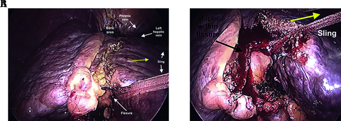
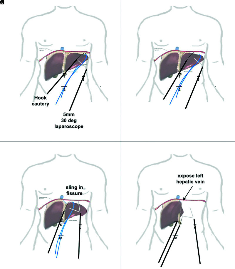
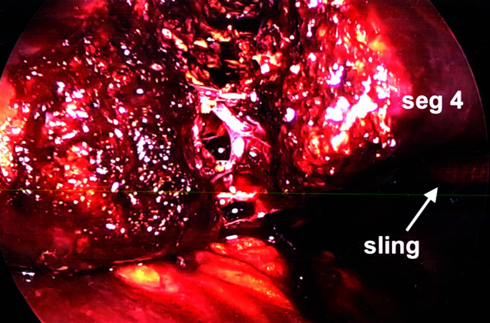
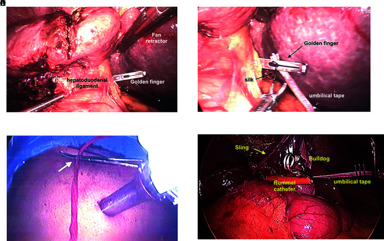
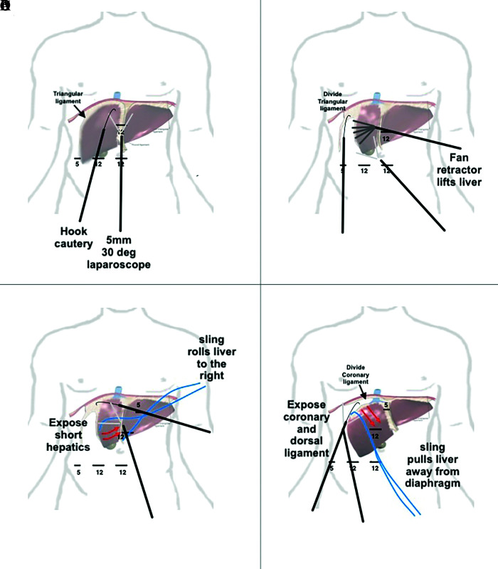
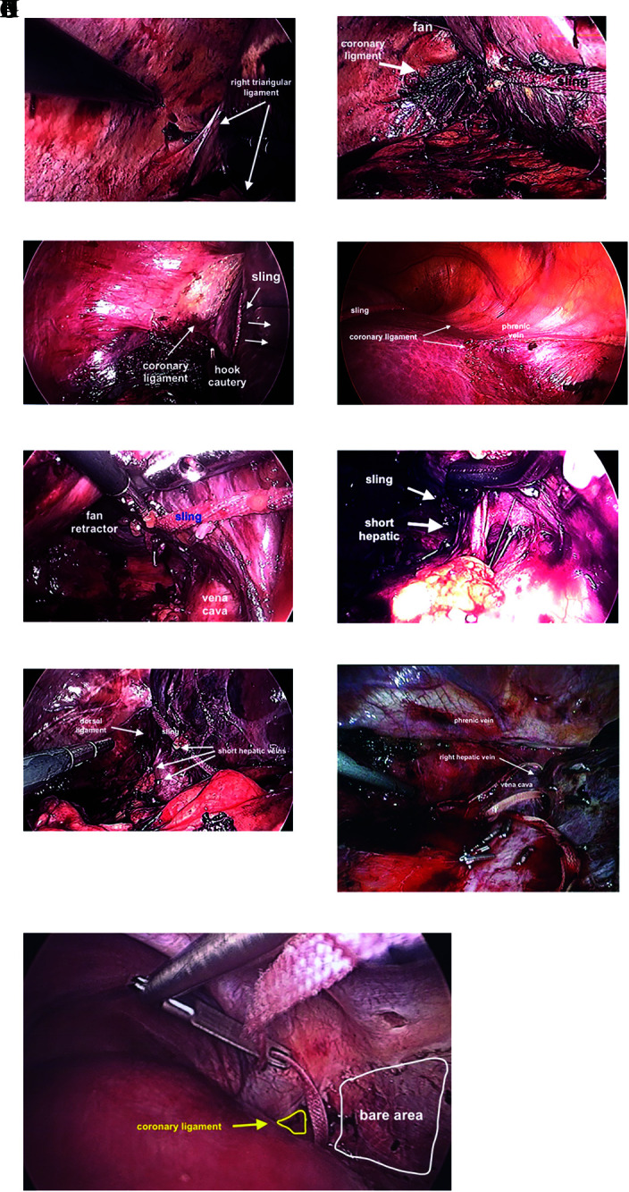
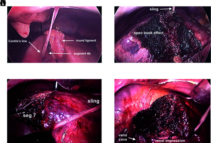
Methods: This retrospective review of over 150 cases performed provides a hands-on approach to laparoscopic hepatic mobilization with the use of an inexpensive technique using a 1" packing tape to "Sling" the liver in-order to divide the ligaments holding the liver in place and optimally position the liver for parenchymal transection.

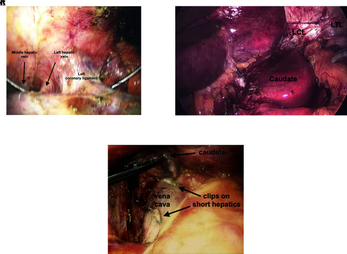
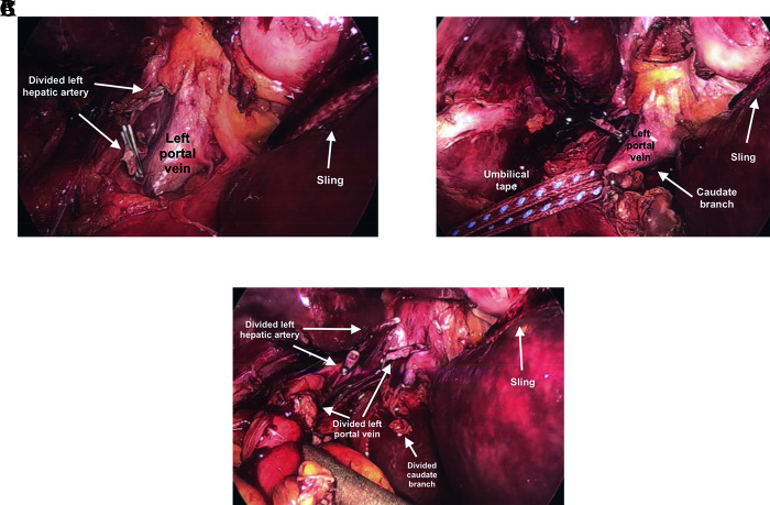
Results: Use of a 1" packing tape to "Sling" the liver intracorporeally is demonstrated to enable mobilization of the liver for tissue sparing non-anatomic, anatomic and major resections.

Conclusion: Use of a 1" packing tape to "Sling" the liver intracorporeally can facilitate mobilization for resection. Surgeons hoping to master minimal access resection should also be well versed in the use of laparoscopic ultrasound and liver transplant "Piggyback" technique.

Keywords: Intracorporial liver mobilization; Laparoscopic liver resection.


Figures

Figure 1.
Figure 1.
Use of 1” sling for left lateral segmentectomy. (A) After division of the left triangular ligament, a 1” sling is passed along the fissure for ligmentum teres, extending through to the fissure for the ligamentum venosum. (B) Using an energy device to transect 1-2 cm of liver tissue, an endoscopic stapling device can be used to divide the structures within the fissure.
Figure 2.
Figure 2.
Stepwise approach to mobilize left lobe of liver. (A) division of the right triangular ligament with a hook cautery visualized using a 30° 5-mm laparoscope. One can also use an umbilical tape or 1-inch packing tape to pull the liver away from the diaphragm. (B) extension of the dissection into the left coronary ligament. (C) exposure of the left hepatic vein. (D) liver being flipped anteriorly to right, dividing posterior coronary ligament.
Figure 3.
Figure 3.
Division of left coronary ligament. (A) Exposure of left hepatic vein adjacent to left coronary ligament with probe between middle and left hepatic vein. (B) Exposure and division of posterior left triangular (LTL) and coronary ligament (LCL). This allows full visualization of the fissure, as well as can be used to mobilize the and resect the caudate lobe. (C) Caudate lobe can be lifted off vena cava after division of short hepatic veins with laparoscopic regular and micro clips.
Figure 4.
Figure 4.
Laparoscopic hilar dissection for left lobectomy. (A) Divided left hepatic artery and exposure of left portal vein. Sling pulling left lateral segment. Traction on hilar plate allows easier dissection of left portal vein. (B) Umbilical tape applying traction to left portal vein prior to dividing. One needs to be mindful of caudate lobe branch off of left portal vein. (C) Divided left portal vein and caudate lobe branch.
Figure 5.
Figure 5.
Parenchymal transection left lobectomy with sling on gentle traction. Gentle traction using sling opens parenchymal transection plane for left lobectomy using ultrasonic irrigator and aspirator. (Misonix)
Figure 6.
Figure 6.
Laparoscopic “Pringle Maneuver.” (A) Golden finger is passed through the Foramen of Winslow. (B) 0 silk is tied to the umbilical tape and passed through the tip of the Golden finger. The umbilical tape is then pulled through the Foramen of Winslow and both ends are externalized. (C) Umbilical tape or sling through externalized through small stab and secured with a Kelly clamp to provide traction. (D) Umbilical tape is fed through a trimmed Rummel catheter, externalized through a stab wound. Sling is set up for right lobectomy (yellow arrow) and laparoscopic “Bulldog” is used to sinch down the Rummel catheter over the umbilical tape tourniqueting the hepatoduodenal ligament, causing inflow control.
Figure 7.
Figure 7.
Stepwise approach to mobilize right lobe of liver. (A) division of round, falciform ligaments, and separation of bare area. (B) lift and rotate liver to the left using 10mm fan retractor and divide right triangular and coronary ligaments. (C) slide sling over right lobe and retract liver to the left exposing right coronary ligament along vena cava. (D) slide sling over dome of liver and retract inferiorly exposing coronary ligament and right hepatic vein.
Figure 8.
Figure 8.
Mobilization of right lobe. (A) Right triangular ligament prior to dividing with a hook cautery. (B) Liver mobilized to the left facilitated with sling, fan retractor and division of coronary ligament. (C) Dome of liver retracted inferiorly opening coronary ligament aided by sling. (D) Division of anterior coronary ligament aided by retraction of liver to towards left foot aided by sling. (E) The “Batcave,” after coronary ligament divided as much as possible, the right lobe can be lifted off the vena cava aided by a fan retractor and sling allowing visualization the division of short hepatic branches. Care should be taken to prevent the sling from sliding into vena cava and tear a short hepatic vein. (F) Exposure short hepatic vein with right angle clamp. (G) Dorsal hepatic ligament prior to division. (H) Dorsal hepatic ligament after division, allowing full release of right lobe of liver. (I) Golden finger used to feed umbilical tape between right and middle hepatic veins.
Figure 9.
Figure 9.
Intracorporeal sling of liver to facilitate tissue transection. (A) Right lobe sling prior to tissue transection within Cantile’s line after gallbladder removed. (B) Right lobe sling during tissue transection, open book effect. (C) Sling over dome, under renal impression, pulled counterclockwise, lifting liver allowing gravity to assist with tissue transection of segment 7. (D) Completed resection of segment 7.

References

    1. Buell JF, Cherqui D, Geller DA, et al. ; World Consensus Conference on Laparoscopic Surgery. The international position on laparoscopic liver surgery: the Louisville Statement, 2008. Ann Surg. 2009;250(5):825–830. - PubMed
    1. Lamadé W, Glombitza G, Fischer L, et al. . The impact of 3-dimensional reconstructions on operation planning in liver surgery. Arch Surg. 2000;135(11):1256–1261. - PubMed
    1. Björnsson B, Lundgren L. A personal computer freeware as a tool for surgeons to plan liver resections. Scand J Surg. 2016;105(3):153–157. - PubMed
    1. Ielpo B, Pittau G, Ciacio O, et al. . Standardized laparoscopic right hepatic lobe mobilization. J Hepatobiliary Pancreat Sci. 2022;29(5):e30–e32. - PubMed
    1. Choi SH, Choi GH, Han DH, Kwon SW, Choi JS. Laparoscopic right hepatectomy: toward protocolization and simplification. Ann Surg Oncol. 2017;24(2):554–555. - PubMed

LinkOut - more resources