. 2024 Jul 6;9(9):2774-2785.
doi: 10.1016/j.ekir.2024.07.005. eCollection 2024 Sep.

Kidney Biopsy Findings After Lung Transplantation

Affiliations

Kidney Biopsy Findings After Lung Transplantation

David de Saint Gilles et al. Kidney Int Rep. .

Abstract

Introduction: The early diagnosis of histological kidney damage after lung transplantation (LT) is of paramount importance given the negative prognostic implications of kidney disease.

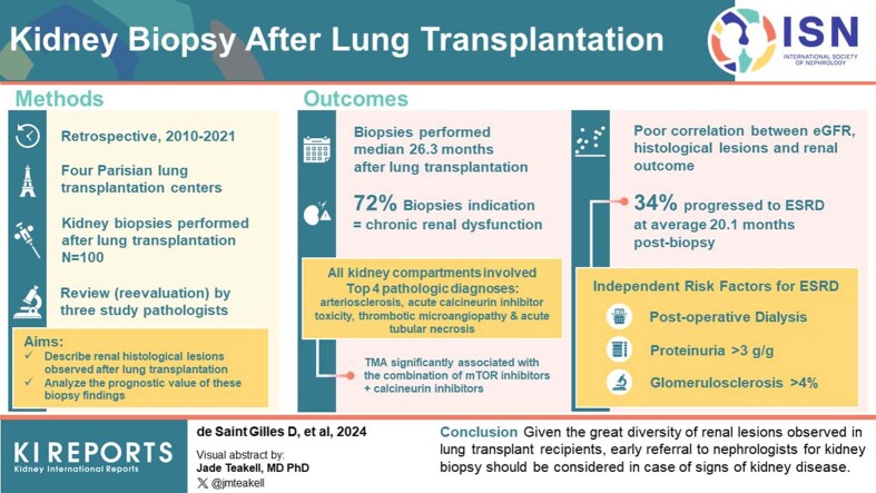
Methods: Three pathologists analyzed all kidney biopsies (KBs) (N = 100) performed from 2010 to 2021 on lung transplant patients in 4 Paris transplantation centers.

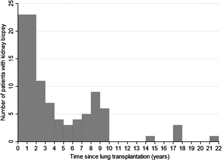
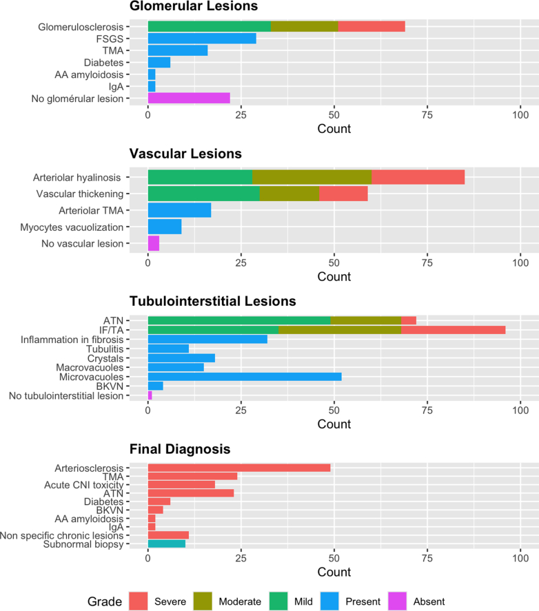
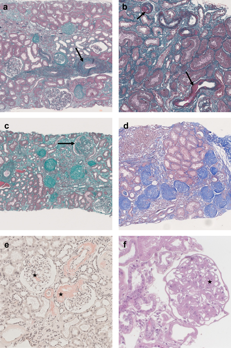
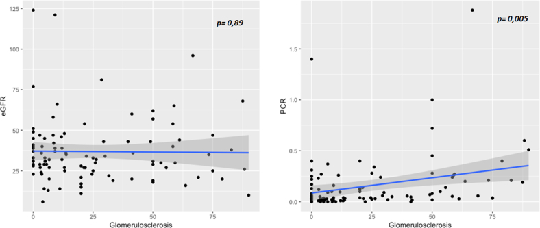
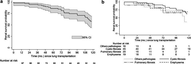
Results: The main indication for biopsy was chronic renal dysfunction (72% of patients). Biopsies were performed at a median of 26.3 months after transplantation and 15 months after a decline in estimated glomerular filtration rate (eGFR) or the onset of proteinuria. Biopsies revealed a wide spectrum of chronic lesions involving the glomerular, vascular, and tubulointerstitial compartments. The 4 most frequent final diagnoses, observed in 18% to 49% of biopsies, were arteriosclerosis, acute calcineurin inhibitor (CNI) toxicity, thrombotic microangiopathy (TMA) and acute tubular necrosis (ATN). TMA was significantly associated with a combination of mTOR inhibitors (mTORi) or CNIs with biological signs present in only 50% of patients. The eGFR was poorly correlated with most lesions, particularly percent glomerulosclerosis, and with the risk of end-stage renal disease (ESRD). Thirty-four patients progressed to ESRD at an average of 20.1 months after biopsy. Three factors were independently associated with the risk of ESRD: postoperative dialysis, proteinuria >3 g/g and percent glomerulosclerosis >4%.

Conclusion: Given the great diversity of renal lesions observed in lung transplant recipients, early referral to nephrologists for KB should be considered for these patients when they present with signs of kidney disease.

Keywords: acute kidney injury; calcineurin inhibitor toxicity; chronic kidney disease; kidney pathology; lung transplantation; thrombotic microangiopathy.


Figures

None
Graphical abstract
Figure 1
Figure 1
Delay between the lung transplantation and the kidney biopsy. Each chart represents the number of patients who had a renal biopsy over 1 year.
Figure 2
Figure 2
Histopathological findings on the kidney biopsy. See Supplementary Table S1 for the details of the scoring. AA, AA amyloidosis; ATN, acute tubular necrosis; BKVN, BK virus nephropathy; CNI, calcineurin inhibitors; FSGS, focal segmental glomerulosclerosis; IF/TA, interstitial fibrosis and tubular atrophy; TMA, thrombotic microangiopathy.
Figure 3
Figure 3
Acute kidney lesions. (a) Glomerular and arteriolar thrombotic microangiopathy with acute fibrin thrombi in the arteriolar lumen (arrow) and clear subendothelial spaces with double contours (star). Masson Trichrome, original magnification ×20. (b) Ischemic glomeruli with retracted flocculus in the Bowman space (star) and numerous tubular macrovacuolizations (arrow) in the cortex. Masson Trichrome, original magnification ×5. (c) Crystal (arrow) within the cytoplasm of an epithelial cell of a proximal tubule, HES, original magnification ×20. This crystal is refringent under polarized light. (d) None refringent crystals found in the tubular lumen of proximal tubules (arrows). Masson Trichrome, original magnification ×10. (e) Ischemic glomerulus and numerous tubular microvacuolizations in proximal tubules (arrows) suggestive of acute CNI toxicity. Masson Trichrome, original magnification ×10. (f) Immunohistochemistry with anti SV40 antibody showing several positive nuclei (arrows), in favor of a polyomavirus nephropathy. Original magnification ×40. CNI, calcineurin inhibitors.
Figure 4
Figure 4
Chronic kidney lesions. (a) Interstitial fibrosis with severe arteriosclerosis (arrow). Masson Trichrome, original magnification ×5. (b) Severe multifocal arteriolar hyalinosis (arrows). Masson Trichrome, original magnification ×10. (c) Severe chronic lesions with several globally sclerotic glomeruli and a focal and segmental glomerulosclerosis (arrow). Masson Trichrome, original magnification ×5. Estimated GFR 96 ml/min per 1.73 m2, massive proteinuria 18.8 g/g. (d) Focal cortical atrophy in the subcapsular cortex containing numerous globally sclerotic glomeruli. Masson Trichrome, original magnification ×5. (e) Glomerular and vascular red Congo positive deposits in favor with the diagnosis of amyloidosis (stars). SAA protein immunohistochemistry was positive, in favor of AA amyloidosis. Congo Red staining, original magnification ×10. (f) Nodular glomerulosclerosis (star) in a diabetic nephropathy. HES, original magnification ×20.
Figure 5
Figure 5
Correlation between estimated glomerular filtration rate (eGFR, ml/min per 1.73 m2) or PCR (protein-to-creatinine ratio, g/mmol) and percent of glomerulosclerosis (%). P-value corresponds to Pearson correlation test.
Figure 6
Figure 6
(a) Kaplan Meier estimate of renal survival after lung transplantation (months). (b) Kaplan Meier estimate of renal survival after lung transplantation by pulmonary diseases. Survivals were compared with the log-rank test (P = NS).

References

    1. Hardy J.D., Webb W.R., Dalton M.L., Walker G.R. Lung homotransplantation in man: report of the initial case. JAMA. 1963;186:1065–1074. doi: 10.1001/jama.1963.63710120001010. - DOI - PubMed
    1. Rapport Agence de la Biomédecine 2021. https://rams.agence-biomedecine.fr/greffe-cardio-pulmonaire-et-pulmonaire-0
    1. Ojo A.O., Held P.J., Port F.K., et al. Chronic renal failure after transplantation of a nonrenal organ. N Engl J Med. 2003;349:931–940. doi: 10.1056/NEJMoa021744. - DOI - PubMed
    1. Lertjitbanjong P., Thongprayoon C., Cheungpasitporn W., et al. Acute kidney injury after lung transplantation: a systematic review and meta-analysis. JCM. 2019;8:1713. doi: 10.3390/jcm8101713. - DOI - PMC - PubMed
    1. Atchade E., Barour S., Tran-Dinh A., et al. Acute kidney injury after lung transplantation: perioperative risk factors and outcome. Transplant Proc. 2020;52:967–976. doi: 10.1016/j.transproceed.2020.01.018. - DOI - PubMed

LinkOut - more resources