

Effects of platelet-rich fibrin on human endometrial stromal cells behavior in comparison to platelet-rich plasma
- PMID: 39291268
- PMCID: PMC11405248
- DOI: 10.3389/fcell.2024.1445928
Effects of platelet-rich fibrin on human endometrial stromal cells behavior in comparison to platelet-rich plasma
Abstract
Introduction: Intrauterine transfusion of platelet-rich plasma (PRP) has become a new treatment for thin endometrium (TE) in recent years, but its low efficacy due to rapid release of growth factors limits its clinical use. Platelet-rich fibrin (PRF) starts the coagulation cascade reaction immediately after the blood comes into contact with the test tube. The natural coagulation process results in stable platelet activation and the slow release of growth factors.
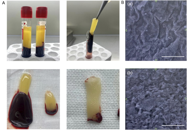
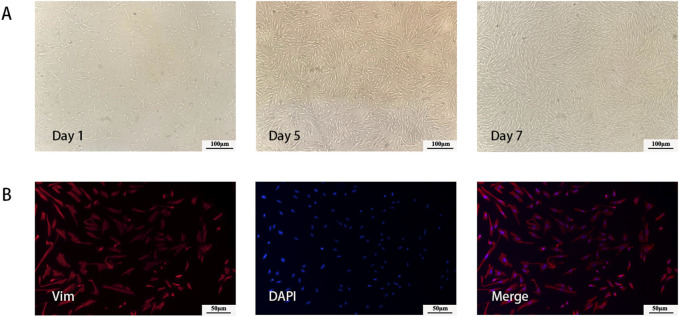
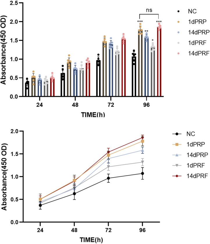
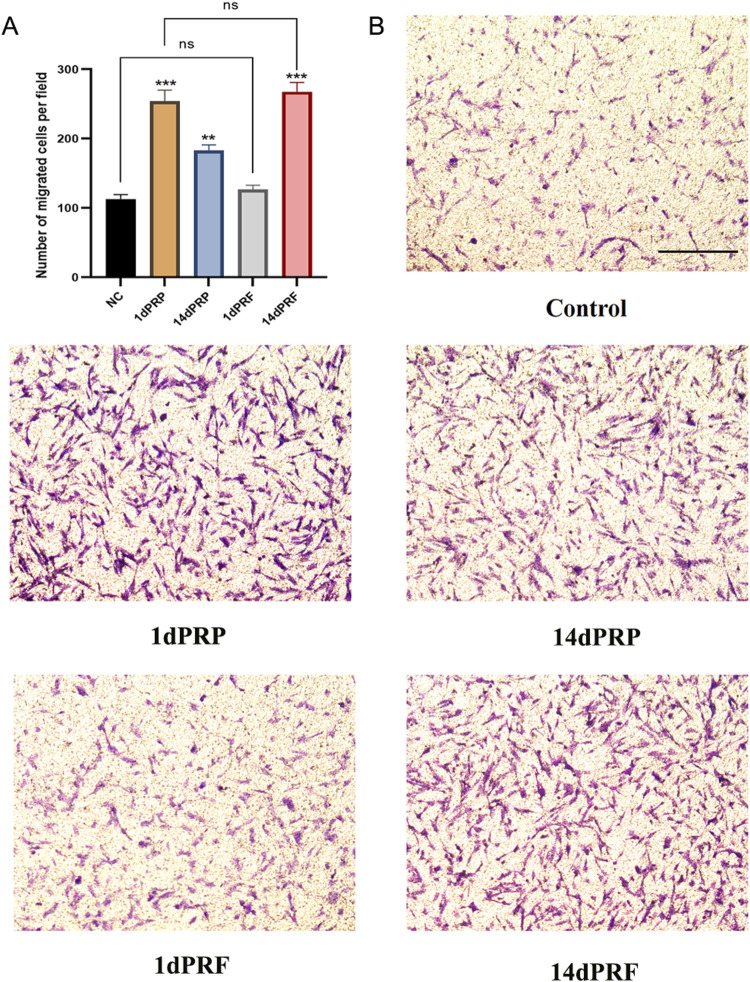
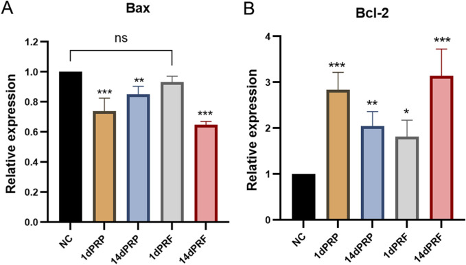
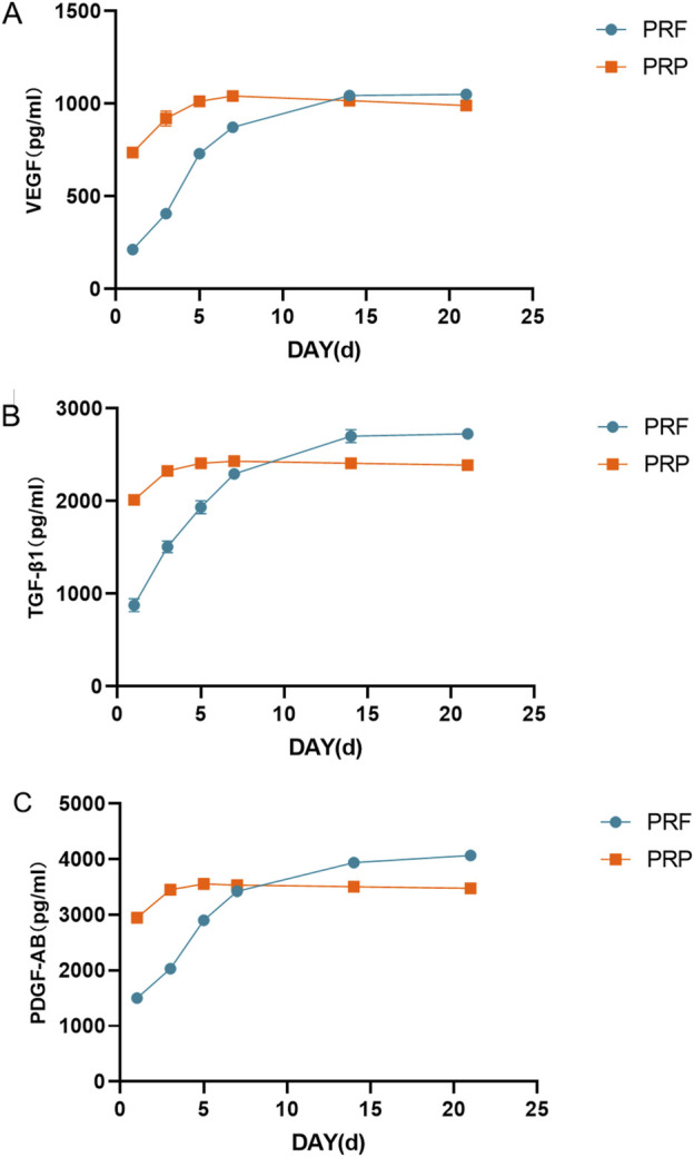
Methods: In our study, primary human endometrial stromal cells (hESCs) were extracted from endometrial tissue. PRP and PRF were prepared from the patient cubital vein blood. Stromal cells were cultured in conditioned medium supplemented with PRP and PRF. Differences in cell behavior were observed by cell proliferation test and cell migration test. The relative expression levels of apoptotic Bax and antiapoptotic Bcl-2 genes were measured by qRT-PCR. The release of growth factors from PRP and PRF was detected by ELISA.
Results: We found that both PRP and PRF inhibited apoptosis of hESCs, which favored cell proliferation and migration. In addition, PRF releases growth factors for a longer period of time compared to PRP.
Discussion: PRF offer a more sustained therapeutic effect compared to PRP, which provides a new idea for endometrial regeneration and repair.
Keywords: cell apoptotic; endometrial stromal cells; platelet-rich fibrin; platelet-rich plasma; thin endometrium.
Copyright © 2024 Yuan, Li, Du, Liu, Wang and Hao.
Conflict of interest statement
The authors declare that the research was conducted in the absence of any commercial or financial relationships that could be construed as a potential conflict of interest.
Figures
Similar articles
-
Platelet-rich concentrates differentially release growth factors and induce cell migration in vitro.Clin Orthop Relat Res. 2015 May;473(5):1635-43. doi: 10.1007/s11999-015-4192-2. Clin Orthop Relat Res. 2015. PMID: 25690170 Free PMC article.
-
Effects of an injectable platelet-rich fibrin on osteoblast behavior and bone tissue formation in comparison to platelet-rich plasma.Platelets. 2018 Jan;29(1):48-55. doi: 10.1080/09537104.2017.1293807. Epub 2017 Mar 29. Platelets. 2018. PMID: 28351189
-
Effect of Liquid Platelet-rich Fibrin and Platelet-rich Plasma on the Regenerative Potential of Dental Pulp Cells Cultured under Inflammatory Conditions: A Comparative Analysis.J Endod. 2019 Aug;45(8):1000-1008. doi: 10.1016/j.joen.2019.04.002. Epub 2019 Jun 24. J Endod. 2019. PMID: 31248700
-
Platelet-rich plasma (PRP) versus injectable platelet-rich fibrin (i-PRF): A systematic review across all fields of medicine.Periodontol 2000. 2025 Mar 24. doi: 10.1111/prd.12626. Online ahead of print. Periodontol 2000. 2025. PMID: 40125556 Review.
-
Current knowledge and perspectives for the use of platelet-rich plasma (PRP) and platelet-rich fibrin (PRF) in oral and maxillofacial surgery part 2: Bone graft, implant and reconstructive surgery.Curr Pharm Biotechnol. 2012 Jun;13(7):1231-56. doi: 10.2174/138920112800624472. Curr Pharm Biotechnol. 2012. PMID: 21740370 Review.
Cited by
-
Platelet secretions exert anti-inflammatory effects in vitro on neutrophils and uterine epithelia in cattle: a possible role in amplifying the uterine immune network toward pregnancy.Front Immunol. 2025 Mar 31;16:1560996. doi: 10.3389/fimmu.2025.1560996. eCollection 2025. Front Immunol. 2025. PMID: 40230844 Free PMC article.
References
-
- Aghajanova L., Houshdaran S., Balayan S., Manvelyan E., Irwin J. C., Huddleston H. G., et al. (2018). In vitro evidence that platelet-rich plasma stimulates cellular processes involved in endometrial regeneration. J. Assist. Reprod. Genet. 35 (5), 757–770. 10.1007/s10815-018-1130-8 - DOI - PMC - PubMed
-
- Anitua E., Prado R., Troya M., Zalduendo M., de la Fuente M., Pino A., et al. (2016). Implementation of a more physiological plasma rich in growth factor (PRGF) protocol: anticoagulant removal and reduction in activator concentration. Platelets 27 (5), 459–466. 10.3109/09537104.2016.1143921 - DOI - PubMed
LinkOut - more resources
Full Text Sources
Research Materials