. 2024 Sep 3:12:1426762.
doi: 10.3389/fcell.2024.1426762. eCollection 2024.

Regulation of TGF-β1-induced fibroblast differentiation of human periodontal ligament stem cells through the mutually antagonistic action of ectonucleotide pyrophosphatase/phosphodiesterase 1 and 2

Affiliations

Regulation of TGF-β1-induced fibroblast differentiation of human periodontal ligament stem cells through the mutually antagonistic action of ectonucleotide pyrophosphatase/phosphodiesterase 1 and 2

Onyou Ju et al. Front Cell Dev Biol. .

Abstract

Human periodontal ligament stem cells (hPDLSCs) differentiate into periodontal ligament (PDL) fibroblasts, osteoblasts, and cementoblasts. To identify inducers of PDL fibroblastic differentiation, monoclonal antibody series were developed a series of against membrane/extracellular matrix (ECM) molecules through decoy immunization. The anti-PDL13 antibody targets ectonucleotide pyrophosphatase/phosphodiesterase 1 (ENPP1), renowned for regulating skeletal and soft tissue mineralization. ENPP1 accumulates in the periodontal ligament region of tooth roots, and specifically localizes to the cell boundaries and elongated processes of the fibroblastic cells. As ENPP1 expression increases during fibroblastic differentiation, mineralization induced by tissue-nonspecific alkaline phosphatase (TNAP), a pyrophosphate-degrading enzyme, is completely inhibited. This is consistent with ENPP1 and TNAP acting in opposition, and TGF-β1-induced ENPP1 expression creates an essential environment for PDL fibroblast differentiation. Representative fibroblastic differentiation markers decrease with endogenous ENPP1 inhibition by siRNA and antibody blocking. ENPP2 generates lipid signaling molecules. In contrast to ENPP1, ENPP2 disappears in TGF-β1-induced PDL fibroblasts. Ectopic expression of ENPP2 hinders TGF-β1-induced PDL fibroblastic differentiation. Suppression of ENPP1 and ENPP2 leads to severe defects in undifferentiated and differentiated cells, demonstrating that these two factors play opposing roles in soft and hard tissue differentiation but can complement each other for cell survival. In conclusion, increased ENPP1 is crucial for TGF-β1-induced PDL differentiation, while ENPP2 and TNAP can inhibit ENPP1. ENPP1 and ENPP2 exhibit complementary functions in the cell survival.

Keywords: ENPP1; ENPP2; TGF-β1; fibroblastic differentiation; periodontal ligament stem cells (PDLSC).


Conflict of interest statement

The authors declare that the research was conducted in the absence of any commercial or financial relationships that could be construed as a potential conflict of interest.

Figures

FIGURE 1
FIGURE 1
The antigenic molecule recognized by anti-PDL13 antibody is highly increased in TGF-β1-induced PDL fibroblastic differentiation of human periodontal ligament cells (hPDLCs). (A) TGF-β1 treatment promotes fibroblastic differentiation of hPDLCs. The primary cultured hPDLCs were treated with 10 ng/mL TGF-β1 (TGF) for 9 days for induction of fibroblastic differentiation. 10 μM SB431542 was treated together with TGF-β1 (TGF/SB). The transcriptional expression of representative markers in cells were analyzed by qRT-PCR. (B) Anti-PDL13 antibody recognizes a PDL fibroblast-specific antigen. Histograms (a) and dot plots (b) in flow cytometry showed the fluorescence intensity of bound antibodies in undifferentiated hPDLCs (no) and hPDLCs treated with TGF-β1 and/or SB431542 (TGF and TGF/SB). In (b), for detection of anti-PDL13-positive cells, Fluorescein isothiocyanate (FITC)-labeled anti-Mouse IgG secondary antibody was used (FITC-PDL13). For detection of PDL fibroblasts, anti-PLAP-1/asporin antibody and Cyanine3 (Cy3)-labeled anti-Rabbit IgG secondary antibody (Cy3-PLAP-1).
FIGURE 2
FIGURE 2
The antigenic molecule recognized by anti-PDL13 antibody is ectonucleotide pyrophosphatase/phosphodiesterase 1 (ENPP1). (A) Identification of the antigenic molecule recognized by anti-PDL13 antibody. Intact PDL fibroblasts were labeled with biotin and used for immunoprecipitation with anti-PDL13 antibody. Lane 1, pre-cleared agarose bead (PCB); lane 2, anti-PDL13 immunoprecipitates (IP). (B) Cross-reactivity between anti-PDL13 antibody and a commercial anti-ENPP1 antibody in HeLa cells expressing ENPP1 cDNA (a) and in TGF-β1-induced PDL fibroblasts (b). Full length cDNA construct tagged by FLAG was expressed in HeLa cells and total cell extract (CE) detected by anti-FLAG and anti-ENPP1 antibodies. Anti-PDL13 immunoprecipitates (α-PDL13 IP) recognized by immunobinding assay (IB) with both anti-FLAG (α-FLAG IB) and anti-ENPP1 (α-ENPP1 IB) antibodies.
FIGURE 3
FIGURE 3
ENPP1 protein is upregulated in TGF-β1-induced PDL fibroblasts and accumulated in human periodontal ligament. (A) The expression level of representative proteins in hPDLCs. hPDLCs were treated with TGF-β1 (TGF) and/or SB431542 (SB) and used for preparation of total cell extract. Lane 1, undifferentiated hPDLCs; lane 2, TGF-β1-induced PDL fibroblasts; lane 3, hPDLCs treated with SB431542 for inhibition of TGF-β1 function. (B) Subcellular localization of ENPP1/PDL13 was examined by immunocytochemistry in PDL fibroblasts. ENPP1/PDL13 was detected by anti-PDL13 antibody and FITC-labeled secondary antibody, and the cell boundary was detected by Cy3-palloidin. Nucleus region in cells was stained by 4',6-diamidino-2-phenylindole (DAPI).
FIGURE 4
FIGURE 4
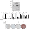
Ectopic expression of ENPP1 inhibits osteo/cementoblastic differentiation and mineralization. (A) The expression level of representative proteins in hPDLCs. hPDLCs were treated with cytokines (TGF or BMP7) and transfected with cDNA construct for ENPP1 overexpression (ENPP1-OE). Total cell extract were prepared and used for immunobinding. (B) Transcriptional expression of the representative differentiation markers. Lane 1, undifferentiated hPDLCs; lane 2, hPDLCs treated with TGF-β1; lane 3, hPDLCs treated with BMP7; lane 4, hPDLCs treated with BMP7 and ENPP1 cDNA (ENPP1-OE). (C) Mineralization was inhibited by ENPP1 overexpression (ENPP1-OE). hPDLCs introduced with ENPP1 cDNA were incubated in osteoinduction media (OM) for 21 days. Mineralization was analyzed by alizarin staining as described in Materials and methods.
FIGURE 5
FIGURE 5
Inorganic pyrophosphate inhibits osteo/cementoblastic differentiation and mineralization. (A) The expression level of representative proteins in hPDLCs. hPDLCs were treated with TGF-β1 (TGF) or BMP7 (B7), and/or treated with inorganic pyrophosphate (PPi). Total cell extract were prepared and used for immunobinding. (B) Transcriptional expression of the representative differentiation markers. Lane 1, undifferentiated hPDLCs; lane 2, undifferentiated hPDLCs with PPi; lane 3, hPDLCs treated with TGF-β1; lane 4, hPDLCs treated with TGF-β1 and PPi; lane 5, hPDLCs treated with BMP7; lane 6, hPDLCs treated with BMP7 and PPi. (C) Mineralization was inhibited by treatment with inorganic pyrophosphate (PPi). hPDLCs treated with BMP7 and/or PPi were incubated in osteoinduction media (OM) for 21 days. Mineralization was analyzed by alizarin staining as described in Materials and Methods.
FIGURE 6
FIGURE 6
Ectopic expression of ENPP2 inhibits PDL fibroblastic differentiation induced by TGF-β1 but has no effect on mineralization. (A) The expression level of representative proteins in hPDLCs. hPDLCs were treated with TGF-β1 (TGF) and/or transfected with ENPP2 cDNA construct for ENPP2 overexpression (ENPP2-OE). Total cell extract were prepared and used for immunobinding. SB431542 was used for as a control for inhibition of TGF-β1-induced PDL fibroblastic differentiation. Lane 1, undifferentiated hPDLCs; lane 2, hPDLCs treated with TGF-β1; lane 3, hPDLCs treated with TGF-β1 and ENPP2 cDNA; lane 4, hPDLCs cotreated with TGF-β1 and SB431542. (B) Transcriptional expression of the representative differentiation markers. (C) Mineralization was not affected by ENPP2 overexpression. For induction of mineralization. cells were incubated in osteoinduction media (OM) for 21 days. Mineralization was analyzed by alizarin staining as described in Materials and methods.
FIGURE 7
FIGURE 7
Depletion of ENPP1 reduced the efficiency of TGF-β1-induced PDL fibroblastic differentiation. (A) Expression amounts of ENPP1 protein in hPDLCs and PDL fibroblasts were dramatically decreased by introducing siRNA to knock out a gene expression (ENPP1-KO). Lane 1, undifferentiated hPDLCs; lane 2, undifferentiated hPDLCs with ENPP1 siRNA; lane 3, TGF-β1-induced PDL fibroblasts; lane 4, PDL fibroblasts with siRNA. (B) Transcriptional expression of the representative PDL fibroblastic and osteo/cementoblastic markers.
FIGURE 8
FIGURE 8
Antibody blocking of ENPP1 by anti-PDL13 antibody. (A) Expression of ENPP1 protein in hPDLCs and PDL fibroblasts. Increased ENPP1 by TGF-β1 (TGF) was not changed by treatment with anti-PDL13 antibody (Ab). Lane 1 and 4, no treated cells; lane 2 and 5, cells treated with 40 μg/mL of antibody; lane 3 and 6, cells treated with 100 μg/mL of antibody. Upper arrowhead in α-actin panel indicated heavy chains (HC) of anti-PDL13 antibody, which were internalized in cells. (B) Changes in elongated spinous cell morphology by antibody blocking of ENPP1. -TGF/-Ab, hPDLCs; -TGF/+Ab, hPDLCs treated with 40 μg/mL of anti-PDL13 antibody; +TGF/-Ab, TGF-β1-induced PDL fibroblasts; +TGF/+Ab, PDL fibroblasts treated with 50 μg/mL of anti-PDL13 antibody. (C) Transcriptional expression of the representative PDL fibroblastic markers. ns, not significant; *, p < 0.05; **, p < 0.01; ***, p < 0.001.
FIGURE 9
FIGURE 9
Suppression of ENPP1 and/or ENPP2 expression in undifferentiated and differentiated hPDLSCs. (A) Depletion of ENPP2 in undifferentiated hPDLCs. (a), Expression amounts of ENPP2; (b), cell viability assay; c, cell morphology. In b, cell proliferation was analyzed using CCK-8 as described in Materials and Methods. Filled and open circle indicated undifferentiated hPDLCs and depletion of ENPP2, respectively. (B) Cell viability of cells depleted of ENPP1 in the absence of ENPP2 and vice versa. (a), Expression amounts of ENPP1 and ENPP2; (b), cell viability assay. In (a), lane 1∼5 indicated undifferentiated hPDLCs, TGF-β1-induced PDL fibroblasts, PDL fibroblasts with depleted ENPP1, PDL fibroblasts treated with SB431542, and PDL fibroblasts treated with SB431542 and ENPP2 siRNA (ENPP2-KO) in that order. (C) Effect of the major enzyme products of ENPP1 and ENPP2 on cell survival.
FIGURE 10
FIGURE 10
Schematic illustration of the roles of ENPP1, ENPP2, and TNAP in differentiations and mineralization of hPDLSCs. In TGF-β1-induced PDL fibroblastic differentiation, ENPP1 expression increases but TNAP and ENPP2 completely decrease. Conversely, ENPP1 is not expressed in BMP7-mediated osteo/cementoblastic differentiation, but TNAP is increased. TNAP expression is essential for induction of mineralization. For maintaining PDL fibroblastic differentiation, ENPP2 expression is blocked as well as TNAP. ENPP1 and ENPP2 act in opposition to each other during the differentiation process of hPDLSCs. In osteo/cementoblasts and undifferentiated hPDLSCs, ENPP2 is important for cell survival. On the other hand, ENPP1 is involved in cell survival during PDL fibroblast differentiation induced by TGF-β1, in which ENPP2 is completely inhibited.

References

    1. Addison W. N., Azari F., Sorensen E. S., Kaartinen M. T., McKee M. D. (2007). Pyrophosphate inhibits mineralization of osteoblast cultures by binding to mineral, up-regulating osteopontin, and inhibiting alkaline phosphatase activity. J. Biol. Chem. 282 (21), 15872–15883. 10.1074/jbc.M701116200 - DOI - PubMed
    1. Arai K., Kasashima Y., Kobayashi A., Kuwano A., Yoshihara T. (2002). TGF-beta alters collagen XII and XIV mRNA levels in cultured equine tenocytes. Matrix Biol. 21 (3), 243–250. 10.1016/s0945-053x(02)00005-7 - DOI - PubMed
    1. Borza R., Salgado-Polo F., Moolenaar W. H., Perrakis A. (2022). Structure and function of the ecto-nucleotide pyrophosphatase/phosphodiesterase (ENPP) family: tidying up diversity. J. Biol. Chem. 298 (2), 101526. 10.1016/j.jbc.2021.101526 - DOI - PMC - PubMed
    1. Brochet X., Lefranc M. P., Giudicelli V. (2008). IMGT/V-QUEST: the highly customized and integrated system for IG and TR standardized V-J and V-D-J sequence analysis. Nucleic Acids Res. 36, W503–W508. 10.1093/nar/gkn316 - DOI - PMC - PubMed
    1. Chen S. C., Marino V., Gronthos S., Bartold P. M. (2006). Location of putative stem cells in human periodontal ligament. J. Periodontal Res. 41 (6), 547–553. 10.1111/j.1600-0765.2006.00904.x - DOI - PubMed

LinkOut - more resources