. 2024 Sep 11;19(1):20241020.
doi: 10.1515/med-2024-1020. eCollection 2024.

Paraquat disrupts the blood-brain barrier by increasing IL-6 expression and oxidative stress through the activation of PI3K/AKT signaling pathway

Affiliations

Paraquat disrupts the blood-brain barrier by increasing IL-6 expression and oxidative stress through the activation of PI3K/AKT signaling pathway

Tao Liu et al. Open Med (Wars). .

Abstract

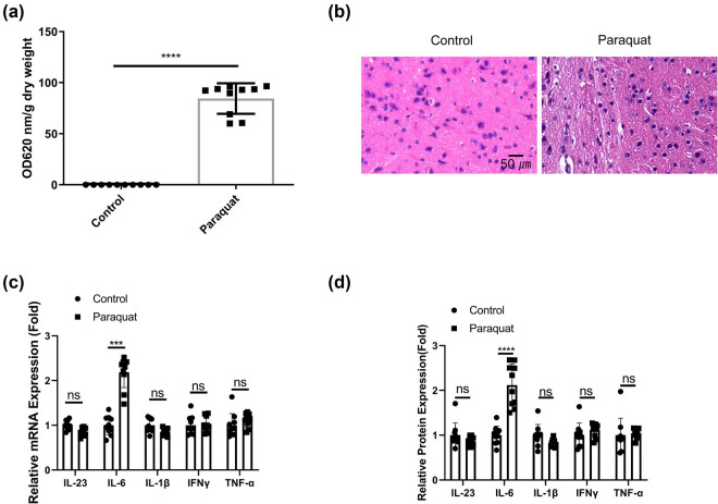
Background: Paraquat (PQ) is a frequently used herbicide with neurotoxic effects after acute or chronic exposure. Although in vitro evidence supports the PQ toxicity to dopamine cells, its in vivo effects (especially the chronic exposure) remain ambiguous. In this study, we investigated the effect of chronic PQ exposure on the blood-brain barrier (BBB) damage and the underlying mechanisms.

Methods: Adult male Sprague Dawley rats and primary human brain microvascular endothelial (PHBME) cells were exposed to PQ as the animal and cell models. Evans Blue staining and hematoxylin & eosin staining were conducted to examine the BBB and brain tissue damages. The inflammatory cytokines were quantified via enzyme linked immunosorbent assay. The changes of PI3K/AKT signaling pathway were detected by western blot.

Results: PQ exposure can cause significant pathological lesions in the brain tissues and the BBB. IL-6 and reactive oxygen species levels were found to be significantly upregulated after PQ exposure in both the animal and cell models. PQ treatment could arrest the cell proliferation and migration in PHBME cells. PQ treatment promoted the phosphorylation of PI3K and AKT, and the application of PI3K inhibitor could attenuate PQ-induced IL-6 production, oxidative stress, BBB disruption, and brain tissue damage.

Conclusion: Our study demonstrated that chronic PQ exposure could impair the BBB function and induce brain tissue damage. The overactivation of the PI3K/AKT pathway, consequent upregulation of IL-6 production, and increased oxidative stress appear to mediate the inflammatory damage resulting from PQ exposure.

Keywords: BBB; IL-6; PI3K/AKT signaling; inflammation.; paraquat.


Conflict of interest statement

Conflict of interest: The authors of this study promise to be free of any conflicts of interest.

Figures

Figure 1
Figure 1
PQ induces IL-6-associated inflammatory damages in the BBB and brain tissues. Rat were administrated with normal saline or PQ for 3 weeks. (a). Evans blue dye penetration analysis and quantification in each group. (b) H&E staining of the brain tissues. (c) qRT-PCR and (d) ELISA analysis of inflammatory cytokines associated with neuroinflammation (IL-23, IL-6, IL-1β, IFNγ, and TNF-α) in the blood samples. *p < 0.05; **p < 0.01; ***p < 0.001; ****p < 0.0001.
Figure 2
Figure 2
PQ impairs the function of PHBME cells at sub-toxic concentration. Brain endothelial vascular cells (PHBME cells) were treated with PQ for indicated duration. (a) LDH cytotoxicity assay after different doses of PQ treatment for 48 h. (b) CCK-8 cell proliferation of PHBME cells after sub-toxic PQ treatment (80 μM) for different durations. (c) Scratch assay in PHBME cells after sub-toxic PQ treatment (80 μM) for 48 h. (d) qRT-PCR and (e) ELISA analysis of inflammatory cytokines associated with neuroinflammation (IL-23, IL-6, IL-1β, IFNγ, and TNF-α) in PHBME cells after sub-toxic PQ treatment (80 μM) for 48 h. *p < 0.05; **p < 0.01; ***p < 0.001; ****p < 0.0001.
Figure 3
Figure 3
PQ activates PI3K/AKT signaling pathway. Western blot analyses of total PI3K, AKT, and STAT3, as well as the phosphorylation levels in the control group and PQ-treated group. (a) Data in PHBME cells. (b) Data in brain tissues of rat model. *p < 0.05; **p < 0.01; ***p < 0.001; ****p < 0.0001.
Figure 4
Figure 4
PI3K inhibition attenuates the effect of PQ on PHBME cells. PHBME cells were treated with sub-toxic dose of PQ in the presence or absence of PIK3 inhibitor (LY294002, 50 μM), or treated with PIK3 inhibitor alone. (a) Western blot analyses of total PI3K, AKT, and STAT3, as well as the phosphorylation levels. (b) CCK-8 cell proliferation. (c) Scratch assay. (d) qRT-PCR and (e) ELISA analysis of the IL-6 expression. (f) Detection of ROS levels. *p < 0.05; **p < 0.01; ***p < 0.001; ****p < 0.0001.
Figure 5
Figure 5
PI3K inhibition ameliorates PQ-induced inflammatory damages in the BBB and brain tissues. Rats were administrated with PQ for 3 weeks in the presence or absence of PI3K inhibitor or treated with PIK3 inhibitor alone. (a) Evans blue dye penetration analysis and quantification in each group. (b) H&E staining of the brain tissues. (c) ELISA analysis of inflammatory cytokine IL-6 in the blood samples. (d) Detection of ROS levels in the brain tissues. *p < 0.05; **p < 0.01; ***p < 0.001; ****p < 0.0001.

References

    1. Song CY, Feng MX, Li L, Wang P, Lu X, Lu YQ. Tripterygium wilfordii Hook. f. ameliorates paraquat-induced lung injury by reducing oxidative stress and ferroptosis via Nrf2/HO-1 pathway. Ecotoxicol Environ Saf. 2023 Mar;252:114575. - PubMed
    1. Zheng Q, Zhang Y, Wang X, Wang F, Zhao H. Isorhapontigenin modulates SOX9/TOLLIP expression to attenuate cell apoptosis and oxidative stress in paraquat-induced acute kidney injury. Oxid Med Cell Longev. 2022 Jun;2022:3328623. - PMC - PubMed
    1. Ren Y, Yang Z, Sun Z, Zhang W, Chen X, Nie S. Curcumin relieves paraquat‑induced lung injury through inhibiting the thioredoxin interacting protein/NLR pyrin domain containing 3‑mediated inflammatory pathway. Mol Med Rep. 2019 Dec;20(6):5032–40. - PMC - PubMed
    1. Liu Z, Huang F, Zhao S, Ma L, Shi Q, Zhou Y. Homicidal paraquat poisoning: poisoned while drinking. J Forensic Sci. 2022 May;67(3):1312–9. - PubMed
    1. See WZ, Naidu R, Tang KS. Cellular and molecular events leading to paraquat-induced apoptosis: mechanistic insights into Parkinson’s disease pathophysiology. Mol Neurobiol. 2022 Jun;59(6):3353–69. - PMC - PubMed

LinkOut - more resources