. 2024 Nov;11(42):e2401573.
doi: 10.1002/advs.202401573. Epub 2024 Sep 18.

Different Biomechanical Cell Behaviors in an Epithelium Drive Collective Epithelial Cell Extrusion

Affiliations

Different Biomechanical Cell Behaviors in an Epithelium Drive Collective Epithelial Cell Extrusion

Lakshmi Balasubramaniam et al. Adv Sci (Weinh). 2024 Nov.

Abstract

In vertebrates, many organs, such as the kidney and the mammary gland form ductal structures based on the folding of epithelial sheets. The development of these organs relies on coordinated sorting of different cell lineages in both time and space, through mechanisms that remain largely unclear. Tissues are composed of several cell types with distinct biomechanical properties, particularly at cell-cell and cell-substrate boundaries. One hypothesis is that adjacent epithelial layers work in a coordinated manner to shape the tissue. Using in vitro experiments on model epithelial cells, differential expression of atypical Protein Kinase C iota (aPKCi), a key junctional polarity protein, is shown to reinforce cell epithelialization and trigger sorting by tuning cell mechanical properties at the tissue level. In a broader perspective, it is shown that in a heterogeneous epithelial monolayer, in which cell sorting occurs, forces arising from epithelial cell growth under confinement by surrounding cells with different biomechanical properties are sufficient to promote collective cell extrusion and generate emerging 3D organization related to spheroids and buds. Overall, this research sheds light on the role of aPKCi and the biomechanical interplay between distinct epithelial cell lineages in shaping tissue organization, providing insights into the understanding of tissue and organ development.

Keywords: Proc Natl Acad Sci USA; cell sorting; collective cell extrusion; dewetting; mechanobiology.


Conflict of interest statement

The authors declare no conflict of interest.

Figures
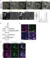
Figure 1
Figure 1
aPKCi overexpression in MDCK cells promotes the expression of epithelial marker keratin 8 and their segregation from the WT counterparts. (A) Phase contrast and fluorescent imaging of MDCKII‐WT mixed 50:50 with MDCKII‐GFP‐aPKCi (top) or with MDCKII‐GFP‐CAAX (bottom) 48 h after seeding. (Scale bar, 100 µm). Extrusions are pointed with pink arrows. On the right, the mixing index was obtained from two independent experiments for PKC and one experiment for GFP‐CAAX. (B) Expression levels of Keratin 8, endogeneous aPKCi, the exogeneous GFP‐aPKCi in MDCKII and MDCKII‐GFP‐aPKCi cells obtained from western blot. Beta‐catenin is the loading control. (C) z‐projection of confocal images of a monolayer of MDCKII‐WT cells and MDCKII‐GFP‐aPKCi cells after 5 days of coculture on glass stained for nuclei (DAPI in blue), keratin 8 (magenta) (Scale bar, 100 µm). (D) Confocal images of a monolayer of MDCKII‐WT cells mixed with MDCKII‐GFP‐aPKCi cells on glass after 3 days of coculture stained for nuclei (DAPI in blue) and actin (phalloidin in magenta). (Scale bar, 100 µm).
Figure 2
Figure 2
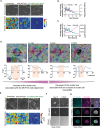
Collective delamination of highly epithelial aPKCi+ cluster is governed by cell sorting and epithelium confinement A) Evolution of cell sorting of MCF‐10A (90%) and MDCKII‐GFP aPKCi (10%) over time represented by phase contrast images (top) and velocity magnitude overlaid with velocity vectors (bottom) at early time points. Marked ROIs represent outline of the aPKCi+ cluster (Scale bar, 50 µm). B) & C) Evolution of aspect ratio B) and cluster area C) (magenta) and cluster velocity magnitude (cyan) over time for 1 example. D) (top) orientation maps overlaid on phase contrast images where the orientation maps refer to the angle of each pixel coarse grained over 10 pixels over different time points (t = 0, 15 h, 30 h and 60 h). (bottom) The angle plotted over the line drawn on the top corresponding image for each image. Orange shaded region indicates the MDCKII‐GFP‐aPKCi cluster and regions outside indicate WT cells. Scale: 100 µm. Color coding of the angles are provided as an inset E) Evolution of cell sorting of MCF‐10A (90%) and MDCKII‐GFP‐aPKCi (10%) over time represented by phase contrast images (top) and velocity magnitude overlaid with velocity vectors (bottom) at late time points. Marked ROIs represent outline of the aPKCi+ cluster (Scale bar, 50 µm). F) Evolution after 10 days of co‐culture of cell sorting of MCF‐10A (90%) and MDCKII‐GFP‐aPKCi (10%) over time represented by phase contrast images (Scale bar, 100 µm) stained for cleaved caspase 3. Delamination of the MDCKII‐GFP‐aPKCi is observed. G) Confocal images of spheroids‐like structure of MDCKII‐GFP‐aPKCi cells on glass stained for nuclei (DAPI in yellow), E‐cadherin (cyan) and actin (phalloidin in magenta). The small inset shows the MDCKII‐ GFP‐aPKCi cells (Scale bar, 100 µm).
Figure 3
Figure 3
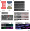
Differential traction forces and stresses govern cell sorting, cluster formation and collective extrusion of MDCKII‐GFP‐aPKCi clusters. A) Phase contrast imaging showing (top layer) cluster evolution with time, traction force magnitude in Pa overlaid with traction force vector (middle layer) and isotropic 2D stress in Pa.µm (bottom layer). B) Evolution of average traction force (Tx, Ty) and area of a single representative cluster (Scale bar, 50 µm). (C/D/G) Confocal images of a monolayer of MCF‐10A cells mixed with 10% MDCKII‐GFP‐aPKCi cells on glass stained for nuclei (DAPI in yellow), E‐cadherin (C/D cyan), and phalloidin ((D/E) magenta, (G) cyan), phospho‐MLC2 (G) and vinculin ((C) magenta) after 3, 6 and 9 days post‐mixing and vinculin ((C) magenta) after 3 and 6 days post‐mixing. Pink arrows in D) show apical actin cables. The height of apical z‐plane from the basal plane is indicated. (Scale bars, 100 µm, zoom scale bars, 25 µm). (E) Mosaic of z‐projection of confocal images of MCF‐10A WT cells mixed with 10% MDCKII‐GFP‐aPKCi cells on glass treated for 2 days with 20 µm blebbistatin or DMSO (control). (Scale bars, 100 µm). F) Quantification of the straightness of the E‐cadherin junctions. n is the number of junctions quantified. (H) Evolution of nuclear movement overlaid with velocity vectors for a cluster of MCF‐10A (90%) and MDCKII‐GFP‐aPKCi (10%) over time. Roi in yellow indicates the GFP‐aPKCi cluster (Scale bar, 50 µm). (I) Kymograph of tangential velocity averaged along the radial direction plotted along the radial direction from center of cluster outward (magenta line in H).
Figure 4
Figure 4
Collision between highly epithelial MDCKII‐GFP‐aPKCi cells with MCF‐10A cells trigger budding of MDCKII‐GFP‐aPKCi cells. A) Schematic of the collision assay. B) Time lapse phase contrast images of the collision assay. (Scale bars, 100 µm). C) Kymograph of the cells before and after collision as a function of time. D) Evolution of the collision between MCF‐10A and MDCKII‐GFP‐aPKCi cells overlaid with orientation vectors over time. Scale bar: 100 µm. E/F) Confocal images of collision between MCF‐10A cells and MDCKII‐GFP‐aPKCi cells stained for nuclei (DAPI in yellow), E‐cadherin (cyan), and phalloidin (magenta) after 5 E) and 7 F) days post‐seeding. (Scale bars, 100 µm).

Similar articles

References

    1. Andrew D. J., Ewald A. J., Developmental Biology 2010, 341, 34. - PMC - PubMed
    1. Saiz N., Grabarek J. B., Sabherwal N., Papalopulu N., Plusa B., Development 2013, 140, 4311. - PMC - PubMed
    1. Foty R. A., Steinberg M. S., Dev. Biol. 2005, 278, 255. - PubMed
    1. Amack J. D., Manning M. L., Science 2012, 338, 212. - PubMed
    1. Chan E. H., Chavadimane Shivakumar P., Clément R., Laugier E., Lenne P. F., eLife 2017, 6, e22796. - PMC - PubMed

LinkOut - more resources