Review
. 2024 Oct;13(19):e036555.
doi: 10.1161/JAHA.124.036555. Epub 2024 Sep 18.

Exercise Alleviates Cardiovascular Diseases by Improving Mitochondrial Homeostasis

Affiliations
Review

Exercise Alleviates Cardiovascular Diseases by Improving Mitochondrial Homeostasis

Huijie Zhang et al. J Am Heart Assoc. 2024 Oct.

Abstract

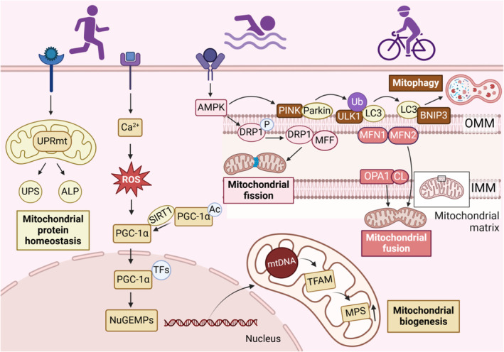
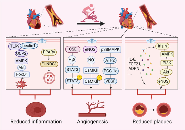
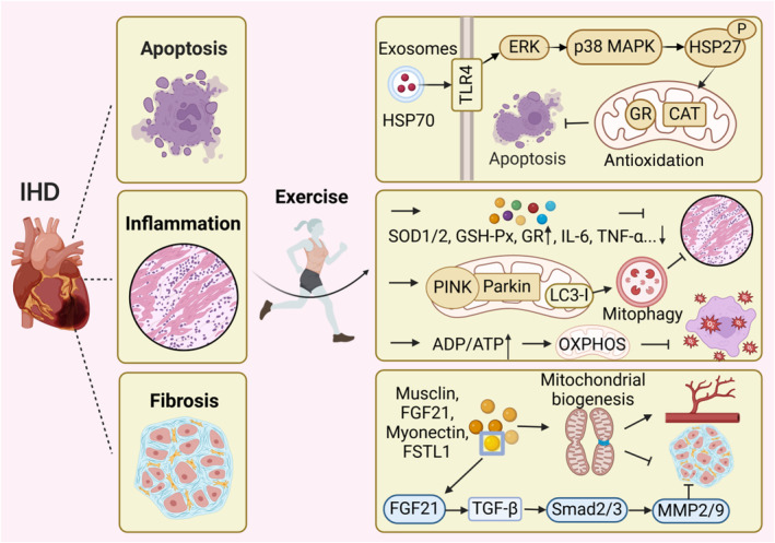
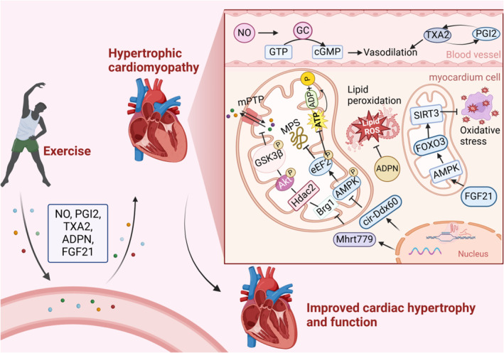
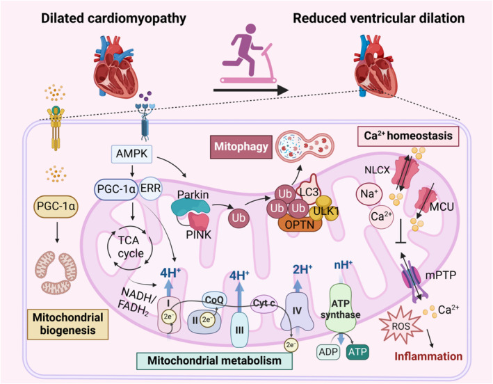
Engaging in regular exercise and physical activity contributes to delaying the onset of cardiovascular diseases (CVDs). However, the physiological mechanisms underlying the benefits of regular exercise or physical activity in CVDs remain unclear. The disruption of mitochondrial homeostasis is implicated in the pathological process of CVDs. Exercise training effectively delays the onset and progression of CVDs by significantly ameliorating the disruption of mitochondrial homeostasis. This includes improving mitochondrial biogenesis, increasing mitochondrial fusion, decreasing mitochondrial fission, promoting mitophagy, and mitigating mitochondrial morphology and function. This review provides a comprehensive overview of the benefits of physical exercise in the context of CVDs, establishing a connection between the disruption of mitochondrial homeostasis and the onset of these conditions. Through a detailed examination of the underlying molecular mechanisms within mitochondria, the study illuminates how exercise can provide innovative perspectives for future therapies for CVDs.

Keywords: cardiovascular diseases; exercise; exerkines; mitochondrial homeostasis.


Figures

Figure 1
Figure 1. Endurance exercise regulates mitochondrial quality control.
Exercise maintains mitochondrial function by promoting mitochondrial biogenesis, dynamic balance, autophagy, and protein homeostasis. Endurance exercise stimulates the PGC‐1α pathway to drive mitochondrial biogenesis. Physical exercise also activates AMPK, which promotes PINK‐dependent mitophagy and facilitates mitochondrial fission through DRP1 and MFF. The fusion of the OMM is mediated by MFN1/2, and OPA1 facilitates the fusion of the IMM, together driving mitochondrial fusion. In addition, exercise training activates the mitochondrial unfolded protein response, which regulates the ubiquitin‐proteasome system and the autophagy‐lysosomal pathway to degrade damaged or misfolded proteins and maintain mitochondrial protein homeostasis. Ac indicates acetylation; ALP, autophagy‐lysosomal pathway; AMPK, adenosine5′‐monophosphate‐activated protein kinase; BNIP3, BCL2 adenovirus E1B 19kDa interacting protein 3; DRP1, dynamin‐related protein 1; IMM, inner mitochondrial membrane; LC3, microtubule‐associated protein 1 light chain 3; MFF, mitochondrial fission factor; MFN1/2, mitochondrial fusion protein 1/2; NuGEMP, nuclear genes encoding mitochondrial protein; OMM, outer mitochondrial membrane; OPA1, optic atrophy protein 1; P, phosphorylation; PGC‐1α, peroxisome proliferator‐activated receptor‐gamma coactivator 1alpha; PINK, PTEN‐induced putative kinase; ROS, reactive oxygen species; SIRT1, sirtuin 1; TFs, transcription factors; TFAM, mitochondrial transcription factor A; Ub, ubiquitin; ULK1, unc‐51‐like autophagy‐activating kinase 1; UPRmt, mitochondrial unfolded protein response; and UPS, ubiquitin‐proteasome system.
Figure 2
Figure 2. Exercise improves mitochondrial homeostasis in atherosclerosis.
Atherosclerosis exhibits vascular inflammation, vascular damage and accumulation of plaques. Regular exercise can reduce atherosclerotic plaques and inflammatory responses, promoting the formation of endothelial vessels through the improvement of mitochondria‐related signaling pathways. Exhaustive exercise activates antioxidant pathways within the mitochondria of skeletal muscle in mice, while endurance swim training induces FUNDC1‐mediated mitophagy to reduce inflammatory responses in aged mice. Moderate‐intensity treadmill exercise and swimming sessions stimulate STAT3/CaMKII signaling pathway within the mitochondria of mice with diabetic cardiomyopathy and PGC‐1α signaling pathway within the skeletal muscle of rats, promoting angiogenesis. Moreover, physical exercise regulates the formation of atherosclerotic plaques by modulating various myokines, such as IL‐6, FGF21, and irisin. In this figure, exerkine irisin is taken as an example to illustrate its function of reducing endothelial damage and plaques in diabetic mice. ADPN indicates adiponectin; Akt, phosphorylated serine/threonine kinase; AMPK, adenosine5′‐monophosphate‐activated protein kinase; ATF2, activating transcription factor 2; CaMKII, calmodulin‐dependent protein kinase II; CSE, cystathionine‐γ‐lyase; eNOS, endothelial nitric oxide synthase; FGF21, fibroblast growth factor 21; FoxO1, forkhead box O1; FUNDC1, FUN14 domain containing 1; IL‐6, interleukin‐6; NO, nitric oxide; p38MAPK, p38 mitogen‐activated protein kinase; PGC‐1α, peroxisome proliferator‐activated receptor‐gamma coactivator 1alpha; PI3K, phosphatidylinositol‐3‐hydroxykinase; PPARγ; peroxisome proliferator‐activated receptor γ; ROS, reactive oxygen species; STAT3, signal transducer and activator of transcription 3; TLR9, toll‐like receptor 9; UCP2, uncoupling protein 2; and VEGF, vascular endothelial growth factor.
Figure 3
Figure 3. Exercise improves IHD by facilitating intercellular communication.
Following ischemia, cell apoptosis ensues, cardiac fibrosis develops, and the inflammatory response is exacerbated during reperfusion. Exercise training can reduce apoptosis, inflammation and myocardial fibrosis following IR and promote vascular regeneration by improving mitochondrial redox balance. During exercise, exerkines are transported via exosomes to specific sites where they exert antiapoptotic functions through the increased expression of mitochondrial antioxidant enzymes, such as glutathione reductase, catalase. After exercise, the increase of antioxidant enzymes and the decrease inflammatory factors can reduce inflammation. Remarkably, swimming training make an elevated expression of PINK/Parkin and LC3‐I, which can modulate mitophagy to exert a similar anti‐inflammatory effect. Endurance exercise also enhances the ADP/ATP ratio, thereby promoting mitochondrial OXPHOS and reducing mitochondrial oxidative stress. The release of exerkines such as musclin, FGF21, FSTL1, and myonectin promote mitochondrial dynamics, biogenesis, and autophagy, which collectively contribute to angiogenesis and inhibit myocardial fibrosis. In this figure, exerkine FGF21 is taken as an example to illustrate its antifibrotic effect. Notably, exerkines play a crucial role in augmenting intercellular communication and ameliorating cardiovascular diseases. CAT indicates catalase; ERK, extracellular signal‐regulated protein kinase; FGF21, fibroblast growth factor 21; FSTL1, follistatin like 1; GR, glutathione reductase; GSH‐Px, glutathione peroxidase; HSP70, heat shock protein 70; IHD, ischemic heart disease; IL‐6, interleukin‐6; IR, ischemia–reperfusion; LC3, light chain 3; MMP2/9, matrix metalloproteinase‐2/9; p38 MAPK, p38 mitogen‐activated protein kinase; OXPHOS, oxidative phosphorylation; P, phosphorylation; PINK, PTEN‐induced putative kinase; Smad2/3, phospho Thr8; SOD, superoxide dismutase; TGF‐β, transforming growth factor‐beta; TLR4, toll‐like receptor 4; and TNF‐α, tumor necrosis factor‐alpha.
Figure 4
Figure 4. Exercise improves mitochondrial function in HCM.
Exerkines are released into the bloodstream through exercise, influencing cardiac vessels and cardiomyocytes. They enhance mitochondrial function, promote vascular relaxation, and contribute to antimyocardial hypertrophy effects. Increased NO activates GC to catalyze the synthesis of GTP into cGMP, promoting vascular relaxation, and the balance between TXA2 and prostacyclin also contributes to this effect.
Figure 5
Figure 5. Exercise mitigates DCM.
In DCM, disruptions in Ca2+ regulation, mitochondrial dysfunction, and inflammation coexist, mutually reinforcing and exacerbating the condition. Exercise promotes mitochondrial biogenesis, enhances mitochondrial metabolism, supports proper mitochondrial oxidative phosphorylation, and ensures energy supply and mitochondrial function. Additionally, exercise stimulates PINK‐dependent mitophagy, reducing inflammation. It also facilitates the normal transport of mitochondrial Ca2+, maintains Ca2+ homeostasis, and helps prevent oxidative stress and inflammation caused by mPTP opening. Through effective exercise intervention, the progression of DCM is significantly inhibited, leading to reduced ventricular dilation and improved cardiac function. AMPK indicates adenosine5′‐monophosphate‐activated protein kinase; CoQ, coenzyme Q; Cyt C, cytochrome C; DCM, dilated cardiomyopathy; ERK, extracellular regulated protein kinase; ERR, estrogen‐related receptor; FADH, flavine adenine dinucleotide; LC3, light chain 3; MCU, mitochondrial calcium uniporter; mPTP, mitochondrial permeability transition pore; NADH, nicotinamide adenine dinucleotide; NLCX, mitochondrial Na+/Ca2+ exchanger; OPTN, optineurin; PGC‐1α, peroxisome proliferator‐activated receptor‐gamma coactivator 1alpha; PINK, PTEN‐induced putative kinase; ROS, reactive oxygen species; TCA cycle, tricarboxylic acid cycle; Ub, ubiquitin; and ULK1, unc‐51‐like autophagy‐activating kinase 1.

Similar articles

Cited by

References

    1. Benjamin EJ, Blaha MJ, Chiuve SE, Cushman M, Das SR, Deo R, de Ferranti SD, Floyd J, Fornage M, Gillespie C, et al. Heart disease and stroke Statistics‐2017 update: a report from the American Heart Association. Circulation. 2017;135:e146–e603. doi: 10.1161/CIR.0000000000000485 - DOI - PMC - PubMed
    1. Chow LS, Gerszten RE, Taylor JM, Pedersen BK, van Praag H, Trappe S, Febbraio MA, Galis ZS, Gao Y, Haus JM, et al. Exerkines in health, resilience and disease. Nat Rev Endocrinol. 2022;18:273–289. doi: 10.1038/s41574-022-00641-2 - DOI - PMC - PubMed
    1. Kokkinos P, Faselis C, Samuel IBH, Lavie CJ, Zhang J, Vargas JD, Pittaras A, Doumas M, Karasik P, Moore H, et al. Changes in cardiorespiratory fitness and survival in patients with or without cardiovascular disease. J Am Coll Cardiol. 2023;81:1137–1147. doi: 10.1016/j.jacc.2023.01.027 - DOI - PubMed
    1. Bull FC, Al‐Ansari SS, Biddle S, Borodulin K, Buman MP, Cardon G, Carty C, Chaput JP, Chastin S, Chou R, et al. World Health Organization 2020 guidelines on physical activity and sedentary behaviour. Br J Sports Med. 2020;54:1451–1462. doi: 10.1136/bjsports-2020-102955 - DOI - PMC - PubMed
    1. Jia D, Tian Z, Wang R. Exercise mitigates age‐related metabolic diseases by improving mitochondrial dysfunction. Ageing Res Rev. 2023;91:102087. doi: 10.1016/j.arr.2023.102087 - DOI - PubMed