Observational Study
. 2024 Oct;13(19):e034351.
doi: 10.1161/JAHA.124.034351. Epub 2024 Sep 18.

Quantitative Electroencephalography for Predication of Neurological Dysfunction in Type A Aortic Dissection: A Prospective Observational Study

Affiliations
Observational Study

Quantitative Electroencephalography for Predication of Neurological Dysfunction in Type A Aortic Dissection: A Prospective Observational Study

Ya-Peng Wang et al. J Am Heart Assoc. 2024 Oct.

Abstract

Background: Type A aortic dissection presents challenges with postoperative cerebral complications, and this study evaluates the predictive value of quantitative electroencephalography for perioperative brain function prognosis.

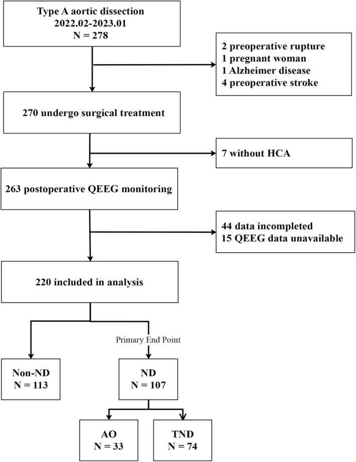
Methods and results: Amplitude-integrated electroencephalography (aEEG) processes raw signals through filtering, amplitude integration, and time compression, displaying the data in a semilogarithmic format. Using this method, postoperative relative band power (post-RBP) α% and dynamic aEEG (ΔaEEG) grade were significantly associated with neurological dysfunction in univariate and multivariable analyses, with area under the receiver operating characteristic curve of 0.876 (95% CI, 0.825-0.926) for the combined model. Postoperative relative band power α% and ΔaEEG were significantly associated with adverse outcomes, with area under the receiver operating characteristic curve of 0.903 (95% CI, 0.835-0.971) for the combined model. Postoperative relative band power α% and ΔaEEG were significantly associated with transient neurological dysfunction and stroke, with areas under the receiver operating characteristic curve of 0.818 (95% CI, 0.760-0.876) and 0.868 (95% CI, 0.810-0.926) for transient neurological dysfunction, and 0.815 (95% CI, 0.743-0.886) and 0.831 (95% CI, 0.746-0.916) for stroke. Among 56 patients, the Alberta Stroke Program Early Computed Tomography score was superior to ΔaEEG in predicting neurological outcomes (area under the receiver operating characteristic curve of 0.872 versus 0.708 [95% CI, 0.633-0.783]; P<0.05).

Conclusions: Perioperative quantitative electroencephalography monitoring offers valuable insights into brain function changes in patients with type A aortic dissection. ∆aEEG grades can aid in early detection of adverse outcomes, while postoperative relative band power and ∆aEEG grades predict transient neurological dysfunction. Quantitative electroencephalography can assist cardiac surgeons in assessing brain function and improving outcomes in patients with type A aortic dissection.

Registration: URL: https://www.chictr.org.cn; Unique identifier: ChiCTR2200055980.

Keywords: amplitude‐integrated electroencephalogram; aortic dissection; cerebral complication; quantitative electroencephalography; relative band power.


Figures

Figure 1
Figure 1. Consolidated Standards of Reporting Trials diagram demonstrating selection of patients undergoing TAAD surgery repair.
AO indicates adverse outcome; HCA, hypothermic circulatory arrest; ND, neurological dysfunction; QEEG, quantitative electroencephalography; TAAD, type A aortic dissection; and TND, transient neurological dysfunction.
Figure 2
Figure 2. QEEG principle schematic diagram.
QEEG collects the raw electroencephalographic signal from the scalp, analyzes it by FFT, and displays the spectrum and power spectrum of the EEG sequence signal in the form of a trend spectrum. FD indicates frequency domain; FFT, fast Fourier transform; L, left cerebral hemisphere; R, right cerebral hemisphere; and TD, time domain.
Figure 3
Figure 3. Intraoperative aEEG.
A, Anesthetic induction and commencement of surgery. B, During cooling and maintenance of the target hypothermic temperature. C, Starting selective cerebral perfusion. D, Rewarming of cardiopulmonary bypass. E, Rewarming to 36.5 °C. The yellow arrow is caused by the interference of electric knife during operation. aEEG indicates amplitude‐integrated electroencephalogram.
Figure 4
Figure 4. QEEG‐based performance metrics for predicting ND.
A, ROC curve for ΔaEEG. B, ROC curve for log(post‐RBP α%). C, ROC curve for model 1 combining ΔaEEG and log(post‐RBP α%). D, Calibration plot for model 1 with a Brier score of 0.133. Post‐RBP α% exhibited a skewed distribution and was transformed using a logarithmic scale to achieve a normal distribution for logistic regression analysis. ΔaEEG indicates dynamic amplitude‐integrated electroencephalogram; AUC, area under the curve; ND, neurological dysfunction; and post‐RBP, postoperative relative band power.
Figure 5
Figure 5. The aEEG transitioned from CNV to DNV, and CT scan suggested extensive cerebral infarction on the right side.
aEEG indicates amplitude‐integrated electroencephalogram; CNV, continuous normal voltage; CT, computed tomography; and DNV, discontinuous normal voltage.
Figure 6
Figure 6. QEEG‐Based Performance Metrics for Predicting AO.
A, ROC curve for ΔaEEG. B, ROC curve for log(post‐RBP α%). C, ROC curve for model 2, combining ΔaEEG and post‐RBP δ%. D, Calibration plot for model 2 with a Brier score of 0.081. Post‐RBP α% exhibited a skewed distribution and was transformed using a logarithmic scale to achieve a normal distribution for logistic regression analysis. ΔaEEG indicates dynamic amplitude‐integrated electroencephalogram; AO, adverse outcome; AUC, area under the curve; post‐RBP, postoperative relative band power; and ROC, receiver operating characteristics.
Figure 7
Figure 7. During TAAD surgery, the patient underwent selective right cerebral perfusion.
Following rewarming to a target temperature of 36.5 °C, there was a noticeable delay in the recovery of the left‐side aEEG when compared with the right side. Subsequently, the patient developed postoperative delirium. aEEG indicates amplitude‐integrated electroencephalogram; CNV, continuous normal voltage; DNV, discontinuous normal voltage; and TAAD, type A aortic dissection.
Figure 8
Figure 8. QEEG‐based performance metrics for predicting TND.
A, ROC curve for ΔaEEG for TND. B, ROC curve for log(post‐RBP α%) for TND. C, ROC curve for model 3 combining ΔaEEG and log(post‐RBP α%) for TND. D, Calibration plot for model 3 with a Brier score of 0.133. Post‐RBP α% exhibited a skewed distribution and was transformed using a logarithmic scale to achieve a normal distribution for logistic regression analysis. aEEG indicates amplitude‐integrated electroencephalogram; post‐RBP, postoperative relative band power; ROC, receiver operating characteristic; and TND, transient neurological dysfunction.
Figure 9
Figure 9. Diagnostic performance of electroencephalography and post‐RBP α% models for stroke prediction: ROC and calibration analyses.
The main results were depicted in 3 panels (A through C), each illustrating different aspects of the model evaluation. D, A Brier score of 0.122 suggested that the predicted probabilities were well‐calibrated with the observed outcomes. Post‐RBP α% exhibited a skewed distribution and was transformed using a logarithmic scale to achieve a normal distribution for logistic regression analysis. aEEG indicates amplitude‐integrated electroencephalogram; ΔaEEG indicates dynamic amplitude‐integrated electroencephalogram; AUC, area under the curve; post‐RBP, postoperative relative band power; and ROC, receiver operating characteristic.
Figure 10
Figure 10. ROC curve analysis: predicting AOs and NOs using ΔaEEG and ASPECT scores.
A, ROC curve analysis showing the AUC of ΔaEEG and ASPECT between the AUCs (DeLong's test) for predicting AOs; (B) for predicting NO. ΔaEEG indicates dynamic amplitude‐integrated electroencephalogram; ASPECT, Alberta Stroke Program Early Computed Tomography; AUC, area under the curve; NO, neurological outcome; and ROC, receiver operating characteristic.

References

    1. Haldenwang PL, Wahlers T, Himmels A, Wippermann J, Zeriouh M, Kroner A, Kuhr K, Strauch JT. Evaluation of risk factors for transient neurological dysfunction and adverse outcome after repair of acute type a aortic dissection in 122 consecutive patients. Eur J Cardiothorac Surg. 2012;42:e115–e120. doi: 10.1093/ejcts/ezs412 - DOI - PubMed
    1. Chikwe J, Cavallaro P, Itagaki S, Seigerman M, Diluozzo G, Adams DH. National outcomes in acute aortic dissection: influence of surgeon and institutional volume on operative mortality. Ann Thorac Surg. 2013;95:1563–1569. doi: 10.1016/j.athoracsur.2013.02.039 - DOI - PubMed
    1. Kruger T, Weigang E, Hoffmann I, Blettner M, Aebert H, Investigators G. Cerebral protection during surgery for acute aortic dissection type a: results of the German registry for acute aortic dissection type a (GERAADA). Circulation. 2011;124:434–443. doi: 10.1161/CIRCULATIONAHA.110.009282 - DOI - PubMed
    1. Dumfarth J, Kofler M, Stastny L, Plaikner M, Krapf C, Semsroth S, Grimm M. Stroke after emergent surgery for acute type a aortic dissection: predictors, outcome and neurological recovery. Eur J Cardiothorac Surg. 2018;53:1013–1020. doi: 10.1093/ejcts/ezx465 - DOI - PubMed
    1. Zierer A, Moon MR, Melby SJ, Moazami N, Lawton JS, Kouchoukos NT, Pasque MK, Damiano RJ Jr. Impact of perfusion strategy on neurologic recovery in acute type a aortic dissection. Ann Thorac Surg. 2007;83:2122–2128. doi: 10.1016/j.athoracsur.2007.01.041 - DOI - PubMed

Publication types

MeSH terms

LinkOut - more resources