

Assessment of central vein sign and paramagnetic rim lesions in pediatric multiple sclerosis
- PMID: 39291789
- PMCID: PMC11572724
- DOI: 10.1002/acn3.52208
Assessment of central vein sign and paramagnetic rim lesions in pediatric multiple sclerosis
Abstract
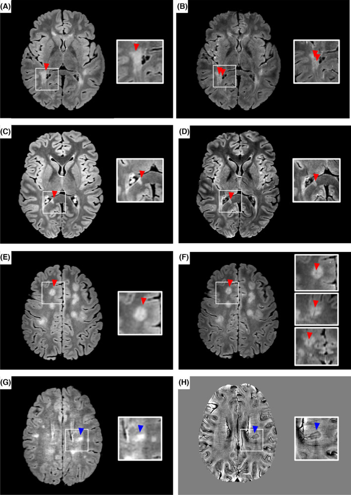
The evaluation of white matter lesions (WMLs) showing the central vein sign (CVS) and paramagnetic rim lesions (PRLs) has been suggested to enhance the diagnostic work-up of adult multiple sclerosis (MS). We aimed to evaluate the fulfillment of different CVS criteria and the added value of PRLs in 22 pediatric MS patients. Eleven patients (50%) fulfilled the 40%-rule threshold. Nineteen (86%) patients had ≥3 CVS+ WMLs or ≥1 PRL, whereas 17 (77%) had ≥6 CVS+ WMLs or ≥1 PRL. A simplified CVS-based approach, with the combined evaluation of ≥1 PRL in patients with ≥6 CVS+ WMLs, may improve MS diagnosis in pediatric patients.
© 2024 The Author(s). Annals of Clinical and Translational Neurology published by Wiley Periodicals LLC on behalf of American Neurological Association.
Conflict of interest statement
The authors have no conflict of interest to declare.
Figures
References
-
- Sati P, Oh J, Constable RT, et al. The central vein sign and its clinical evaluation for the diagnosis of multiple sclerosis: a consensus statement from the north American imaging in multiple sclerosis cooperative. Nat Rev Neurol. 2016;12:714‐722. - PubMed
-
- Margoni M, Preziosa P, Storelli L, et al. Paramagnetic rim and core sign lesions in paediatric multiple sclerosis patients. J Neurol Neurosurg Psychiatry. 2023;94:873‐876. - PubMed
MeSH terms
LinkOut - more resources
Full Text Sources
Medical