. 2024 Nov 5;12(11):e0094224.
doi: 10.1128/spectrum.00942-24. Epub 2024 Sep 18.

A bacteriophage cocktail targeting Yersinia pestis provides strong post-exposure protection in a rat pneumonic plague model

Affiliations

A bacteriophage cocktail targeting Yersinia pestis provides strong post-exposure protection in a rat pneumonic plague model

Paul B Kilgore et al. Microbiol Spectr. .

Abstract

Yersinia pestis, one of the deadliest bacterial pathogens ever known, is responsible for three plague pandemics and several epidemics, with over 200 million deaths during recorded history. Due to high genomic plasticity, Y. pestis is amenable to genetic mutations as well as genetic engineering that can lead to the emergence or intentional development of pan-drug-resistant strains. Indeed, antibiotic-resistant strains (e.g., strains carrying multidrug-resistant or MDR plasmids) have been isolated in various countries and endemic areas. Thus, there is an urgent need to develop novel, safe, and effective treatment approaches for managing Y. pestis infections. This includes infections by antigenically distinct strains for which vaccines (none FDA approved yet) may not be effective and those that cannot be managed by currently available antibiotics. Lytic bacteriophages provide one such alternative approach. In this study, we examined post-exposure efficacy of a bacteriophage cocktail, YPP-401, to combat pneumonic plague caused by Y. pestis CO92. YPP-401 is a four-phage preparation effective against a panel of at least 68 genetically diverse Y. pestis strains. Using a pneumonic plague aerosol challenge model in gender-balanced Brown Norway rats, YPP-401 demonstrated ~88% protection when delivered 18 h post-exposure for each of two administration routes (i.e., intraperitoneal and intranasal) in a dose-dependent manner. Our studies provide proof-of-concept that YPP-401 could be an innovative, safe, and effective approach for managing Y. pestis infections, including those caused by naturally occurring or intentionally developed multidrug-resistant strains.IMPORTANCECurrently, there are no FDA-approved plague vaccines. Since antibiotic-resistant strains of Y. pestis have emerged or are being intentionally developed to be used as a biothreat agent, new treatment modalities are direly needed. Phage therapy provides a viable option against potentially antibiotic-resistant strains. Additionally, phages are nontoxic and have been approved by the FDA for use in the food industry. Our study provides the first evidence of the protective effect of a cocktail of four phages against pneumonic plague, the most severe form of disease. When treatment was initiated 18 h post infection by either the intranasal or intraperitoneal route in Brown Norway rats, up to 87.5% protection was observed. The phage cocktail had a minimal impact on a representative human microbiome panel, unlike antibiotics. This study provides strong proof-of-concept data for the further development of phage-based therapy to treat plague.

Keywords: Tier-1 select agent; Yersinia pestis; aerosol challenge; bacteriophage; biodefense; pneumonic plague; rat model; therapeutic.


Conflict of interest statement

The YPP-401 cocktail was developed by Intralytix, Inc. The following authors report conflicts of interest with respect to Intralytix, Inc.: A.S. (co-founder, employee, shareholder); J.A.S. (employee, shareholder); J.W. (employee, shareholder); and L.H. (employee).

Figures
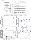
Fig 1
Fig 1
YPP-401 provides post-exposure efficacy against Y. pestis in rats when delivered intraperitoneally or intranasally at 18 hpi. COHORT 1: (A) Schematic of study design and dosing regimens. Shown are (B) survival, (C) body weight, and (D) Y. pestis burden in the blood at 42 hpi (taken immediately prior to 42 h treatment timepoint). Other measures were (E) terminal organ bacterial burden (i.e., lung, liver, heart, spleen) and clinical score (i.e., appearance, activity, respirations, facial expression; Table S2). Brown Norway rats (M/F; 10–12 wk) received a full body aerosol challenge (0 h) of WT virulent Y. pestis strain CO92 at Dp ~ 9.56 × 10⁶ to 1.24 × 10⁷ CFU. At 18 hpi, cohort 1 received either 2 days (total four doses, BID, ~q6–12 h) of i.p. vehicle (white/open circles; PBS; n = 4), i.p. YPP-401 (blue squares; ~2 × 10¹⁰ PFUs; n = 8), i.n. YPP-401 (red triangles; ~4 × 10⁹ PFUs; n = 8), oral YPP-401 (gray diamonds; ~2 × 10¹⁰ PFUs; n = 8), or 10 oral QD doses of levofloxacin (levo) (black circles; 50 mg/kg; n = 4). Body weight graph (B) shows the arithmetic mean with the standard error of the mean (SEM) depicted as error bars. Bacteremia (D) and organ burden (E) show the geometric mean with geometric standard deviation (GSD) shown as error bars. For statistical comparisons, Kaplan-Meier analysis with Logrank (Mantel-Cox) test was used for survival (B), one-way analysis of variance (ANOVA) with Kruskal-Wallis test and Dunn’s multiple comparison test were used for (D) while two-way ANOVA with Tukey’s post-hoc test was used for (E). Limit of detection (LOD) is depicted by the dotted horizontal line. Studies were performed at UTMB under ABSL-3. The asterisk indicates a significant difference compared to vehicle control or between the two groups indicated by a horizontal line. *P < 0.05; **P < 0.01; ***P < 0.001; ****P < 0.0001.
Fig 2
Fig 2
YPP-401 provides dose-dependent post-exposure efficacy against Y. pestis in rats when delivered intranasally at 18 hpi. COHORT 2: Animals were challenged as described in Fig. 1A. Similar dosing regimens starting at 18 hpi were used with the following changes: cohort 2 received either 2 days (total four doses, BID, ~q6–12 h) of i.p. vehicle (white/open circles; PBS; n = 2), i.n. YPP-401 (red triangles; ~2 × 10¹⁰ PFUs; n = 8), or 10 oral QD doses of levo (black circles; 50 mg/kg; n = 2). Shown are (A) survival, (B) body weights, and (C) Y. pestis burden in the blood at 42 hpi (taken immediately prior to 42 h treatment timepoint). Other measures were (D) terminal organ bacterial burden (i.e., lung, liver, heart, spleen) and clinical score (i.e., appearance, activity, respirations, facial expression; Table S3). Body weight graph (B) shows the arithmetic mean with the SEM depicted as error bars. Bacteremia (C) and organ burden (D) show the geometric mean with GSD shown as error bars. For statistical comparisons, Kaplan-Meier analysis with Logrank (Mantel-Cox) test was used for survival (A), one-way ANOVA with Kruskal-Wallis test and Dunn’s multiple comparison test were used for (C) while two-way ANOVA with Tukey’s post-hoc test was used for (D). LOD is depicted by the dotted horizontal line. Studies were performed at UTMB under ABSL-3. The asterisk indicates a significant difference compared to vehicle control or between the two groups indicated by a horizontal line. *P < 0.05; **P < 0.01.
Fig 3
Fig 3
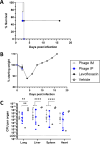
YPP-401 dosing regimens protective at 18 hpi are insufficient when initiated at 42 hpi. COHORT 3: Animals were challenged as described in Fig. 1A. Similar dosing regimens were started at 42 hpi with the following changes: cohort 3 received either one or two doses (BID, ~q6–12 h) of i.p. vehicle (white/open circles; PBS; n = 2), i.m. YPP-401 (gray circles; ~5 × 10¹⁰ PFUs; n = 8), i.p. YPP-401 (blue squares; ~5 × 10¹⁰ PFUs; n = 8), or 1 or 10 QD doses of levo (black circles; 50 mg/kg; n = 2). Shown are (A) survival, (B) body weights, and (C) Y. pestis burden in terminal organs (i.e., lung, liver, heart, spleen) and clinical score (i.e., appearance, activity, respirations, facial expression; Table S4). Body weight graph (B) shows the arithmetic mean with the SEM depicted as error bars. Organ burden (C) shows the geometric mean with GSD shown as error bars. For statistical comparisons, Kaplan-Meier analysis with Logrank (Mantel-Cox) test was used for survival (A), two-way ANOVA with Tukey’s post-hoc test was used for (D). LOD is depicted by the dotted horizontal line. Studies were performed at UTMB under ABSL-3. The asterisk indicates a significant difference between the two groups indicated by a horizontal line. *P < 0.05; **P < 0.01; ***P < 0.001; ****P < 0.0001.

Update of

References

    1. Rollins SE, Rollins SM, Ryan ET. 2003. Yersinia pestis and the plague. Am J Clin Pathol 119 Suppl:S78–S85. doi:10.1309/DQM9-3R8Q-NQWB-FYU8 - DOI - PubMed
    1. Sun W, Singh AK. 2019. Plague vaccine: recent progress and prospects. NPJ Vaccines 4:11. doi:10.1038/s41541-019-0105-9 - DOI - PMC - PubMed
    1. Derbes VJ. 1966. De Mussis and the great plague of 1348. A forgotten episode of bacteriological warfare. JAMA 196:59–62. doi:10.1001/JAMA.1966.03100140113030 - DOI - PubMed
    1. Wheelis M. 2002. Biological warfare at the 1346 siege of Caffa. Emerg Infect Dis 8:971–975. doi:10.3201/eid0809.010536 - DOI - PMC - PubMed
    1. McGovern TW, Friedlander AM. 1997. Plague, p 479–502. In Frederick R, Sidell MD, Ernest T, Takafuji MD (ed), Medical aspects of chemical and biological warfare. Office of The Surgeon General at TMM Publications, Borden Institute, Walter Reed Army Medical Center, Washington, DC.

LinkOut - more resources