. 2024 Oct 24;206(10):e0014224.
doi: 10.1128/jb.00142-24. Epub 2024 Sep 18.

Modulation of Vibrio cholerae gene expression through conjugative delivery of engineered regulatory small RNAs

Affiliations

Modulation of Vibrio cholerae gene expression through conjugative delivery of engineered regulatory small RNAs

Pilar Menendez-Gil et al. J Bacteriol. .

Abstract

The increase in antibiotic resistance in bacteria has prompted the efforts in developing new alternative strategies for pathogenic bacteria. We explored the feasibility of targeting Vibrio cholerae by neutralizing bacterial cellular processes rather than outright killing the pathogen. We investigated the efficacy of delivering engineered regulatory small RNAs (sRNAs) to modulate gene expression through DNA conjugation. As a proof of concept, we engineered several sRNAs targeting the type VI secretion system (T6SS), several of which were able to successfully knockdown the T6SS activity at different degrees. Using the same strategy, we modulated exopolysaccharide production and motility. Lastly, we delivered an sRNA targeting T6SS into V. cholerae via conjugation and observed a rapid knockdown of the T6SS activity. Coupling conjugation with engineered sRNAs represents a novel way of modulating gene expression in V. cholerae opening the door for the development of novel prophylactic and therapeutic applications.

Importance: Given the prevalence of antibiotic resistance, there is an increasing need to develop alternative approaches to managing pathogenic bacteria. In this work, we explore the feasibility of modulating the expression of various cellular systems in Vibrio cholerae using engineered regulatory sRNAs delivered into cells via DNA conjugation. These sRNAs are based on regulatory sRNAs found in V. cholerae and exploit its native regulatory machinery. By delivering these sRNAs conjugatively along with a real-time marker for DNA transfer, we found that complete knockdown of a targeted cellular system could be achieved within one cell division cycle after sRNA gene delivery. These results indicate that conjugative delivery of engineered regulatory sRNAs is a rapid and robust way of precisely targeting V. cholerae.

Keywords: T6SS; V. cholerae; conjugation; modulation gene expression; sRNAs.


Conflict of interest statement

The authors declare no conflict of interest.

Figures
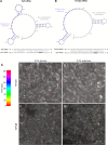
Fig 1
Fig 1
TarVipA sRNA shuts down the T6SS activity of V. cholerae. (A) TarB secondary structure predicted with Vienna RNAfold web server (23) and TarB and tcpF mRNA predicted interaction as stated by Bradley et al. (22). (B) TarVipA secondary structure predicted with Vienna RNAfold web server (23) and INTaRNAv2 (25) predicted interaction of TarVipA and vipA mRNA. The recognition sequences of TarB to tcpF mRNA and TarVipA to vipA mRNA are highlighted in purple. tcpF and vipA start codons are highlighted in bold. The numbering of tcpF and vipA mRNAs is relative to the start of translation whereas TarB and TarVipA numbering is relative to the start of transcription. (C) Spectrum temporal-colored code from ImageJ-Fiji software (26) used to analyze the microscopy in D–G. The temporal code assigns a different color to foci appearing in each time point of the time-lapse. White is assigned to non-dynamic foci/aggregates. (D - G) Time-lapse fluorescence microscopy imagining of V. cholerae 2740-80 clpV::clpV-mCherry carrying pBAD33-TarVipA (D, E) or pBAD33-TarFlgB (F, G). Cells were either grown in glucose (D, F) or in arabinose (E, G) to repress or express pBAD33 expression, respectively. Images were taken every 20 s for 2 min. Scale bar is 10 mm for all images shown.
Fig 2
Fig 2
T6SS activity modulation differs depending on the gene being targeted. (A) Genetic organization of the T6SS genes (32) with genes targeted by the engineered sRNAs highlighted in white. (B) Representative image of the competition assay between E. coli MG1655 (prey) and V. cholerae 2740-80 clpV::clpV-mCherry strains carrying the sRNAs (predators). As a negative control, the predator V. cholerae 2740-80 clpV::clpV-mCherry (T6SS+) was included. As a positive control, V. cholerae 2740-80 clpV::clpV-mCherry ∆vipA (T6SS−) was used. (C) Quantification of the E. coli CFU recovery after competition with V. cholerae strains shown in panel B. Data represent the average of at least three independent replicates, each one done in technical duplicate. Error bars represent the standard deviation (SD) of these three replicates. Asterisks represent statistical significance when compared with the T6SS+ no sRNA control (*P value < 0.05 and ***P value < 0.001; ns not significant).
Fig 3
Fig 3
Engineered sRNAs can modulate EPS production and motility in V. cholerae. (A) Quantification of prey CFUs recovered after the competition assay between different V. cholerae strains carrying pBAD33 with sRNAs targeting EPS production (preys) and the V. cholerae 2740-80 clpV::clpV-mCherry (predator T6SS+). Data represent the average of at least three independent replicates, each one done in technical duplicate. Error bars represent the SD. E.V., empty vector. (B) Representative image of the motility assay of different V. cholerae strains carrying sRNAs targeting the flagellum apparatus expressed from pBAD33 backbones. (C) Quantification of the motility halos (diameter) shown in panel C. Data are represented as a ratio between the halo of a sample in relation to the halo diameter of the empty vector sample (normalized diameter). Data represent the average of at least four independent replicates, each one done in technical duplicate. Error bars represent the SD of these replicates. Asterisks represent statistical significance when compared with the empty vector sample (paired two-tail t-test, *P value < 0.05, **P value < 0.01, and ***P value < 0.001; ns not significant).
Fig 4
Fig 4
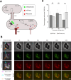
TarVipA rapidly inhibits V. cholerae T6SS activity when delivered via conjugation. (A) Illustrative image of the assay developed to track conjugation events and T6SS activity with fluorescence microscopy. E. coli MFDpir donor cells, carrying either pBAD33-TarVipA-tetO array or pBAD33-tetO array, were mixed with V. cholerae 2740-80 clpV::clpV-mCherry pBAD33-J23103-tetR-mNeonGreen recipient cells 10:1 and incubated for 30, 60, or 90 min before imaging . First, donors transfer the plasmid carrying the tetO array and TarVipA or the tetO array only into a V. cholerae strain through the type IV secretion system (T4SS) (1). This V. cholerae strain has a ClpV-mCherry fusion to detect T6SS activity. It also expresses a TetR-mNeonGreen fusion, which binds to the tetO array, shifting the green fluorescence from being uniformly distributed in the cell to being coalesced into a fluorescent focus (2). Once the plasmid is in the recipient cell, it transcribes the sRNA (3) that will bind to its target mRNA, inhibiting its translation. The sRNA function will be observed by a reduction in the T6SS activity (4). Three biological replicates were conducted for each sRNA delivered. (B) Representative image of a 2-min time-lapse microscopy showing the delivery of TarVipA sRNA. An E. coli donor can be found surrounded by several V. cholerae cells, four of them in direct contact with the E. coli cell. Of those four, two were transconjugants (presence of green foci). The red channel shows the presence of active T6SS foci. Merge images are the combination of the red and green channels. Temporal code image analysis of phase, red, and green channels was performed to detect dynamic foci. (C) Quantification of cells with an active T6SS after receiving TarVipA or the tetO array only. Statistical significance was determined using student’s t-test, ***P < 0.00001, ns not significant.

Similar articles

References

    1. Moore S, Thomson N, Mutreja A, Piarroux R. 2014. Widespread epidemic cholera caused by a restricted subset of Vibrio cholerae clones. Clin Microbiol Infect 20:373–379. doi:10.1111/1469-0691.12610 - DOI - PubMed
    1. Ali M, Nelson AR, Lopez AL, Sack DA. 2015. Updated global burden of cholera in endemic countries. PLoS Negl Trop Dis 9:e0003832. doi:10.1371/journal.pntd.0003832 - DOI - PMC - PubMed
    1. Peterson KM, Gellings PS. 2018. Multiple intraintestinal signals coordinate the regulation of Vibrio cholerae virulence determinants. Pathog Dis 76. doi:10.1093/femspd/ftx126 - DOI - PubMed
    1. Kanungo S, Azman AS, Ramamurthy T, Deen J, Dutta S. 2022. Cholera. Lancet 399:1429–1440. doi:10.1016/S0140-6736(22)00330-0 - DOI - PubMed
    1. Sakib SN, Reddi G, Almagro-Moreno S. 2018. Environmental role of pathogenic traits in Vibrio cholerae. J Bacteriol 200:e00795-17. doi:10.1128/JB.00795-17 - DOI - PMC - PubMed

Publication types

MeSH terms

LinkOut - more resources