. 2024 Sep 24;121(39):e2411352121.
doi: 10.1073/pnas.2411352121. Epub 2024 Sep 18.

Transdifferentiation occurs without resetting development-specific DNA methylation, a key determinant of full-function cell identity

Affiliations

Transdifferentiation occurs without resetting development-specific DNA methylation, a key determinant of full-function cell identity

Ahmed Radwan et al. Proc Natl Acad Sci U S A. .

Abstract

A number of studies have demonstrated that it is possible to directly convert one cell type to another by factor-mediated transdifferentiation, but in the vast majority of cases, the resulting reprogrammed cells are unable to maintain their new cell identity for prolonged culture times and have a phenotype only partially similar to their endogenous counterparts. To better understand this phenomenon, we developed an analytical approach for better characterizing trans-differentiation-associated changes in DNA methylation, a major determinant of long-term cell identity. By examining various models of transdifferentiation both in vitro and in vivo, our studies indicate that despite convincing expression changes, transdifferentiated cells seem unable to alter their original developmentally mandated methylation patterns. We propose that this blockage is due to basic developmental limitations built into the regulatory sequences that govern epigenetic programming of cell identity. These results shed light on the molecular rules necessary to achieve complete somatic cell reprogramming.

Keywords: development; epigenetics; metaplasia; plasticity; regulation.


Conflict of interest statement

Competing interests statement:The authors declare no competing interest.

Figures

Fig. 1.
Fig. 1.
Direct reprogramming from MEFs to muscle—morphology, expression, and DNA methylation. (A) Representative bright field images showing cell morphology during direct reprogramming process from MEF to muscle. (B) Expression heat map showing genes that are up-regulated (Left) and down-regulated (Right) in normal muscle cells compared to MEFs together with the expression level seen in reprogrammed cells. (C) Heat map and histogram (average methylation for each sample) of regions specifically undermethylated in muscle (Left) or in MEFs (Right) compared to methylation patterns seen in reprogrammed cells and in other tissues (OT) by RRBS.
Diagram 1.
Diagram 1.
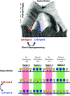
Dynamics of DNA methylation during development. Conrad Waddington’s epigenetic landscape. The ball represents a cell and the bifurcating system of valleys represents trajectories of cell state. This diagram by C.H. Waddington neatly encapsulates the developmental pathways and progressive divergence of cells as they differentiate in the embryo. Reproduced from Waddington © (1) George Allen and Unwin (London). The diagram shows regions of DNA from a developmental perspective. The upper row shows the methylation state of each region at the time of implantation (E6.5) and this same pattern is preserved in most tissues of the adult organism. In other words, the pattern established in the early embryo is then maintained in all tissues, with only some undergoing tissue-specific changes in cell type A or B. Methylated (Yellow), undermethylated (Blue). Region 1 is specifically demethylated in Cell-type B during development. If it does not undergo demethylation following transdifferentiation, this indicates that these cells have failed to adopt a B cell identity probably because the induction factors were unable to activate the site-specific demethylation machinery originally employed during earlier B cell development. Region 2 appears to behave in a very similar manner, but from the developmental perspective, this region is actually set up as unmethylated in the embryo and remains that way in all cells. During Cell-type A development, it becomes specifically de novo methylated—a completely different event. Thus, if the transdifferentiation event failed to demethylate this segment, it is probably because the factors and motifs necessary for specifically demethylating this region do not exist. Indeed, during normal development, this sequence never undergoes specific demethylation—because it is already unmethylated in all cell types of the body. Region 3 is specifically demethylated in Cell-type A. If it fails to become remethylated when A is transdifferentiated to B, this is probably because this type of specific event never occurs in vivo during normal development and the molecular components needed for this do not exist. Region 4 has the exact same A/B differential methylation pattern as that of region 3, but in this case, it is a result of B cell-specific de novo methylation. If this region fails to become methylated in the transdifferentiation process, it is probably because the factors used to convert A to B were unable to activate this specific de novo methylation machinery. It should be noted that, for the sake of simplicity, the analysis in this paper only includes regions of tissue-specific demethylation (e.g., regions 1 and 3).
Fig. 2.
Fig. 2.
Direct reprogramming from MEFs to NPC—DNA methylation. Heatmap and boxplot of regions specifically unmethylated in NPCs (A and B) as compared to those seen in MEFs reprogrammed to NPCs following direct reprogramming for various times using either the Ascl1 or BAM inducers in forebrain (E10–E16) and in OT by WGBS. Their state of methylation is also shown for different stages of forebrain development (E10–E16), as well as in OT.
Fig. 3.
Fig. 3.
Direct reprogramming from hepatocytes to cholangiocytes in vivo. Heatmap and histogram (average RRBS methylation for each sample) of specifically unmethylated regions in normal (Hep) or DDC-treated (Inj Hep) hepatocytes but not in cholangiocytes (A) or in normal (Chol) or DDC-treated (Inj Chol) cholangiocytes but not in hepatocytes (B) as compared to the pattern seen in reprogrammed cells (Direct Rep). (C) ATAC-Seq of cholangiocyte-specific undermethylated regions as determined by RRBS as a function of distance from their center.
Fig. 4.
Fig. 4.
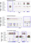
Examples of genes with specifically undermethylated enhancer regions in cholangiocytes. Genome browser tracks showing RNA-Seq, ATAC-Seq, and DNA methylation of genes specifically expressed in cholangiocytes for DDC-treated hepatocytes (Inj Hep) or cholangiocytes (Inj Chol) as compared to reprogrammed cells (Direct Rep). The undermethylated regions are outlined in gray.

References

    1. Waddington C. H., The Strategy of Genes (George Allen and Unwin, London, UK, 1957).
    1. Cedar H., Bergman Y., Programming of DNA methylation patterns. Annu. Rev. Biochem. 81, 97–117 (2012). - PubMed
    1. Kafri T., et al. , Developmental pattern of gene-specific DNA methylation in the mouse embryo and germ line. Genes Dev. 6, 705–714 (1992). - PubMed
    1. Monk M., Boubelik M., Lehnert S., Temporal and regional changes in DNA methylation in the embryonic, extraembryonic and germ cell lineages during mouse embryo development. Development 99, 371–382 (1987). - PubMed
    1. Brandeis M., et al. , Sp1 elements protect a CpG island from de novo methylation. Nature 371, 435–438 (1994). - PubMed

LinkOut - more resources