. 2024 Sep 18;16(765):eadk0845.
doi: 10.1126/scitranslmed.adk0845. Epub 2024 Sep 18.

A deep intronic splice-altering AIRE variant causes APECED syndrome through antisense oligonucleotide-targetable pseudoexon inclusion

Affiliations

A deep intronic splice-altering AIRE variant causes APECED syndrome through antisense oligonucleotide-targetable pseudoexon inclusion

Sebastian Ochoa et al. Sci Transl Med. .

Abstract

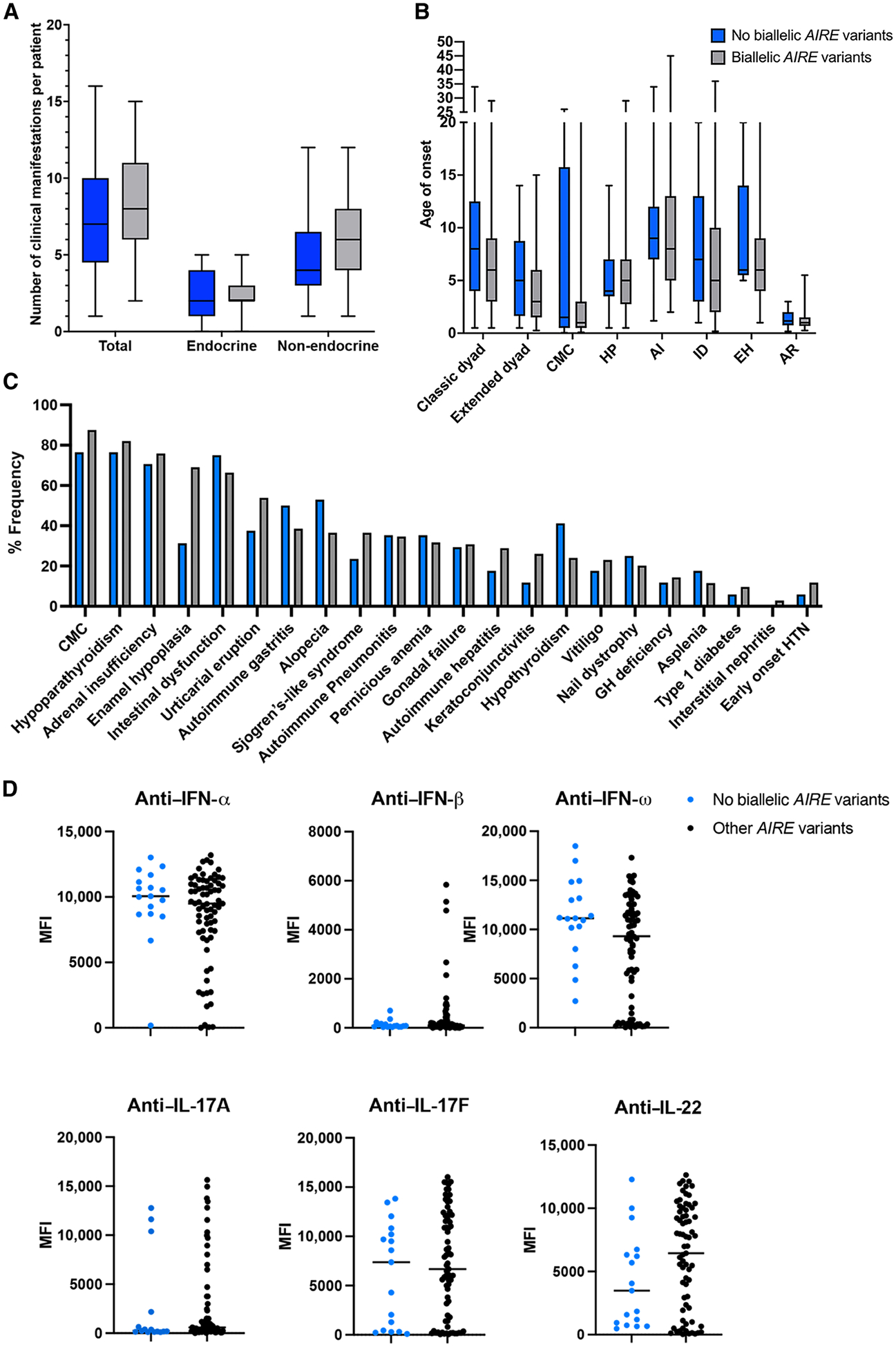
Autoimmune polyendocrinopathy-candidiasis-ectodermal dystrophy (APECED) is a life-threatening monogenic autoimmune disorder primarily caused by biallelic deleterious variants in the autoimmune regulator (AIRE) gene. We prospectively evaluated 104 patients with clinically diagnosed APECED syndrome and identified 17 patients (16%) from 14 kindreds lacking biallelic AIRE variants in exons or flanking intronic regions; 15 had Puerto Rican ancestry. Through whole-genome sequencing, we identified a deep intronic AIRE variant (c.1504-818 G>A) cosegregating with the disease in all 17 patients. We developed a culture system of AIRE-expressing primary patient monocyte-derived dendritic cells and demonstrated that c.1504-818 G>A creates a cryptic splice site and activates inclusion of a 109-base pair frame-shifting pseudoexon. We also found low-level AIRE expression in patient-derived lymphoblastoid cell lines (LCLs) and confirmed pseudoexon inclusion in independent extrathymic AIRE-expressing cell lines. Through protein modeling and transcriptomic analyses of AIRE-transfected human embryonic kidney 293 and thymic epithelial cell 4D6 cells, we showed that this variant alters the carboxyl terminus of the AIRE protein, abrogating its function. Last, we developed an antisense oligonucleotide (ASO) that reversed pseudoexon inclusion and restored the normal AIRE transcript sequence in LCLs. Thus, our findings revealed c.1504-818 G>A as a founder APECED-causing AIRE variant in the Puerto Rican population and uncovered pseudoexon inclusion as an ASO-reversible genetic mechanism underlying APECED.


Conflict of interest statement

Competing interests: J.L.M. is on the advisory board of Ipsen Inc. The other authors declare that they have no competing interests.

Figures

Fig. 1.
Fig. 1.. Characteristics of patients with clinical APECED lacking biallelic AIRE pathogenic variants in exons or flanking intronic regions.
(A) Number of total, endocrine, and nonendocrine APECED-related clinical manifestations per patient. (B) Mean age of developing a clinical APECED diagnosis based on a classic dyad or expanded dyad, and mean age of developing the indicated individual classic and adjunct triad clinical manifestations. CMC, chronic mucocutaneous candidiasis; HP, hypoparathyroidism; AI, adrenal insufficiency; ID, intestinal dysfunction; EH, enamel hypoplasia; AR, APECED rash. (C) Percent frequency of the indicated clinical manifestations. GH, growth hormone; HTN, hypertension. (D) Mean fluorescence intensity (MFI) of autoantibodies to the indicated type 1 interferons (IFNs) and T helper 17 cytokines. For (A) to (D), patients with clinical APECED lacking biallelic pathogenic AIRE variants in exons or flanking intronic regions (n = 17) were compared with patients with APECED with biallelic pathogenic AIRE variants (n = 87). Student’s t tests or Mann-Whitney U tests with Holm’s correction for multiple comparisons.
Fig. 2.
Fig. 2.. Identification of AIRE c.1504-818 G>A as a candidate variant.
(A) Genome-wide plot showing shared, overlapping regions of AOH in chromosome 21 from patients lacking pathogenic AIRE variants in exons or flanking intronic regions (P1 to P7, P11, P13, and P14 to 16). (B) Magnified view of overlapping AOH regions showing genes contained within the shared region. Source: UCSC Genome Browser (http://genome.ucsc.edu) (55). (C) Representation of 45-variant haplotype (spanning 1439 kb) shared among patients. Maternal (M) and paternal (P) inherited alleles are represented for each patient, except P17, where parental sequencing was unavailable and thus represented as alleles 1 and 2. One representative homozygous patient (P1) is shown, and the remaining (P8 to P10, P12, and P17) are compound heterozygous for AIRE c.1504-818 G>A and previously identified pathogenic exonic AIRE variants. The consensus sequence for the haplotype encompassing AIRE c.1504-818 G>A is shown in nonhighlighted text, and gray highlighting represents a mismatch with the consensus. Light green is used to highlight the synonymous marker variant p.G227= and the AIRE intronic variant c.1504-818 G>A. Each variant’s location and protein-coding genes are labeled on the chromosome map. The underlined region in the P12 M allele represents a subhaplotype resembling the consensus haplotype found in AIRE intronic variant c.1504-818 G>A homozygous patients, except lacking the c.1504-818 G>A variant. (D) Rare variants (mean allele frequency less than 0.1% in gnomAD) encompassed within the overlapping region of an AOH, demonstrating a deep intronic variant in AIRE and other rare variants in ICOSLG and CFAP410. GRCh37 (Chr 21) was used for genomic coordinates. (E) Schematic representation of AIRE exons 1 to 14 depicting intron 12 and the position of the variant of interest Chr21:45715448 G>A (GRCh37), with equivalent descriptions g.44295565G>A (NC_000021.9) and c.1504-818 G>A (NM_000383.4). Representative sequencing chromatograms from an unaffected healthy control, patients lacking known pathogenic AIRE variants (P1 to P7, P11, and P13 to P16), and patients with exonic monoallelic pathogenic AIRE variants in compound heterozygosity with AIRE c.1504-818 G>A (P8 to P10, P12, and P17). (F) Familial segregation of c.1504-818 G>A. Previously known pathogenic AIRE variants are annotated in the patients where c.1504-818 G>A was found in compound heterozygosity (P8 to P10, P12, and P17).
Fig. 3.
Fig. 3.. AIRE c.1504-818 G>A results in pseudoexon inclusion.
(A) Schematic representation of extrathymic AIRE expression system using moDCs and EBV-transformed LCLs, followed by quantification of AIRE transcript by RT-PCR and PCR amplification for Sanger sequencing (created with BioRender.com). (B) Relative AIRE expression by RT-PCR in healthy volunteer PBMCs compared with that in LCLs and day 8 (D8) moDCs (n = 5 to 7 per cell type). (C) Sequencing chromatograms of cDNA demonstrating the boundary between exon 12 and exon 13 in a healthy donor (WT), the 109-bp pseudoexon inclusion and exon 13 in P3 who is homozygous for c.1504-818 G>A, and a mixed sequence (in P10 who is compound heterozygous for c.1504-818 G>A and c.132+1_132+3delinsCT). (D) Partial AIRE cDNA sequence depicting a 109-bp pseudoexon between AIRE exons 12 and 13 found in patients carrying c.1504-818 G>A. The Mu nucleotide position is noted as A. (E) cDNA sequence demonstrating a 109-bp pseudoexon inclusion in two independent clones of a healthy donor LCL CRISPR-modified to carry c.1504-818 G>A, which is not present in the parental cell line. *P < 0.05, **P < 0.01, and ****P < 0.0001; unpaired t tests.
Fig. 4.
Fig. 4.. AIRE c.1504-818 G>A is predicted in silico to alter the AIRE C-terminal region, and the net charge and is associated with impaired formation of nuclear foci.
(A) Schematic representation of AIRE domains and motifs. The partial AIRE amino acid sequence (NP_000374.1) corresponding to AIRE exons 12 to 14 is shown, highlighting the differences between WT AIRE (545 amino acids) and the predicted sequence of the c.1504-818 G>A Mu protein (538 amino acids). The LxxLL-4 and PxxPxP motifs are shown in red. (B) Predicted structure of the WT full-length AIRE protein from AlphaFold2. The C-terminal LxxLL-4 motif, potentially responsible for nuclear receptor binding, and the PxxPxP domain are shown in red. Except for the two helices, the C-terminal region of AIRE is predicted to be highly unstructured. (C) The starting (left) and final (right) structures of the WT-AIRE and Mu-AIRE C-terminal peptides from molecular dynamics are depicted. Different colors are used to represent each sample used in the simulation. (D) The charge on each residue is shown in red, along with the residue numbers given on the x axis. The cumulative charge is shown in black as a function of the residue number. The total charge on the WT AIRE is +1e, whereas the Mu AIRE has a positive charge of +11e. The C-terminal 55-residue tail has a net negative (−5e) charge in the WT AIRE, whereas it carries a net positive (+5e) for the equivalent 48-residue C-terminal segment of the Mu AIRE. (E) Representative images depicting the nuclear (4′,6-diamidino-2-phenylindole) localization of AIRE (green) in Alexa Fluor (AF)488 DDK-tagged WT (left) or Mu AIRE (right) single-transfected HEK293 cells (also shown in fig. S8D).
Fig. 5.
Fig. 5.. AIRE c.1504-818 G>A abrogates AIRE function.
(A and B) Relative expression of AIRE-dependent and AIRE-independent transcripts in (A) HEK293 cells and (B) TEC4D6 cells transfected with WT or Mu AIRE DDK-tagged vectors. (C) GSEA plots showing expression of AIRE-induced and AIRE-repressed genes in TEC4D6 cells expressing WT or Mu AIRE. NES, normalized enrichment score. (D) Heatmap showing expression of leading-edge genes from (C) across WT AIRE, Mu AIRE, and GFP control. The positions of KRT14 and IGFL1 are indicated. (E) Relative expression of AIRE-dependent and AIRE-independent transcripts in HEK293 cells cotransfected with various ratios of WT and Mu AIRE, demonstrating a lack of negative dominance. Data were obtained from n = 6 transfections and two independent experiments. *P < 0.05, **P < 0.01, ***P < 0.001, and ****P < 0.0001; ANOVA or Kruskal-Wallis with post hoc Dunn’s tests.
Fig. 6.
Fig. 6.. ASO treatment reverses pseudoexon inclusion and restores the WT AIRE sequence in vitro.
(A) Sequence and alignment of five ASOs relative to the pseudoexon location within AIRE intron 12 (2′-MOE notation removed for simplicity; see Materials and Methods for complete sequences). (B) Representative sequencing chromatograms from c.1504-818 G>A–carrying CRISPR-modified LCL (clone 2) with no electroporation, ASO4 electroporation, or electroporation alone (no cargo). Shown are data from two experiments with similar results. EP, electroporation.

References

    1. Anderson MS, Venanzi ES, Klein L, Chen Z, Berzins SP, Turley SJ, von Boehmer H, Bronson R, Dierich A, Benoist C, Mathis D, Projection of an immunological self shadow within the thymus by the aire protein. Science 298, 1395–1401 (2002). - PubMed
    1. Constantine GM, Lionakis MS, Lessons from primary immunodeficiencies: Autoimmune regulator and autoimmune polyendocrinopathy-candidiasis-ectodermal dystrophy. Immunol. Rev 287, 103–120 (2019). - PMC - PubMed
    1. Oftedal BE, Hellesen A, Erichsen MM, Bratland E, Vardi A, Perheentupa J, Kemp EH, Fiskerstrand T, Viken MK, Weetman AP, Fleishman SJ, Banka S, Newman WG, Sewell WA, Sozaeva LS, Zayats T, Haugarvoll K, Orlova EM, Haavik J, Johansson S, Knappskog PM, Lovas K, Wolff AS, Abramson J, Husebye ES, Dominant mutations in the autoimmune regulator AIRE are associated with common organ-specific autoimmune diseases. Immunity 42, 1185–1196 (2015). - PubMed
    1. Sansom SN, Shikama-Dorn N, Zhanybekova S, Nusspaumer G, Macaulay IC, Deadman ME, Heger A, Ponting CP, Hollander GA, Population and single-cell genomics reveal the Aire dependency, relief from Polycomb silencing, and distribution of self-antigen expression in thymic epithelia. Genome Res. 24, 1918–1931 (2014). - PMC - PubMed
    1. LaFlam TN, Seumois G, Miller CN, Lwin W, Fasano KJ, Waterfield M, Proekt I, Vijayanand P, Anderson MS, Identification of a novel cis-regulatory element essential for immune tolerance. J. Exp. Med 212, 1993–2002 (2015). - PMC - PubMed

Publication types