. 2024 Nov 1;35(11):br21.
doi: 10.1091/mbc.E24-03-0113. Epub 2024 Sep 18.

Loss of intermicrovillar adhesion factor CDHR2 impairs basolateral junctional complexes in transporting epithelia

Affiliations

Loss of intermicrovillar adhesion factor CDHR2 impairs basolateral junctional complexes in transporting epithelia

Caroline S Cencer et al. Mol Biol Cell. .

Abstract

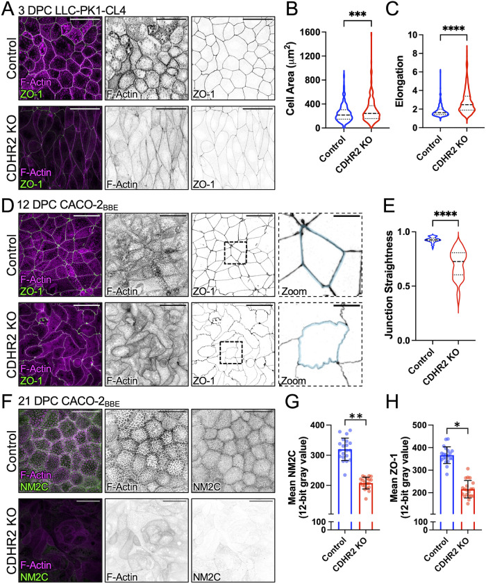
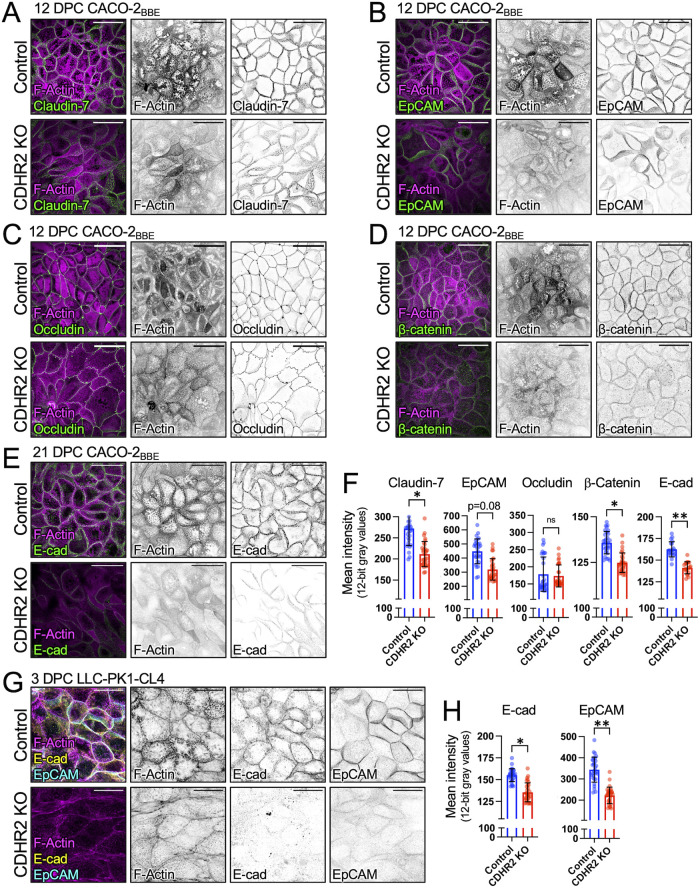
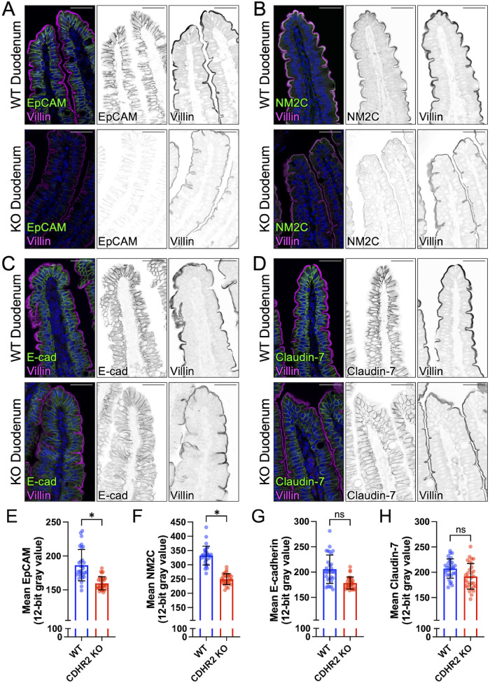
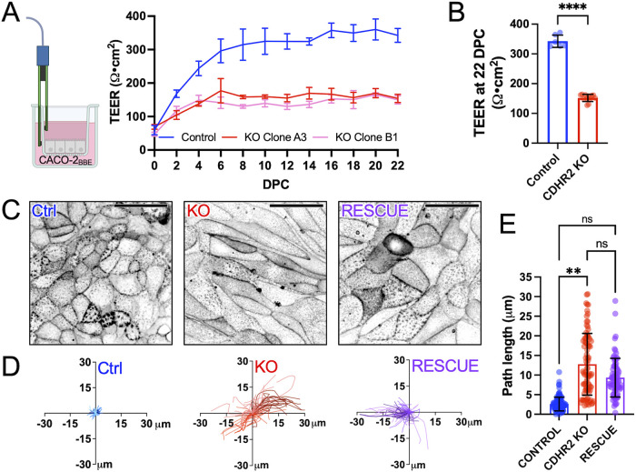
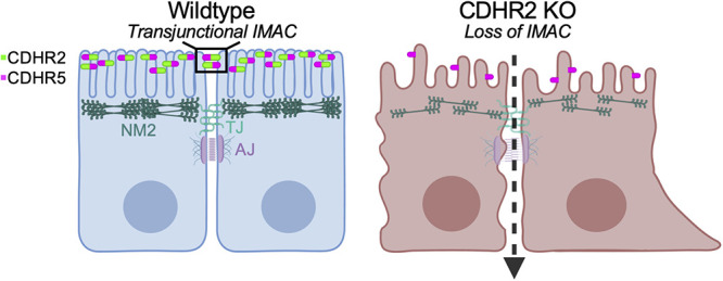
Transporting epithelial cells in the gut and kidney rely on protocadherin-based apical adhesion complexes to organize microvilli that extend into luminal space. In these systems, CDHR2 and CDHR5 localize to the distal ends of microvilli, where they form an intermicrovillar adhesion complex (IMAC) that links the tips of these structures, promotes the formation of a well-ordered array of protrusions, and thus maximizes apical membrane surface area. Recently, we discovered that IMACs can also form between microvilli that extend from neighboring cells, across cell-cell junctions. As an additional point of physical contact between cells, transjunctional IMACs are well positioned to impact the integrity of canonical tight and adherens junctions that form more basolaterally. To begin to test this idea, we examined cell culture and mouse models that lacked CDHR2 expression and were unable to form IMACs. CDHR2 knockout perturbed cell and junction morphology, reduced key components from tight and adherens junctions, impaired barrier function, and increased the motility of single cells within established monolayers. These results support the hypothesis that, in addition to organizing apical microvilli, IMACs provide a layer of cell-cell contact that functions in parallel with canonical tight and adherens junctions to promote epithelial functions.


Conflict of interest statement

Conflicts of interest: The authors declare no financial conflict of interest.

Figures

FIGURE 1:
FIGURE 1:
CDHR2 KO cells exhibit aberrant cell morphologies and decreased apical junction markers. (A) Three DPC Control and CDHR2 KO CL4 cells stained for F-actin (magenta) and ZO-1 (green). (B) Cell area measured in Control cells (n = 384) and CDHR2 KO cells (n = 259) from three experimental replicates. (C) Cell elongation (max Feret/min Feret ratio) measured from the cells analyzed in B. (D) A total of 12 DPC Control and CDHR2 KO CACO-2BBE cells stained for F-actin (magenta) and ZO-1 (green). Zooms show area in the dashed boxes, in this case highlighting straight versus ruffled junctions in Control and CDHR2 KO, respectively. (E) Junctional straightness (see Materials and Methods) from Control and KO cell junctional segments where 1.0 is a straight line; n = 62 segments from three experimental replicates per condition. (F) 21 DPC Control and CDHR2 KO CACO-2BBE cells stained for F-actin (magenta) and NM2C (green). (G) Mean NM2C and (H) ZO-1 intensities measured in CACO-2BBE cells from two experimental replicates, 10 fields per replicate, 20 fields total. Break in Y axis represents level of background fluorescence. In G and H, points represent individual image fields with mean ± SD. Significance levels from unpaired t tests calculated for experimental replicates are also shown above each plot (*p < 0.05, **p < 0.005). Scale bars: 40 µm (A, D, F), 10 µm (D, Zooms).
FIGURE 2:
FIGURE 2:
CDHR2 KO cells exhibit defects in junctional composition. Confocal images of (A) 12 DPC Control and CDHR2 KO CACO-2BBE cells stained for F-actin (magenta) and Claudin-7 (green), (B) F-actin (magenta) and EpCAM (green), (C) F-actin (magenta) and Occludin (green), and (D) F-actin (magenta) and β-catenin (green). (E) Confocal images of 21 DPC Control and CDHR2 KO CACO-2BBE cells stained for F-actin (magenta) and E-cadherin (green). (F) Mean Control and CDHR2 KO CACO-2 BBE junctional protein fluorescence intensities (12-bit gray values) for Claudin-7 (n = 3), EpCAM (n = 3), Occludin (n = 2), β-catenin (n = 3), and E-cadherin (n = 2), where n = number of experimental replicates and each replicate consists of 10 image fields at 60X. (G) Confocal images of three DPC Control and CDHR2 KO CL4 cells stained for F-actin (magenta), E-cadherin (yellow), and EpCAM (cyan). (H) Mean Control and CDHR2 KO CL4 fluorescence intensities (12-bit gray values) for E-cadherin (n = 3) and EpCAM (n = 3), where n = number of experimental replicates and each replicate consists of 10 image fields at 60X. For plots in F and H, points represent individual 60X fields with mean ± SD. Significance levels from unpaired t tests calculated for experimental replicates are shown above each plot (*p < 0.05, **p < 0.005). Break in Y axes represents level of background fluorescence. Scale bars: 40 µm (A–E) and 20 µm (G).
FIGURE 3:
FIGURE 3:
CDHR2 KO mouse duodenum exhibits reduced levels of some junctional components. Confocal images of wildtype (WT) and CDHR2 KO duodenal sections stained for villin (magenta) and the following markers in green: (A) EpCAM, (B) NM2C, (C) E-cadherin, and (D) Claudin-7. Mean intensities (12-bit gray values) measured for (E) EpCAM, (F) NM2C, (G) E-cadherin, and (H) Claudin-7. Nuclei are marked by DRAQ5 (blue). Intensity measurements for each marker were derived from two WT and CDHR2 KO littermate pairs and 15 image fields at 40X were analyzed from each animal. For plots in E–H, points represent individual 40X image fields with mean ± SD. Significance levels from unpaired t tests calculated for experimental replicates are shown above each plot (*p < 0.05). Break in Y axes represents level of background fluorescence. Scale bars: 40 µm (A–D).
FIGURE 4:
FIGURE 4:
CDHR2 KO monolayers exhibit decreased TEER and increased motility. (A) CACO cells were seeded on Transwell inserts and TEER was measured every other day postseeding to 22 DPC. (B) Mean TEER values from n = 6 Control and n = 12 CDHR2 KO (6 per CDHR2 KO clone) Transwells at 22 DPC: 342.6 ± 20.8 Ω·cm2 (Control) versus 152.1 ± 12.2 Ω·cm2 (CDHR2 KO clones). (C) Confocal images of live 3 DPC Control, CDHR2 KO, and Halo-CDHR2 Rescue CL4 cell monolayers labeled with membrane marker CellBriteSteady650. (D) Single-cell trajectories derived from Control, CDHR2 KO, and Halo-CDHR2 Rescue CL4 monolayers imaged for over 8 h. For each condition, three experimental replicates were imaged, and trajectories were calculated from 20 cells in each replicate (60 tracks in total per condition). (E) Total path lengths from trajectories shown in D; points represent individual trajectories shown with mean ± SD. Significance levels from pairwise comparisons using ANOVA calculated for experimental replicates are shown (**p < 0.005).
FIGURE 5:
FIGURE 5:
Loss of CDHR2-dependent intermicrovillar adhesion impairs basolateral junctions: a working model. Features of the CDHR2 KO phenotype include decreased microvillar clustering, aberrant cell morphology, reduced junctional tension, and a loss of key junctional proteins. These defects also give rise to increased junctional permeability and impaired collective cell migration during wound healing.

Update of

References

    1. Aman A, Piotrowski T (2008). Wnt/beta-catenin and Fgf signaling control collective cell migration by restricting chemokine receptor expression. Dev Cell 15, 749–761. - PubMed
    1. Bondow BJ, Faber ML, Wojta KJ, Walker EM, Battle MA (2012). E-cadherin is required for intestinal morphogenesis in the mouse. Dev Biol 371, 1–12. - PMC - PubMed
    1. Buckley CD, Tan J, Anderson KL, Hanein D, Volkmann N, Weis WI, Nelson WJ, Dunn AR (2014). Cell adhesion. The minimal cadherin-catenin complex binds to actin filaments under force. Science 346, 1254211. - PMC - PubMed
    1. Buckley CE, St Johnston D (2022). Apical-basal polarity and the control of epithelial form and function. Nat Rev Mol Cell Biol 23, 559–577. - PubMed
    1. Cencer CS, Silverman JB, Meenderink LM, Krystofiak ES, Millis BA, Tyska MJ (2023). Adhesion-based capture stabilizes nascent microvilli at epithelial cell junctions. Dev Cell 58, 2048–2062.e7. - PMC - PubMed