. 2024 Dec 3;147(12):4033-4042.
doi: 10.1093/brain/awae299.

Biallelic EPB41L3 variants underlie a developmental disorder with seizures and myelination defects

Affiliations

Biallelic EPB41L3 variants underlie a developmental disorder with seizures and myelination defects

Elizabeth A Werren et al. Brain. .

Abstract

Erythrocyte membrane protein band 4.1 like 3 (EPB41L3: NM_012307.5), also known as DAL1, encodes the ubiquitously expressed, neuronally enriched 4.1B protein, part of the 4.1 superfamily of membrane-cytoskeleton adaptors. The 4.1B protein plays key roles in cell spreading, migration and cytoskeletal scaffolding that support oligodendrocyte axon adhesions essential for proper myelination. We herein describe six individuals from five unrelated families with global developmental delay, intellectual disability, seizures, hypotonia, neuroregression and delayed myelination. Exome sequencing identified biallelic variants in EPB41L3 in all affected individuals: two nonsense [c.466C>T, p.(R156*); c.2776C>T, p.(R926*)] and three frameshift [c.666delT, p.(F222Lfs*46); c.2289dupC, p.(V764Rfs*19); c.948_949delTG, p.(A317Kfs*33)]. Quantitative-real time PCR and western blot analyses of human fibroblasts harbouring EPB41L3:c.666delT, p.(F222Lfs*46) indicated ablation of EPB41L3 mRNA and 4.1B protein expression. Inhibition of the nonsense mediated decay (NMD) pathway led to an upregulation of EPB41L3:c.666delT transcripts, supporting NMD as a pathogenic mechanism. Epb41l3-deficient mouse oligodendroglia cells showed significant reduction in mRNA expression of key myelin genes, reduced branching and increased apoptosis. Our report provides the first clinical description of an autosomal recessive disorder associated with variants in EPB41L3, which we refer to as EPB41L3-associated developmental disorder (EADD). Moreover, our functional studies substantiate the pathogenicity of EPB41L3 hypothesized loss-of-function variants.

Keywords: 4.1B; delayed myelination; loss-of-function; neurodevelopmental disorder; oligodendroglia.


Conflict of interest statement

Q.S.P. receives a professional services fee from Merck as part of editorial duties for the Merck manual. All other authors report no competing interests.

Figures
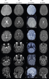
Figure 1
Figure 1
Neurological phenotypes in individuals with biallelic EPB41L3 variants. MRI findings of affected individuals with EPB41L3-associated developmental disorder (EADD). Axial T2-weighted images of Patients P1–P3 and P6 showing delayed myelination. Sagittal T1-weighted images of Patient P1 showing normal midline structures, Patient P2 showing mild cerebellar atrophy, and Patients P3 and P6 showing thin corpus callosum.
Figure 2
Figure 2
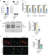
Biallelic variants EPB41L3 are loss-of-function. (A) EPB41L3 (4.1B) linear protein map with biallelic variants identified in individuals with EPB41L3-associated developmental disorder (EADD). Squares denote nonsense and circles denote frameshift variants. All variants localize to the FERM (blue), SAB (yellow) or C-terminal (green) protein domains of 4.1B, based on the Uniprot Q9Y2J2 map. (B) Cross-species evaluation of conservation at affected residues. (C) Relative mRNA expression of EPB41L3 in untreated EPB41L3F222Lfs*46/F222Lfs*46 affected (EPB41L3fs/fs) and control fibroblasts (EPB41L3+/+) measured by quantitative reverse transcription (RT-qPCR) and normalized to ACTB expression (far left) across two independent primer sets (Supplementary Table 5). EPB41L3 mRNA expression relative to ACTB control in fibroblasts treated with DMSO and cycloheximide (CHX) for 5 and 15 h (right). n = 3 independent experiments. Error bars represent standard error of the mean (±SEM). Significance measured by two-way ANOVA (***P < 0.001). (D) Representative (colour inverted) image of western blots probed with anti-EPB41L3 (125 kD) and control anti-GAPDH (36 kD) in affected EPB41L3F222Lfs*46/F222Lfs*46 (EPB41L3fs/fs) and control EPB41L3+/+ fibroblasts. (E) Publicly available RNA sequencing datasets, showing cell-type expression of Epb41l3 in mouse (top) and EPB41L3 in human (bottom) brain. EN = endothelial; fAS = fetal astrocytes; mAS = mature astrocytes; MG/MP = microglia/macrophage; NFO = newly formed oligodendrocytes; OL = oligodendrocytes; OPC = oligodendrocyte precursor cells.
Figure 3
Figure 3
Epb41l3 knockdown is detrimental to mouse oligodendrocyte function and survival. (A) Percentage of apoptotic transfected mouse Oli-neu (left) and Neuro2A (right) cells, as measured by cleaved caspase-3 (Casp3). n = >200 cells per condition across three independent experiments. Significance measured by Student’s t-test (***P < 0.001). (B) Expression of myelin specific genes (Plp1, Mbp and Cnp) by reverse transcription quantitative PCR (RT-qPCR) in mouse Oli-neu cells transfected with a scrambled control (pshScrb) and Epb41l3 shRNA (pshEpb41l3) plasmids relative to untransfected (UT) cells. Expression was normalized to internal control Actb and plotted relative to expression in untransfected control cells (n = 3 replicates). Error bars represented as ± standard error of the mean. Significance measured by Student’s t-test relative to controls (*P < 0.05, **P < 0.01, ***P < 0.001). (C) Left: Representative (colour inverted) images of Western blots probed with anti-CNP (myelin protein) and anti-GAPDH control, from transfected (UT), pshScrb-transfected and pshEpb41l3-transfected mouse Oli-neu cells. Right: Quantification of CNP protein abundance (normalized to GAPDH) and relative to normalized CNP abundance in untransfected cells (n = 3 independent experiments). (D) Immunostaining of CNP in pshEpb41l3-transfected mouse Oli-neu cells (bottom) compared to pshScrb-transfected control (top). Plasmid expression visualized with anti-GFP (middle). Scale bar = 50 µm. (E) Percentage of mouse Oli-neu cells with more than two processes. n = >200 cells per condition across three independent experiments. Error bars represent ± standard error of the mean. Significance measured by Student’s t-test (***P < 0.001).

References

    1. Wang J, Song J, An C, et al. A 130-kDa protein 4.1B regulates cell adhesion, spreading, and migration of mouse embryo fibroblasts by influencing actin cytoskeleton organization. J Biol Chem. 2014;289:5925–5937. - PMC - PubMed
    1. Baines AJ, Lu HC, Bennett PM. The protein 4.1 family: Hub proteins in animals for organizing membrane proteins. Biochim Biophys Acta. 2014;1838:605–619. - PubMed
    1. Peters LL, Weier H-UG, Walensky LD, et al. Four paralogous protein 4.1 genes map to distinct chromosomes in mouse and human. Genomics. 1998;54:348–350. - PubMed
    1. Sun C-X, Robb VA, Gutmann DH. Protein 4.1 tumor suppressors: Getting a FERM grip on growth regulation. J Cell Sci. 2002;115:3991–4000. - PubMed
    1. Parra M, Gee S, Chan N, et al. Differential domain evolution and complex RNA processing in a family of paralogous EPB41 (protein 4.1) genes facilitate expression of diverse tissue-specific isoforms. Genomics. 2004;84:637–646. - PubMed

Substances