

A multi-institutional study to investigate the sparing effect after whole brain electron FLASH in mice: Reproducibility and temporal evolution of functional, electrophysiological, and neurogenic endpoints
- PMID: 39293721
- PMCID: PMC11588524
- DOI: 10.1016/j.radonc.2024.110534
A multi-institutional study to investigate the sparing effect after whole brain electron FLASH in mice: Reproducibility and temporal evolution of functional, electrophysiological, and neurogenic endpoints
Abstract
Background and purpose: Ultra-high dose-rate radiotherapy (FLASH) has been shown to mitigate normal tissue toxicities associated with conventional dose rate radiotherapy (CONV) without compromising tumor killing in preclinical models. A prominent challenge in preclinical radiation research, including FLASH, is validating both the physical dosimetry and the biological effects across multiple institutions.
Materials and methods: We previously demonstrated dosimetric reproducibility of two different electron FLASH devices at separate institutions using standardized phantoms and dosimeters. In this study, tumor-free adult female mice were given 10 Gy whole brain FLASH and CONV irradiation at both institutions and evaluated for the reproducibility and temporal evolution of multiple neurobiological endpoints.
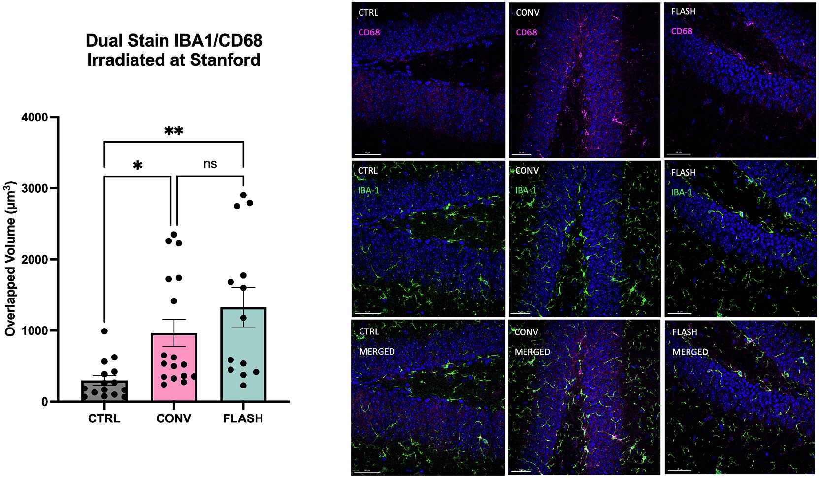
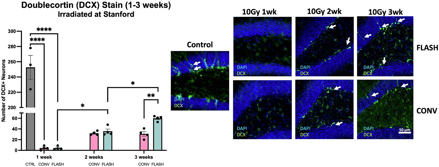
Results: FLASH sparing of behavioral performance on novel object recognition (4 months post-irradiation) and of electrophysiologic long-term potentiation (LTP, 5 months post-irradiation) was reproduced between institutions. Differences between FLASH and CONV on the endpoints of hippocampal neurogenesis (Sox2, doublecortin), neuroinflammation (microglial activation), and electrophysiology (LTP) were not observed at early times (48 h to 2 weeks), but recovery of immature neurons by 3 weeks was greater with FLASH.
Conclusion: In summary, we demonstrated reproducible FLASH sparing effects on the brain between two different beams at two different institutions with validated dosimetry. FLASH sparing effects on the endpoints evaluated manifested at later but not the earliest time points.
Keywords: Electrophysiology; FLASH; Intercomparison; Neurobehavior; Neurogenesis; Neuroinflammation; Radiotherapy.
Copyright © 2024 The Author(s). Published by Elsevier B.V. All rights reserved.
Conflict of interest statement
Declaration of competing interest The authors declare that they have no known competing financial interests or personal relationships that could have appeared to influence the work reported in this paper.
Figures


Similar articles
-
Ex vivo brain MRI to assess conventional and FLASH brain irradiation effects.Radiother Oncol. 2025 Jul;208:110894. doi: 10.1016/j.radonc.2025.110894. Epub 2025 Apr 14. Radiother Oncol. 2025. PMID: 40233872
-
Dosimetric and biologic intercomparison between electron and proton FLASH beams.Radiother Oncol. 2024 Jan;190:109953. doi: 10.1016/j.radonc.2023.109953. Epub 2023 Oct 13. Radiother Oncol. 2024. PMID: 37839557
-
FLASH Effects Induced by Orthovoltage X-Rays.Int J Radiat Oncol Biol Phys. 2023 Nov 15;117(4):1018-1027. doi: 10.1016/j.ijrobp.2023.06.006. Epub 2023 Jun 25. Int J Radiat Oncol Biol Phys. 2023. PMID: 37364800 Free PMC article.
-
Ultra-high dose rate electron beams and the FLASH effect: From preclinical evidence to a new radiotherapy paradigm.Med Phys. 2022 Mar;49(3):2082-2095. doi: 10.1002/mp.15442. Epub 2022 Jan 19. Med Phys. 2022. PMID: 34997969 Free PMC article. Review.
-
FLASH Radiotherapy: What Can FLASH's Ultra High Dose Rate Offer to the Treatment of Patients With Sarcoma?Semin Radiat Oncol. 2024 Apr;34(2):218-228. doi: 10.1016/j.semradonc.2024.02.001. Semin Radiat Oncol. 2024. PMID: 38508786 Review.
Cited by
-
Effectiveness of FLASH vs. Conventional Dose Rate Radiotherapy in a Model of Orthotopic, Murine Breast Cancer.Cancers (Basel). 2025 Mar 25;17(7):1095. doi: 10.3390/cancers17071095. Cancers (Basel). 2025. PMID: 40227580 Free PMC article.
-
FLASH radiotherapy at a crossroads: a bibliometric perspective on progress and challenges.Discov Oncol. 2025 Aug 17;16(1):1570. doi: 10.1007/s12672-025-03400-7. Discov Oncol. 2025. PMID: 40819335 Free PMC article.
-
Navigating the Critical Translational Questions for Implementing FLASH in the Clinic.Semin Radiat Oncol. 2024 Jul;34(3):351-364. doi: 10.1016/j.semradonc.2024.04.008. Semin Radiat Oncol. 2024. PMID: 38880544 Free PMC article. Review.
References
-
- Vozenin MC, Bourhis J, Durante M. Towards clinical translation of FLASH radiotherapy. Nat Rev Clin Oncol 2022;19:791–803. - PubMed
-
- Montay-Gruel P, Petersson K, Jaccard M, Boivin G, Germond JF, Petit B, et al. Irradiation in a flash: Unique sparing of memory in mice after whole brain irradiation with dose rates above 100Gy/s. Radiother Oncol 2017;124:365–9. - PubMed
-
- Montay-Gruel P, Bouchet A, Jaccard M, Patin D, Serduc R, Aim W, et al. X-rays can trigger the FLASH effect: Ultra-high dose-rate synchrotron light source prevents normal brain injury after whole brain irradiation in mice. Radiother Oncol 2018; 129:582–8. - PubMed
-
- Simmons DA, Lartey FM, Schuler E, Rafat M, King G, Kim A, et al. Reduced cognitive deficits after FLASH irradiation of whole mouse brain are associated with less hippocampal dendritic spine loss and neuroinflammation. Radiother Oncol 2019;139:4–10. - PubMed
