. 2024 Sep 18;14(1):21810.
doi: 10.1038/s41598-024-66781-9.

Unveiling Lobophytum sp. the neuroprotective potential of Parkinson's disease through multifaceted mechanisms, supported by metabolomic analysis and network pharmacology

Affiliations

Unveiling Lobophytum sp. the neuroprotective potential of Parkinson's disease through multifaceted mechanisms, supported by metabolomic analysis and network pharmacology

Hussain T Bakhsh et al. Sci Rep. .

Abstract

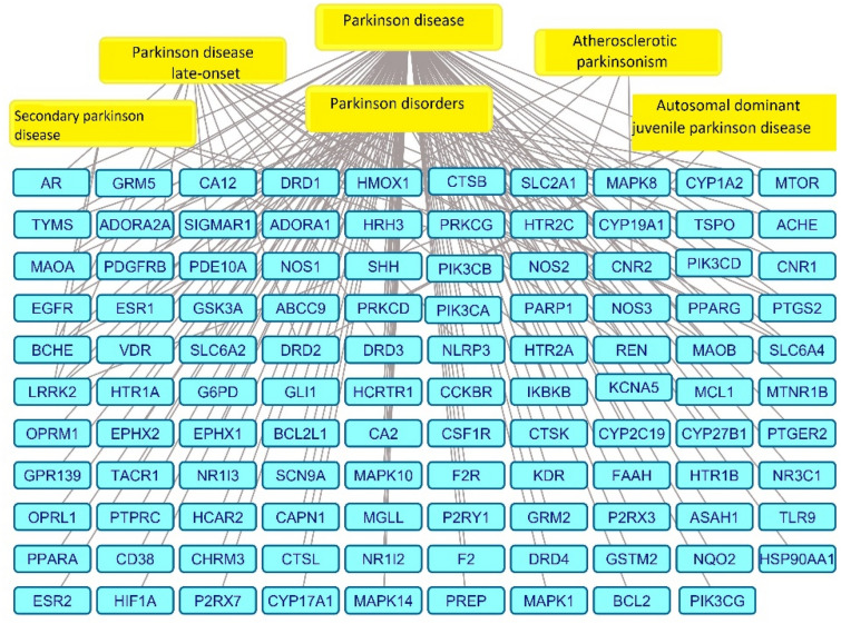
A main feature of neurodegenerative diseases is the loss of neurons. One of the most prevalent neurodegenerative illnesses is Parkinson disease (PD). Although several medications are already approved to treat neurodegenerative disorders, most of them only address associated symptoms. The main aim of the current study was to examine the neuroprotective efficacy and underlying mechanism of Lobophytum sp. crude extract in a rotenone-induced rat model of neurodegeneration mimicking PD in humans. The influence of the treatment on antioxidant, inflammatory, and apoptotic markers was assessed in addition to the investigation of TH (tyrosine hydroxylase) immunochemistry, histopathological changes, and α-synuclein. Metabolomic profiling of Lobophytum sp. crude extract was done by using High-Resolution Liquid Chromatography coupled with Mass Spectrometry (HR-LC-ESI-MS), which revealed the presence of 20 compounds (1-20) belonging to several classes of secondary metabolites including diterpenoids, sesquiterpenoids, steroids, and steroid glycosides. From our experimental results, we report that Lobophytum sp. extract conferred neuroprotection against rotenone-induced PD by inhibiting ROS formation, apoptosis, and inflammatory mediators including IL-6, IL-1β, and TNF-α, NF-кB, and subsequent neurodegeneration as evidenced by decreased α-synuclein deposition and enhanced tyrosine hydroxylase immunoreactivity. Moreover, a computational network pharmacology study was performed for the dereplicated compounds from Lobophytum sp. using PubChem, SwissTarget Prediction, STRING, DisGeNET, and ShinyGO databases. Among the studied genes, CYP19A1 was the top gene related to Parkinson's disease. Dendrinolide compounds annotated a high number of parkinsonism genes. The vascular endothelial growth factor (VEGF) pathway was the top signaling pathway related to the studied genes. Therefore, we speculate that Lobophytum sp. extract, owing to its pleiotropic mechanisms, could be further developed as a possible therapeutic drug for treating Parkinson's disease.

Keywords: Lobophytum sp.; Network pharmacology; Neurodegenerative diseases; Parkinson's disease; Rotenone (ROT); VEGF pathway.


Conflict of interest statement

The authors declare no competing interests.

Figures

Figure 1
Figure 1
A photo-micrograph showing the brain tissue (corpus striatum, CS) of all studied groups: Group I, (A); Control Group; showing cluster of branched multipolar neurons (thick arrows) with moderate-dense basophilic cytoplasm and large pale nuclei. Normal blood vessels (arrows) and slight neuroglia cells infiltration (g). (B1,2,3); Parkinsonism Group; showing many neurons appear shrunken surrounded by perineuronal hallows (tailed arrows). Notice the increased interstitial spaces, dilated blood vessels (arrows) and infiltration by excess neuroglia cells (g). The neuropil has many vacuoles (V) and acidophilic aggregation of Lewy bodies (striped arrow) and gliosis (star) and a group of macrophages with pigmented material (macrophages). Group III, (C); (Lobophytum-treated Group); showing neuronal normal picture with intense basophilic cytoplasm and vesicular nuclei (arrows). Group IV, (D); (Rotenone + Lobophytum-treated Group); showing less apoptotic neuronal cells but excess infiltration of neuroglia cells (g). Some cells still shrunken and surrounded with empty hallows (arrows) (H&E staining ×200 and 400).
Figure 2
Figure 2
A photo-micrograph of the brain tissue (substantia nigra pars compacta, SNC) of all studied groups, to show a closer view of the darkly pigmented dopaminergic neurons. GI, (A) Control Group; showing cluster of closely packed neurons (red arrows) and black cells are the neuromelanin-containing dopaminergic neurons (yellow arrows). Group II, (B) Parkinsonism Group; showing severe loss of dopaminergic cells. Cells showing prominent aggregation of acidophilic plaques “Lewy bodies” (black arrows). GIII, (C); (Lobophytum-treated Group); have apparently the same picture of the control group. GIV, (D) (rotenone + Lobophytum-treated Group); show mild cell dropout accompanied by moderate aggregation of Lewy bodies (black arrows). (H&E staining ×200 and 400).
Figure 3
Figure 3
Histological findings in the brain corpus striatum “CS” and subtantia nigra pars compacta “SNC” immune-stained with tyrosine hydroxylase “TH” of all studied groups: “CS” (A), Control Group; showing densely packed strong positive immune-reactive neurons (thick arrows) and dense positive TH immunoreactive neuropil (stars) “CS” (B1 and 2), Parkinsonism Group; showing less numerous TH immune-positive neurons, peri-nuclear aggregates of dense inclusions (tailed arrows) and beaded or swollen processes (linear arrow). B2; showing immune-negative areas of neuropil (black star) and other areas are immune-positive (yellow star). “CS” (C), (lobophytum-treated Group); have apparently the same picture of the Control Group; strong cytoplasmic TH immune-reaction (thick arrows) and strong immune-reactive neuropil (star). “CS” (D), (Rotenone + Lobophytum-treated Group); neurons showing strong cytoplasmic immune-reaction (arrows) and mild “TH” immune-reactive neuropil (star). “SNC” (E); Control Group; showing densely packed strong positive immune-reactive neurons (thick arrows) and dense positive TH immunoreaction “SNC” (F); Parkinsonism Group; showing few “TH” immune-positive cells (thick arrows) and the neuropil shows beaded or swollen processes (linear arrows) “SNC” (G); (Lobophytum-treated Group); showing have apparently the same picture of the control group. Strong immune-positive neuronal cytoplasmic expression (thick arrows) and strong immune-reactive neuropil. “SNC” (H); (Rotenone + Lobophytum-treated Group); shows strong immune-reactive cytoplasmic expression (arrows) but mild immune-reactive neuropil (tyrosine hydroxylase “TH” immune-staining × 200 and 400).
Figure 4
Figure 4
The impact of the Lobophytum sp. extract on oxidative stress parameters was assessed in a rat model of Parkinson disease. (A): SOD activity, (B): MDA levels. The values are expressed as the mean ± SD. A one-way ANOVA was utilized, followed by a Tukey’s test for conducting multiple comparisons. *p < 0.05 as compared to the control group; #p < 0.05 as compared to the ROT-alone group.
Figure 5
Figure 5
Effect of extract on cholinesterase activity. Values were represented as mean ± SD. A one-way ANOVA test was employed followed by Tukey’s test for multiple comparisons. ****p < 0.0001 as compared to the control group; ###p < 0.001 as compared to the ROT-alone group.
Figure 6
Figure 6
Gene expression of (A): Bcl-2-associated X protein (BAX), (B): Bcl-2 in all study groups. To analyze gene expression in injured tissues, qRT-PCR was used. Following normalization to GAPDH, the data were displayed as fold change compared to the control group. The bars on the graph depict the mean ± SD. One-way ANOVA test was done to calculate statistical difference followed by Tukey’s test for multiple comparisons. *p < 0.05 as compared to the control group; #p < 0.05 as compared to the rotenone-alone group.
Figure 7
Figure 7
Gene expression analysis of pro-inflammatory cytokines in all study groups. (A): TNF-α, (B): IL-6, (C): IL-1β and (D): NF-ƘB. To analyze gene expression in injured tissues, qRT-PCR was used. Following normalization to GAPDH, the data were  displayed as fold change compared to the control group. The bars on the graph depict the mean ± SD. One-way ANOVA test was done to calculate statistical difference followed by Tukey’s test for multiple comparisons. *p < 0.05 as compared to the control group; #p < 0.05 as compared to the ROT-alone group.
Figure 8
Figure 8
Effect of the Lobophytum sp. extract on protein expression of α-synuclein. The bars indicate the mean ± SD. Statistical significance between groups was calculated using one-way ANOVA test and Tukey’s test, where *p < 0.05 indicated significance when compared to the control group, and #p < 0.05 when compared to the ROT group.
Figure 9
Figure 9
Gene-Parkinsonism’s disease network; a network describing the identified genes related to Parkinsonism’s disease, the blue rectangles represent genes, and the yellow rectangles represent Parkinsonism’s disease.
Figure 10
Figure 10
Metabolites-Parkinsonism Genes; a network describing the metabolites links to genes related to parkinsonism; yellow circles are the identified metabolites from Lobophytum sp. The bigger the circle, the more edges, green circles represent the genes related to parkinsonism, and the bigger the circle, the more edges.
Figure 11
Figure 11
(A) PPI Network of the identified genes associated with parkinsonism; (B) the top 10 proteins arranged according to the node degree.
Figure 12
Figure 12
Complete pharmacology network: a network describing the interaction between the Lobophytum sp. extract, the identified metabolites, and the genes related to parkinsonism and parkinsonism conditions. The orange rectangle is Lobophytum sp., the yellow oval shapes represent the metabolites, green diamonds represent genes related to parkinsonism, and blue rectangles represent parkinsonism conditions.
Figure 13
Figure 13
The top 20 KEGG biological pathways.
Figure 14
Figure 14
VEGF signaling pathway (the top identified signaling pathway).
Figure 15
Figure 15
Chemical structures of dereplicated compounds from Lobophytum sp.

Similar articles

Cited by

References

    1. Marras, C. et al. Dihydropyridine calcium channel blockers and the progression of parkinsonism. Ann. Neurol.71(3), 362–369 (2012). - PubMed
    1. Levin, J. et al. The differential diagnosis and treatment of atypical parkinsonism. Dtsch. Arztebl. Int.113(5), 61 (2016). - PMC - PubMed
    1. Roy, S. P. et al. Antiparkinsonian activity of Tabebuia impetiginosa bark and biochemical analysis of dopamine in rat brain homogenates. In Annales Pharmaceutiques Françaises. (Elsevier, 2022). - PubMed
    1. Yahr, M. D. et al. Treatment of parkinsonism with levodopa. Arch. Neurol.21(4), 343–354 (1969). - PubMed
    1. Roy, P. K. et al. New Casbane and cembrane diterpenoids from an Okinawan soft coral, Lobophytum sp. Molecules21(5), 679 (2016). - PMC - PubMed

LinkOut - more resources