

A comprehensive dataset of pattern electroretinograms for ocular electrophysiology research
- PMID: 39294170
- PMCID: PMC11410942
- DOI: 10.1038/s41597-024-03857-1
A comprehensive dataset of pattern electroretinograms for ocular electrophysiology research
Abstract
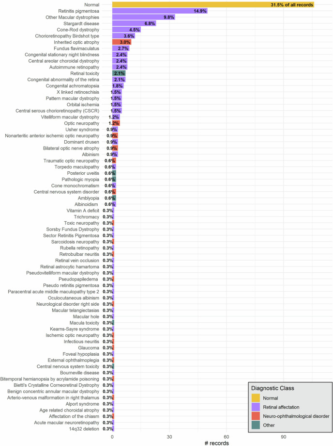
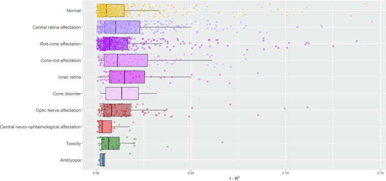
The Pattern Electroretinogram (PERG) is an essential tool in ophthalmic electrophysiology, providing an objective assessment of the central retinal function. It quantifies the activity of cells in the macula and the ganglion cells of the retina, assisting in the differentiation of macular and optic nerve conditions. In this study, we present the IOBA-PERG dataset, an extensive collection of 1354 transient PERG responses accessible on the PhysioNet repository. These recordings were conducted at the Institute of Applied Ophthalmobiology (IOBA) at University of Valladolid, over an extended period spanning nearly two decades, from 2003 to 2022. The dataset includes 336 records, ensuring at least one PERG signal per eye. The dataset thoughtfully includes demographic and clinical data, comprising information such as age, gender, visual acuity measurements, and expert diagnoses. This comprehensive dataset fills a gap in ocular electrophysiological repositories, enhancing ophthalmology research. Researchers can explore a broad range of eye-related conditions and diseases, leading to enhanced diagnostic accuracy, innovative treatment strategies, methodological advancements, and a deeper understanding of ocular electrophysiology.
© 2024. The Author(s).
Conflict of interest statement
The authors declare no competing interests. The funder had no role in the design of the study; in the collection of the data; in the writing of the manuscript, or in the decision to publish the results.
Figures


References
-
- Yu, M., Creel, D. J. & Iannaccone, A. Handbook of Clinical Electrophysiology of Vision, 10.1007/978-3-030-30417-1 (Springer, 2019).
Publication types
MeSH terms
Grants and funding
LinkOut - more resources
Full Text Sources
Miscellaneous
