. 2024 Oct;3(10):1236-1248.
doi: 10.1038/s44161-024-00542-9. Epub 2024 Sep 18.

Cardiac NAD+ depletion in mice promotes hypertrophic cardiomyopathy and arrhythmias prior to impaired bioenergetics

Affiliations

Cardiac NAD+ depletion in mice promotes hypertrophic cardiomyopathy and arrhythmias prior to impaired bioenergetics

Khanh V Doan et al. Nat Cardiovasc Res. 2024 Oct.

Abstract

Nicotinamide adenine dinucleotide (NAD+) is an essential co-factor in metabolic reactions and co-substrate for signaling enzymes. Failing human hearts display decreased expression of the major NAD+ biosynthetic enzyme nicotinamide phosphoribosyltransferase (Nampt) and lower NAD+ levels, and supplementation with NAD+ precursors is protective in preclinical models. Here we show that Nampt loss in adult cardiomyocytes caused depletion of NAD+ along with marked metabolic derangements, hypertrophic remodeling and sudden cardiac deaths, despite unchanged ejection fraction, endurance and mitochondrial respiratory capacity. These effects were directly attributable to NAD+ loss as all were ameliorated by restoring cardiac NAD+ levels with the NAD+ precursor nicotinamide riboside (NR). Electrocardiograms revealed that loss of myocardial Nampt caused a shortening of QT intervals with spontaneous lethal arrhythmias causing sudden cardiac death. Thus, changes in NAD+ concentration can have a profound influence on cardiac physiology even at levels sufficient to maintain energetics.


Conflict of interest statement

Competing interest statement

J.A.B is a consultant to Pfizer and Cytokinetics and has received research funding and materials from Elysium Health and Metro International Biotech, both of which have an interest in NAD+ precursors, as well as Calico. D.P.K is a consultant to Pfizer and Amgen. J.D.R. is an advisor and stockholder in Colorado Research Partners, L.E.A.F. Pharmaceuticals, Bantam Pharmaceuticals, Barer Institute, and Rafael Pharmaceuticals; a paid consultant of Pfizer and Third Rock Ventures; a founder, director, and stockholder of Farber Partners, Serien Therapeutics, and Sofro Pharmaceuticals; a founder and stockholder in Empress Therapeutics; inventor of patents held by Princeton University; and a director of the Princeton University-PKU Shenzhen collaboration. The remaining authors have nothing to declare.

Figures

Extended Data Figure 1.
Extended Data Figure 1.. Cardiomyocyte-specific deletion of Nampt in adult mouse hearts.
a, Western blots of protein lysates from heart ventricles verifying Nampt deletion at 2 and 4 weeks after tamoxifen treatment. Groups include Nampt flox/flox (NamptF/F or WT), Nampt flox/Wildtype × αMHC-MerCreMer (NamptF/+:MCM), Nampt flox/flox × αMHC-MerCreMer (NamptF/F:MCM). The experiment was repeated twice independently with similar results. b-d, Myocardial NAD+ (b), NADH (c) and NAD+/NADH ratio (d) measurements from left ventricular tissues. Data are represented as mean ± s.e.m. P values (two-sided) were determined by ordinary one-way ANOVA with Tukey’s post-hoc tests. Parentheses indicate number of mice.
Extended Data Figure 2.
Extended Data Figure 2.. Cardiac phenotypes of cardiomyocyte-specific Nampt KO mice.
a, b, NADP+ (a) and NADPH (b) relative levels in ventricular tissues. c, d, Blood glucose (c) and lactate (d) levels of WT and cNKO mice at 8 weeks post-TAM. e-g, Echocardiography measures of LV mass (e), LVPW thicknesses (f), and ejection fraction (EF) (g) of WT and cNKO mice before tamoxifen administration. h, i, LV mass (h) and LVPW thicknesses (i) of WT, cardiac Nampt heterozygous (NamptF/+:MCM), and cNKO mice at 4–6 weeks post-TAM. j-m, LV mass (j), LVPW thicknesses (k), LV volume (l), and EF (m) of WT and cNKO females at 8 weeks post-TAM. n, Wet/dried lung weight ratios. o, Representative trichrome staining images (top) and quantification of fibrosis area (bottom) for heart tissues from female mice (scale bar, 1mm at low-magnification and 50μm at high-magnification). p, Representative images of the hearts of cNKO mice following sudden cardiac death (SCD). Data are represented as mean ± s.e.m. P values (two-sided) were determined by two-tailed Student t-tests or ordinary one-way ANOVA with Tukey’s post-hoc tests for comparison of two or multiple groups, respectively. Parentheses indicate number of mice.
Extended Data Figure 3.
Extended Data Figure 3.. Cardiac metabolism and bioenergetics of Nampt-KO hearts.
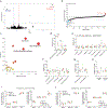
a, Volcano plot of differential gene expression (DGE) analysis showing genes with statistical significance in cNKO hearts at 2 weeks post-TAM. b, Metabolomics analysis of myocardial tissues harvested at 2 weeks post-TAM. Metabolites that were significantly changed with a P-value less than 0.05 were colored in red (increased) or blue (decreased) (n=5 mice/group). c, Metabolic pathway analysis from myocardial metabolite profiles of cNKO hearts versus control at 8-weeks post-TAM. d-g, Relative levels of metabolic intermediates of pentose phosphate pathway (d), tricarboxylic acid (TCA) cycle (e), branched-chain amino acids (f) and ketone bodies (g) in myocardial tissues at 8-weeks post-TAM measured by LC-MS (n=4 mice/group for WT and cNKO, n=8 mice for cNKO-NR group). h, Relative myocardial ATP levels in isolated mitochondria and heart ventricular tissues at 8-weeks post-TAM measured by LC-MS (n=4 mice/group for WT and cNKO, n=8 mice for cNKO-NR group). i, j, Respiration of isolated mitochondria from heart ventricles at 8 weeks post-TAM (i) and at 12 weeks post-TAM (j). Data are represented as mean ± s.e.m. P values (two-sided) were determined by two-tailed Student t-tests or ordinary one-way ANOVA with Tukey’s post-hoc tests for comparison of two or multiple groups, respectively. Parentheses indicate number of mice.
Extended Data Figure 4.
Extended Data Figure 4.. Characterization of sudden cardiac arrest and death in the cNKO mice.
a, Schematic of heart rate variability (HRV) analysis. b, Heart rate recorded from echocardiography. c-g, Parameters of heart rate variability (HRV) analysis using the ECG recordings from echocardiography performed at 1-week post-TAM including c, mean of normal-to-normal R-R intervals (avNN), d, square root of the mean squared differences of successive R-R intervals (RMSSD), e, percentage of successive R-R interval differences greater than 5 milliseconds (pNN5), f, high-frequency (HF) bands power, and g, very low-frequency (VLF) bands power. Data was represented as mean ± s.e.m. P values (two-sided) were determined by ordinary one-way ANOVA with Tukey’s post-hoc tests for comparison of two or multiple groups, respectively. Parentheses indicate number of mice.
Figure 1.
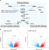
Figure 1.. Nampt mRNA expression is decreased in patients with cardiomyopathy.
a, Schematic of key NAD+ biosynthesis pathways and enzymes. NAD+ is primarily synthesized from tryptophan, nicotinic acid (NA), or nicotinamide (NAM). The NAM salvage pathway involves synthesis nicotinamide mononucleotide (NMN) from NAM through the rate-limiting enzyme nicotinamide phosphoribosyltransferase (Nampt). NMN is subsequently converted to NAD+ through the action of Nicotinamide mononucleotide adenylyl transferases (Nmnat1–3). Nicotinic acid mononucleotide (NAMN) is synthesized from tryptophan through multiple steps ending in quinolinate phosphoribosyltransferase (Qaprt)-catalyzed reaction or from NA via nicotinate phosphoribosyltransferase (Naprt). NAMN is converted to nicotinic acid adenine dinucleotide (NAAD) via Nmnat1–3 and subsequently to NAD+ through NAD+ synthetase (Nadsyn1). Nicotinamide riboside (NR) provides an alternative path to NMN by feeding into the salvage pathway via nicotinamide riboside kinase 1 or 2 (Nmrk1–2), bypassing Nampt. b, Volcano plots showing differential gene expression (DGE) analysis in dilated cardiomyopathy (DCM, left, n=161) and hypertrophic cardiomyopathy (HCM, right, n=28) patients versus the non-failing group (NF, n=161). P-values (two-sided) were determined by the Limma statistical analysis with DESeq2 package between groups and the Benjamini-Hochberg method was used to correct for multiple comparisons.
Figure 2.
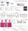
Figure 2.. Loss of cardiac NAD+ induces hypertrophic cardiomyopathy and sudden deaths.
a, Schematic of the conditional cardiac-specific inducible Nampt knockout (cNKO) mouse line. Conditional Nampt floxed mice were crossed with aMHC-MerCreMer (MCM) mice to allow inducible deletion of Nampt in the adult heart (5–6-months old) with TAM treatment (30 mg/kg/day for 3-consecutive days by I.P. injection). b, Experimental scheme for studying cardiac phenotypes of Nampt floxed (WT) and cNKO mice. A group of cNKO mice were administered nicotinamide riboside (NR) supplementation via oral delivery at the dose of 500 mg/kg/day, starting from the time of TAM treatment. c, Western blots of heart ventricle tissues at 8 weeks after TAM treatment. The experiment was repeated three times independently with similar results. d-f, Myocardial NAD+ (d), NADH (e) and NAD+/NADH ratio (f) in heart ventricular tissues. g, Body weight changes of WT and cNKO mice with or without NR treatment. h-l, Echocardiography measurement of left ventricle dimensions and contraction including representative M-mode images (h), left ventricular posterior wall (LVPW) thicknesses (i), LV mass (j), LV volume (k), and ejection fraction (EF) (l). m, Representative images of trichrome staining (top) and quantification of fibrosis area (bottom) for heart tissues from male mice (scale bar, 1mm at low-magnification and 50μm at high-magnification). n, Heart weight (HW) to body weight (BW) ratios. o, mRNA expression of genes related to hypertrophy (left) and fibrosis (right) in heart tissues. p, Kaplan-Meier survival analysis of cardiac Nampt knockout (cNKO) males (left) and females (right) with or without NR treatment. Data are represented as mean ± s.e.m. P values (two-sided) were determined by ordinary one-way ANOVA with Tukey’s post-hoc tests. For survival analysis, the Log-rank (Mantel-Cox) test was used. Parentheses indicate number of mice.
Figure 3.
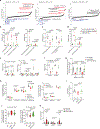
Figure 3.. Loss of NAD+ alters cardiac metabolism and mildly impacts myocardial bioenergetics.
a, Two-independent metabolomic datasets from myocardial tissues harvested at 8 weeks after TAM. Metabolites that were significantly changed with a P-value less than 0.05 were colored in red (increased) or blue (decreased). Dataset 2 included cNKO mice with NR rescue. (n=4–5 mice/group for WT and cNKO each dataset, n=8 mice for cNKO-NR group, dataset 2 only). b-f, Pooled data showing relative levels of indicated glycolytic intermediates (b), fatty acyl-carnitines (c), metabolites of the pentose phosphate pathway (d), metabolites of NAD+ metabolism (e), and ADP-ribose/cyclic ADP-ribose (f) in myocardial tissues measured by LC-MS (n=8–9 mice/group). g, Cardiac mitochondrial NAD+ levels at 8- and 12-weeks after TAM. h,i, Mitochondrial respiration of myocardial tissue lysates (h) and cardiac ATP levels (i) at 12 weeks after TAM. j, Time to exhaustion on a treadmill at indicated time points after TAM (parentheses indicate number of mice tested). k, Gene expression of Nppa, Nppb peptides, and Nmrk2 in myocardial tissues at 12 weeks after TAM. Data are represented as mean ± s.e.m. P values (two-sided) were determined by two-tailed Student t-tests or ordinary one-way ANOVA with Tukey’s post-hoc tests for comparison of two or multiple groups, respectively. Parentheses indicate number of mice.
Figure 4.
Figure 4.. Characterization of the electrical effects of cardiac NAD+ depletion prior to and at sudden death.
a, Schematic of telemetry study using implantable telemetric probes for ECG and blood pressure monitoring in Nampt floxed (WT) and cNKO mice. b-g, Changes of heart rate (b), systolic (c) and diastolic (d) blood pressure, QT intervals (inset: ECG cycle) (e), QRS complex (f), and ST intervals (g) of WT and cNKO mice before and at indicated time points after TAM treatment. h, The last 10 seconds of ECGs recorded from cNKO mice that suffered from a sudden cardiac death (SCD). Data are represented as mean ± s.e.m. P values (two-sided) were determined by two-tailed Student t-tests. Parentheses indicate number of mice.

References

    1. Belenky P, Bogan KL & Brenner C NAD+ metabolism in health and disease. Trends Biochem Sci 32, 12–9 (2007). - PubMed
    1. Goodman RP, Calvo SE & Mootha VK Spatiotemporal compartmentalization of hepatic NADH and NADPH metabolism. J Biol Chem 293, 7508–7516 (2018). - PMC - PubMed
    1. Xu P & Sauve AA Vitamin B3, the nicotinamide adenine dinucleotides and aging. Mechanisms of Ageing and Development 131, 287–298 (2010). - PubMed
    1. Cantó C, Menzies KJ & Auwerx J NAD+ Metabolism and the Control of Energy Homeostasis: A Balancing Act between Mitochondria and the Nucleus. Vol. 22 31–53 (2015). - PMC - PubMed
    1. Mouchiroud L, Houtkooper RH & Auwerx J NAD+ metabolism: a therapeutic target for age-related metabolic disease. Critical reviews in biochemistry and molecular biology 48, 397–408 (2013). - PMC - PubMed

MeSH terms

Grants and funding