. 2025 Aug;16(4):1156-1172.
doi: 10.1007/s12975-024-01295-0. Epub 2024 Sep 19.

12/15-Lipooxygenase Inhibition Reduces Microvessel Constriction and Microthrombi After Subarachnoid Hemorrhage in Mice

Affiliations

12/15-Lipooxygenase Inhibition Reduces Microvessel Constriction and Microthrombi After Subarachnoid Hemorrhage in Mice

Ari Dienel et al. Transl Stroke Res. 2025 Aug.

Abstract

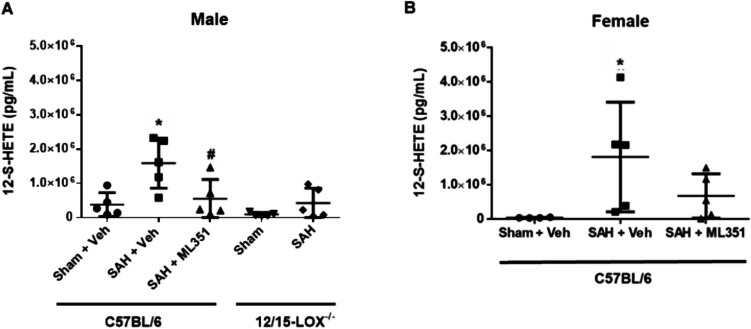
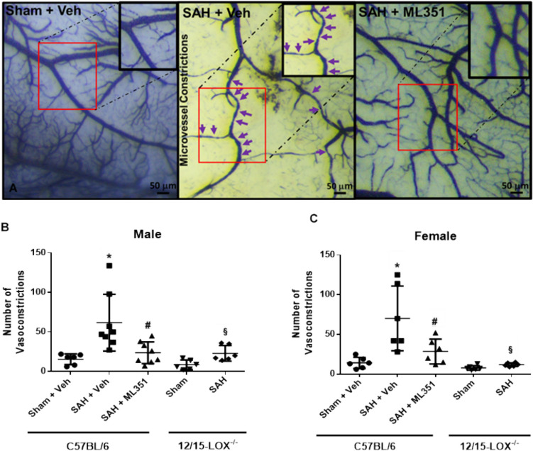
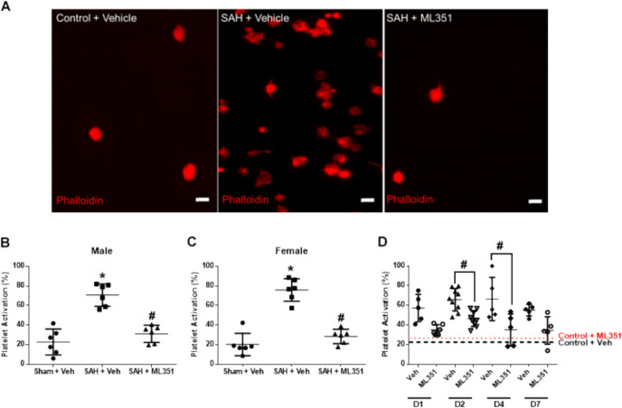
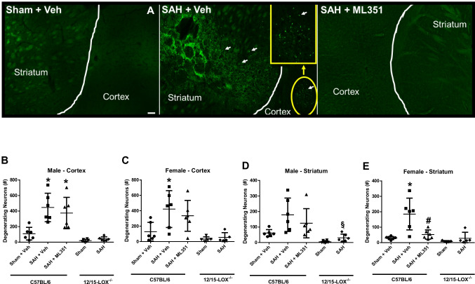
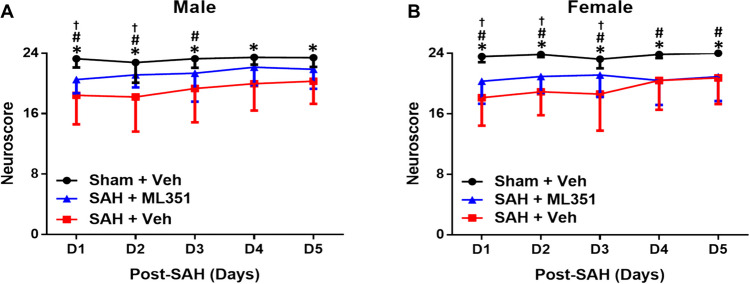
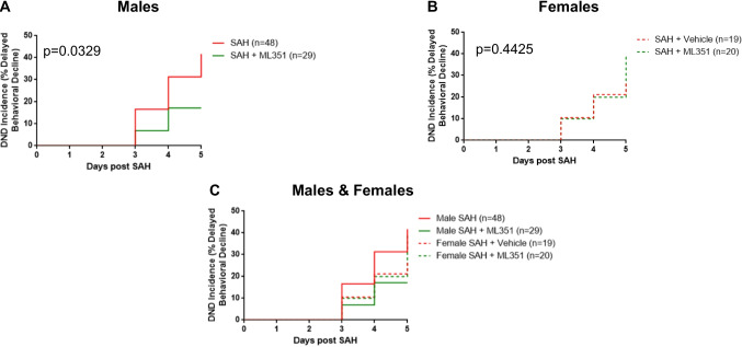
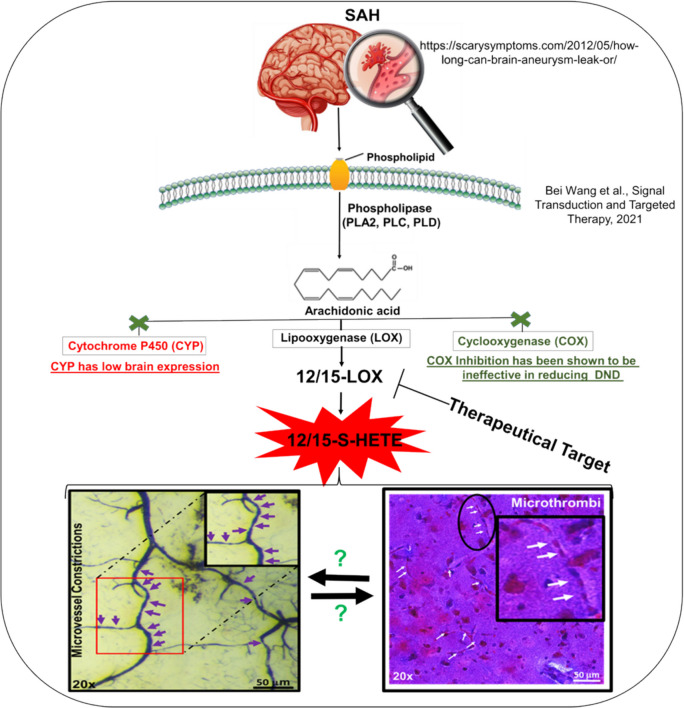
Impaired cerebral circulation, induced by blood vessel constrictions and microthrombi, leads to delayed cerebral ischemia after subarachnoid hemorrhage (SAH). 12/15-Lipooxygenase (12/15-LOX) overexpression has been implicated in worsening early brain injury outcomes following SAH. However, it is unknown if 12/15-LOX is important in delayed pathophysiological events after SAH. Since 12/15-LOX produces metabolites that induce inflammation and vasoconstriction, we hypothesized that 12/15-LOX leads to microvessel constriction and microthrombi formation after SAH, and thus, 12/15-LOX is an important target to prevent delayed cerebral ischemia. SAH was induced in C57BL/6 and 12/15-LOX-/- mice of both sexes by endovascular perforation. Expression of 12/15-LOX was assessed in brain tissue slices and in vitro. C57BL/6 mice were administered either ML351 (12/15-LOX inhibitor) or vehicle. Mice were evaluated for daily neuroscore and euthanized on day 5 to assess cerebral 12/15-LOX expression, vessel constrictions, platelet activation, microthrombi, neurodegeneration, infarction, cortical perfusion, and development of delayed deficits. Finally, the effect of 12/15-LOX inhibition on platelet activation was assessed in SAH patient samples using a platelet spreading assay. In SAH mice, 12/15-LOX was upregulated in brain vascular cells, and there was an increase in 12-S-HETE. Inhibition of 12/15-LOX improved brain perfusion on days 4-5 and attenuated delayed pathophysiological events, including microvessel constrictions, microthrombi, neuronal degeneration, and infarction. Additionally, 12/15-LOX inhibition reduced platelet activation in human and mouse blood samples. Cerebrovascular 12/15-LOX overexpression plays a major role in brain dysfunction after SAH by triggering microvessel constrictions and microthrombi formation, which reduces brain perfusion. Inhibiting 12/15-LOX may be a therapeutic target to improve outcomes after SAH.

Keywords: 12/15-Lipooxygenase; Arterioles; Delayed neurological deficit; Microthrombi; Microvessel constrictions; Platelets; Subarachnoid hemorrhage.


Conflict of interest statement

Declarations. Competing Interests: The authors declare no competing interests.

Figures

Fig. 1
Fig. 1
Expression of 12-LOX. A Quantification of 12-LOX protein in brain lysat. n = 6/group/sex and (B) its representative figure of Western blot. One-way ANOVA with Tukey. *p < 0.05 vs Sham, #p < 0.05 vs SAH D1. C–E Immunostaining for 12- and 15-LOX in brains from male mice 5 days after SAH. 12/15-LOX is green, laminin (vasculature) is red. Scale bar = 50 µm. F Quantification of 12-LOX in human brain pericytes, human brain microvascular endothelial cells, and mouse brain microvascular endothelial cells. n = 4/group, unpaired t-test, *p < 0.05, control vs Hb. G Protein expression (Western blot) of 12-LOX and β-Actin in human brain pericytes, human brain microvascular endothelial cells, and mouse brain microvascular endothelial cells
Fig. 2
Fig. 2
Plasma 12-S-HETE levels after SAH. n = 5/group/sex. Kruskal–Wallis with Dunn’s test. *p < 0.05 vs Sham, #p < 0.05 vs SAH D1, §p < 0.05 vs SAH D5
Fig. 3
Fig. 3
12/15-LOX inhibition reduces plasma 12-S-HETE on post-SAH day 5. A Male mice. B Female mice. n = 5/group/strain/sex, except for female Sham + Veh which n = 4. One-way ANOVA test with Tukey (male) and Dunn’s multiple comparison test (female), unpaired t-test for 12/15-LOX−/− mice. *p < 0.05 vs Sham + Veh or Sham, #p < 0.05 vs SAH + Veh. One data point for the Sham + Veh group was excluded since it was more than 7800% from the mean
Fig. 4
Fig. 4
12/15-LOX inhibition reduces microvessel constrictions on post-SAH day 5. A Representative images of microvessel constrictions in the brain. B, C Quantification of constrictions in arterioles. n = 6–8/group/sex. One-way ANOVA with Tukey post hoc. *p < 0.05 vs Sham + Veh, #p < 0.05 vs SAH + Veh. Unpaired t-test for 12/15-LOX−/− mice: §p < 0.05 vs 12/15-LOX−/−-Sham
Fig. 5
Fig. 5
12/15-LOX inhibition reduces platelet activation after SAH in mice and humans. A Representative images of mouse platelet morphology. Scale bar = 5 µm. B, C Quantification of platelet activation for mice. One-way ANOVA with Tukey post hoc. n = 6/group. *p < 0.05 vs Sham + Veh and #p < 0.05 vs SAH + Veh. D Quantification of platelet activation for SAH patients. Lines show the mean values of platelet activation from the control patient’s blood treated with vehicle (black dashed) or ML351 (red dotted). Unpaired t-test. n = 5–9/group/time-point. #p < 0.05
Fig. 6
Fig. 6
12/15-LOX inhibition reduces microthrombi on Day 5 after SAH. A Representative image of microthrombi in the brain of a SAH mouse. Scale bar 50 µm. B, C Quantification of microthrombi in mice. n = 6/group/sex/strain. One-way ANOVA with Tukey post hoc. *p < 0.05 vs Sham + Veh. Unpaired t-test for 12/15-LOX−/− mice
Fig. 7
Fig. 7
12/15-LOX inhibition with ML351 reduces neurodegeneration on day 5 after SAH. A Representative image of neurodegeneration. BE Quantification of neuronal degeneration in the brain cortex (B, C) and striatum (D, E). n = 6/group/sex/strain. One-way ANOVA with Tukey post hoc. *p < 0.05 vs Sham + Veh, #p < 0.05 vs SAH + Veh. Unpaired t-test for 12/15-LOX−/− mice, §p < 0.05 vs 12/15-LOX−/−-Sham
Fig. 8
Fig. 8
Inhibition of 12/15-LOX improves brain perfusion in the watershed area. A Representative image of the contralateral cortical surface. Images show the perfusion of MCA (white box), watershed (black box), and ACA (purple box) regions; BD Graphical representation of brain perfusion at the MCA, watershed, and ACA region. n = 6/group/sex/strain. Two-way ANOVA with Sidak’s multiple comparison test. *p < 0.05 vs SAH + Veh
Fig. 9
Fig. 9
12/15-LOX inhibition ameliorates neurological deficits in SAH mice. A Graphical Representation of the neuroscore in males, n = 25–41 male/group/time-point, and B in females, n = 25–30 female/group/time point. Friedman ANOVA followed by Wilcoxon signed-rank post hoc then Bonferroni correction. *p < 0.05 vs Sham + Veh vs SAH + Veh, #p < 0.05 vs Sham + Veh vs SAH + ML351, p < 0.05 vs SAH + Veh vs SAH + ML351
Fig. 10
Fig. 10
DND incidence. Data from all mice of the same sex were combined for DND analysis. SAH and SAH + Vehicle male mice were combined to increase power since there was no difference in DND incidence between these two injury control groups (Supplemental Fig. 6). Log-rank (Mantel-Cox) test
Fig. 11
Fig. 11
Day 5 infarct area after SAH. A Representative images of infarction for Sham + Veh, SAH + Veh, and SAH + ML351. B, C Graphical representation of infarct size in males and females. n = 6–8/group/sex/strain. One-way ANOVA with Tukey post hoc. *p < 0.05 vs Sham + Veh, #p < 0.05 vs SAH + Veh. Unpaired t-test for 12/15-LOX−/− mice: §p < 0.05 vs 12/15-LOX−/−-Sham
Fig. 12
Fig. 12
Schematic of the role of 12/15-lipooxygenase in inducing microthrombi and microvessel constrictions after SAH

Update of

References

    1. Rowland MJ, Hadjipavlou G, Kelly M, Westbrook J, Pattinson KTS. Delayed cerebral ischaemia after subarachnoid haemorrhage: looking beyond vasospasm. Br J Anaesth. 2012;109(3):315–29. 10.1093/BJA/AES264. - PubMed
    1. Macdonald RL. Delayed neurological deterioration after subarachnoid haemorrhage. Nat Rev Neurol. 2014;10(1):44–58. 10.1038/NRNEUROL.2013.246. - PubMed
    1. Gaetani P, Marzatico F, Y Baena RR, et al. Arachidonic acid metabolism and pathophysiologic aspects of subarachnoid hemorrhage in rats. Stroke. 1990;21(2):328–332. 10.1161/01.STR.21.2.328. - PubMed
    1. Palta S, Saroa R, Palta A. Overview of the coagulation system. Indian J Anaesth. 2014;58(5):515–23. 10.4103/0019-5049.144643. - PMC - PubMed
    1. Clarke JV, Suggs JM, Diwan D, et al. Microvascular platelet aggregation and thrombosis after subarachnoid hemorrhage: a review and synthesis. J Cereb Blood Flow Metab. 2020;40(8):1565. 10.1177/0271678X20921974. - PMC - PubMed

MeSH terms

Substances

LinkOut - more resources