. 2024 Sep 18;24(1):318.
doi: 10.1186/s12935-024-03501-3.

Enhanced anti-tumor efficacy of S3I-201 in breast cancer mouse model through Wharton jelly- exosome

Affiliations

Enhanced anti-tumor efficacy of S3I-201 in breast cancer mouse model through Wharton jelly- exosome

Masoomeh Hosseini et al. Cancer Cell Int. .

Abstract

Objective: Exosomes, membrane-enveloped vesicles found in various cell types, including Wharton's jelly mesenchymal stem cells, play a crucial role in intercellular communication and regulation. Their use as a cell-free nanotechnology and drug delivery system has attracted attention. Triple-negative breast cancer (TNBC) is a major global health problem and is characterized by a high mortality rate. This study investigates the potential of Wharton's Jelly mesenchymal stem cell-derived exosomes (WJ-Exo) as carriers of S3I-201 and their effects on STAT3 expression in breast cancer cell lines, and evaluates whether these exosomes can enhance the anti-tumor effect of S3I-201.

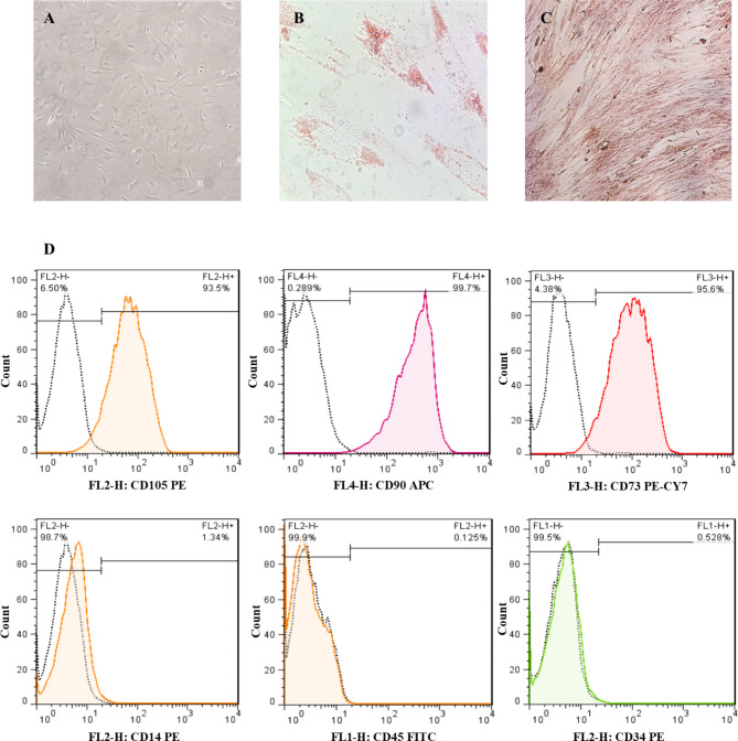
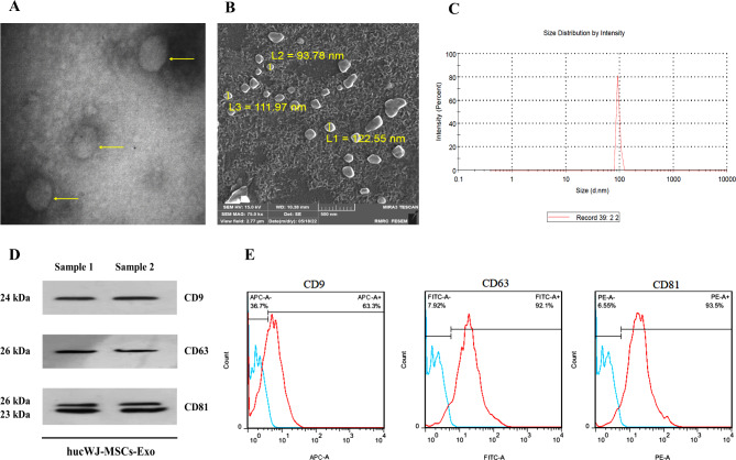
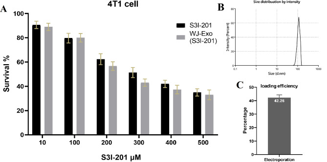
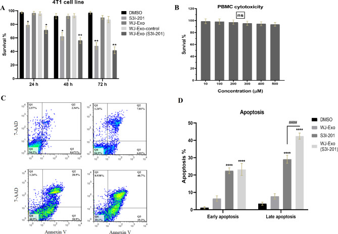
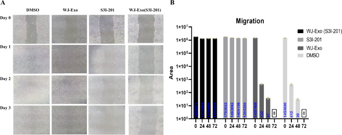
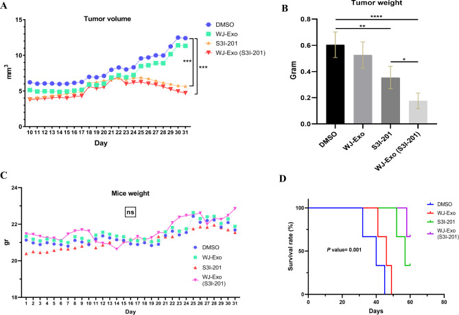
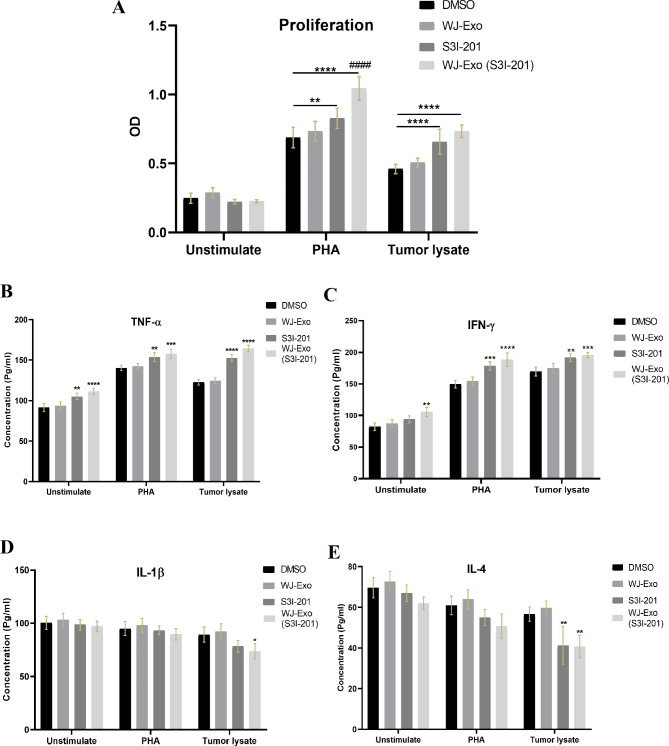
Methods: The filtered WJ-Exos were analyzed by Transmission Electron Microscopy (TEM), Scanning electron microscopy (SEM), Dynamic Light Scattering (DLS), flow cytometry, and Western blotting. These exosomes were then used for loading with S3I-201, resulting in the nano-formulation WJ-Exo(S3I-201). The effect of WJ-Exo(S3I-201) on 4T1 cancer cells was investigated in vitro using MTT assay, flow cytometry, wound healing assay, Western blotting and Quantitative Real-Time Polymerase chain reaction (qPCR) analysis. Finally, the therapeutic efficacy of the nano-formulation was investigated in vivo using a tumor-bearing mouse model.

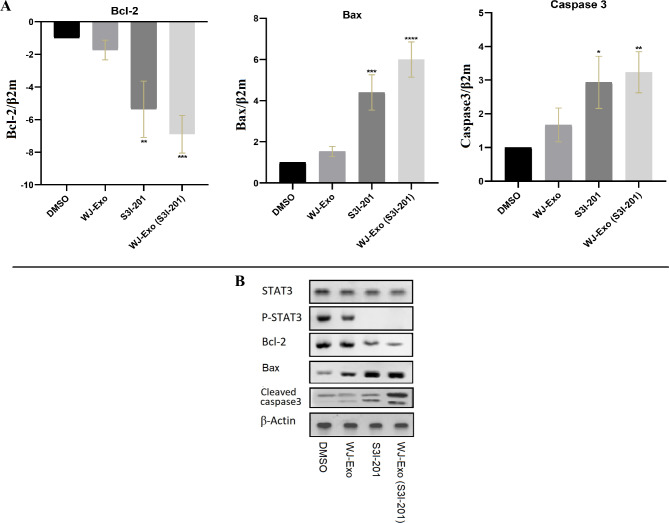
Results: In vitro experiments showed that co-incubation of 4T1 cells with the nano-formulation resulted in a significant reduction in p-STAT3 levels, induction of apoptosis, modulation of Bcl-2, Bax and caspase-3 protein and gene expression, and inhibition of migration. In vivo, treatment of tumor-bearing mice with WJ-Exo(S3I-201) showed a strong antitumor effect that exceeded the efficacy observed in the S3I-201 group.

Conclusion: Our results demonstrate that WJ-Exo is an effective carrier for targeting S3I-201 to tumor cells and enhances the therapeutic efficacy of S3I-201 in tumor-bearing mice.

Keywords: Breast cancer; Drug delivery; Exosome; STAT3 inhibitor; WJ-mesenchymal stem cells.


Conflict of interest statement

The authors declare no competing interests.

Figures

Fig. 1
Fig. 1
Characterization of WJ-MSCs. (A) WJ-MSCs cultures in the third passage show monolayer growth, adherent properties and fibroblast-like morphology under 100x magnification. The white bars represent 50 μm. (B) Adipogenic differentiation of WJ-MSCs into adipocytes visualized by staining with Oil Red O for lipid droplets. (C) Osteogenic differentiation of WJ-MSCs in osteocytes visualized by Alizarin Red S staining for calcium phosphate accumulation. (D) Flow cytometric analysis of surface marker expression of WJ-MSCs with positive markers such as anti-CD105-PE, anti-CD90-APC and anti-CD73-PE-CY7 antibodies and negative markers such as anti-CD14-PE, anti-CD45-FITC and anti-CD34-PE antibodies. The labeled cells were analyzed for marker expression by flow cytometry
Fig. 2
Fig. 2
Characterization of WJ-Exo: (A) Transmission electron microscopy (TEM) shows exosomes with a characteristic round morphology (yellow arrows), with a scale bar of 200 nm. (B) Scanning electron microscope (SEM) image of WJ-Exo, with a scale of 5 μm. (C) Size distribution by dynamic light scattering (DLS). (D) Western blot analysis of WJ-Exo showing the expression of CD9, CD63 and CD81. Equal amounts of total exosomal proteins were immunoblotted. (E) Flow cytometric plots showing the expression of CD9, CD63 and CD81 in WJ-Exo
Fig. 3
Fig. 3
Determination of S3I-201 loading efficiency into exosomes and evaluation of cell viability. (A) 4T1 cells were exposed to 1 µg exosome with different concentrations of WJ-Exo (S3I-201) and free S3I-201 (10, 100, 200, 300, 400, 500 µM) for 48 h in a medium-free environment. Cell viability was measured by MTT assay, and WJ-Exo(S3I-201) showed increased cytotoxicity with an IC50 of 301.4 µM, exceeding the cytotoxicity of free S3I-201 with an IC50 of 337.1 µM. (B) The dynamic light scattering (DLS) determined size of WJ-Exo after loading was measured to be 117.3 ± 5.6 nm. (C) The loading efficiency of S3I-201, calculated from the HPLC area under the curve and the standard curve, was 42.26%
Fig. 4
Fig. 4
In vitro anti-tumor effects of S3I-201-loaded WJ-Exos. (A) Cell viability of 4T1 cells was examined after treatment with DMSO, S3I-201 (IC50: 337.1), WJ-Exo, WJ-Exo control and WJ-Exo(S3I-201, IC50: 301.4 µM) for 24, 48 and 72 h using the MTT assay. WJ-Exo-control means incubation of WJ-Exo with S3I-201 without electroporation to test passive loading of drug to exosomes. (B) The viability of healthy cells remained unaffected after 48 h of incubation with different concentrations of free S3I-201. (C) 4T1 cells were cultured in a 24-well plate with 5 µg of WJ-Exo (S3I-201) loaded with S3I-201 (IC50: 301.4 µM) and IC50: 337.1 µM of free S3I-201. Flow cytometry was used to evaluate cell apoptosis in each group after 48 h, and (D) shows the results of statistical analysis. The data shown are mean ± SD (n = 3). (*P < 0.05, **P < 0.001, ***P < 0.03, ****P < 0.0001 vs. the control group) and (# compare between free S3I-201 vs. WJ-Exo(S3I-201)). Statistical significances indicated with * are compared with the DMSO group. Data underwent statistical analysis using One-Way and Two-Way ANOVA followed by Tukey post hoc test for multiple comparisons
Fig. 5
Fig. 5
Effects of WJ-Exo(S3I-201) treatment on gene and protein expression in 4T1 cells. (A) 4T1 cells (1 × 105 cells/96-well plate) were cultured with 1 µg of WJ-Exo(S3I-201, 301.4 µM), S3I-201 (337.1 µM), 1 µg of WJ-Exo and DMSO for 48-hour. RT-qPCR was used to measure the mRNA expression of Bcl-2, Bax and caspase-3 in 4T1 cells. (*P < 0.05, **P < 0.001, ***P < 0.03, ****P < 0.0001 vs. control group). Statistical significances indicated with * are compared with the DMSO group. (B) 4T1 cells (7 × 105 cells/well) were treated with free S3I-201 (337.1 µM), WJ-Exo(S3I-201) (5 µg exosome loaded with 301.4 µM of S3I-201), WJ-Exo and DMSO. Western blot analysis was performed in the study to analyze the expression levels of STAT3, P-STAT3 (Y705), Bcl-2, Bax, and cleaved caspase-3 in 4T1 cells, with β-actin serving as a control for protein loading. Data underwent statistical analysis using One-Way ANOVA followed by Tukey post hoc test for multiple comparisons
Fig. 6
Fig. 6
WJ-Exo(S3I-201) inhibits the migration of 4T1 cells. (A) A wound healing assay visually shows the initial and final scratch conditions in the 4T1 cell line treated with free S3I-201 (337.1 µM), WJ-Exo(S3I-201) (5 µg exosome loaded with 301.4 µM of S3I-201), WJ-Exo and DMSO. (B) Quantitative results for the wound area analysis are presented. (*P < 0.05, **P < 0.001, ***P < 0.03, ****P < 0.0001 vs. the control group). Data underwent statistical analysis using Two-Way ANOVA followed by Tukey post hoc test for multiple comparisons
Fig. 7
Fig. 7
Evaluation of the anti-tumor effects of WJ-Exo, S3I-201 and WJ-Exo(S3I-201) in tumor-bearing mice. (A) Determination of tumor volume (n = 5/group) and (B) excision of tumors for weight measurement after 21 days of treatment. (C) Monitoring of daily body weight changes in each group. (D) Determination of survival rates (n = 3). The mice received intraperitoneal injections of DMSO, S3I-201 (56 µg/dose), WJ-Exo (10 µg of exosome) and WJ-Exo (S3I-201) (10 µg of exosome loaded with 56 µg/dose S3I-201) on days 10, 12 and 14. Values represent mean ± SD (n = 8; *P < 0.05, **P < 0.001, ***P < 0.03, ****P < 0.0001 vs. control). Data underwent statistical analysis using One-Way and Two-Way ANOVA followed by Tukey post hoc test for multiple comparisons, along with Kaplan-Meier estimation for survival analysis
Fig. 8
Fig. 8
Evaluation of splenocyte proliferation and cytokine levels. (A) Splenocyte proliferation: Mouse splenocytes (106 cells/ml) were stimulated with phytohemagglutinin (PHA) and tumor lysate for 72 h at 37 °C and 5% CO2. Non-stimulated splenocytes under identical conditions were used as controls. Statistical significances indicated with * are compared with the DMSO group. Statistical comparisons between the S3I-201 group and WJ-Exo (S3I-201) were labeled with ####. (B-E) Cytokine Production: Cytokine levels in cells cultured with PHA and tumor lysate were measured. Murine splenocytes (106 cells/ml) were stimulated for 72 h at 37 °C in 5% CO2. Controls comprised non-stimulated splenocytes under the same conditions. Data, representative of at least three (n = 3) independent experiments per group, were analyzed via Student’s t-test (n = 3; *p < 0.05, **p < 0.001, ***p < 0.03, ***p < 0.0001 vs. control). Data underwent statistical analysis using Two-Way ANOVA followed by Tukey post hoc test for multiple comparisons (A-E)

Similar articles

Cited by

References

    1. Sung H, et al. Global cancer statistics 2020: GLOBOCAN estimates of incidence and mortality worldwide for 36 cancers in 185 countries. Cancer J Clin. 2021;71(3):209–49. - PubMed
    1. Sirkisoon SR, et al. Interaction between STAT3 and GLI1/tGLI1 oncogenic transcription factors promotes the aggressiveness of triple-negative breast cancers and HER2-enriched breast cancer. Oncogene. 2018;37(19):2502–14. - PMC - PubMed
    1. Byun WS, et al. Antitumor activity of pulvomycin via targeting activated-STAT3 signaling in docetaxel-resistant triple-negative breast cancer cells. Biomedicines. 2021;9(4):436. - PMC - PubMed
    1. Qin J-J, et al. STAT3 as a potential therapeutic target in triple negative breast cancer: a systematic review. J Experimental Clin Cancer Res. 2019;38:1–16. - PMC - PubMed
    1. Cetinkaya BD. Targeting the SH2 Domain of STAT3 Proteins in Breast Cancer Treatment. Current Researches in Health Sciences-II, 2023: p. 143.

LinkOut - more resources