. 2024 Sep 18;21(1):228.
doi: 10.1186/s12974-024-03219-z.

iPLA2β loss leads to age-related cognitive decline and neuroinflammation by disrupting neuronal mitophagy

Affiliations

iPLA2β loss leads to age-related cognitive decline and neuroinflammation by disrupting neuronal mitophagy

Li Jiao et al. J Neuroinflammation. .

Abstract

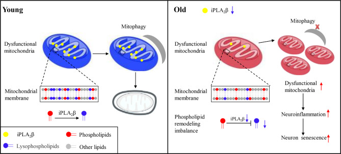
Background: During brain aging, disturbances in neuronal phospholipid metabolism result in impaired cognitive function and dysregulation of neurological processes. Mutations in iPLA2β are associated with neurodegenerative conditions that significantly impact brain phospholipids. iPLA2β deficiency exacerbates mitochondrial dysfunction and abnormal mitochondrial accumulation. We hypothesized that iPLA2β contributes to age-related cognitive decline by disrupting neuronal mitophagy.

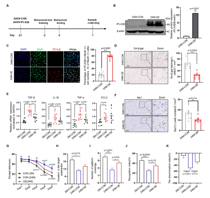
Methodology: We used aged wild-type (WT) mice and iPLA2β-/- mice as natural aging models to assess cognitive performance, iPLA2β expression in the cortex, levels of chemokines and inflammatory cytokines, and mitochondrial dysfunction, with a specific focus on mitophagy and the mitochondrial phospholipid profile. To further elucidate the role of iPLA2β, we employed adeno-associated virus (AAV)-mediated iPLA2β overexpression in aged mice and re-evaluated these parameters.

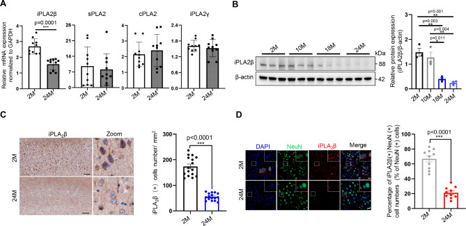
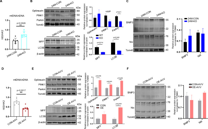
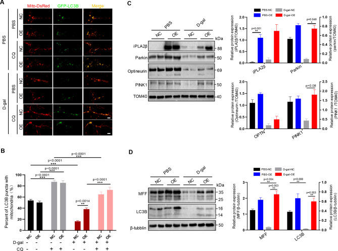
Results: Our findings revealed a significant reduction in iPLA2β levels in the prefrontal cortex of aged brains. Notably, iPLA2β-deficient mice exhibited impaired learning and memory. Loss of iPLA2β in the PFC of aged mice led to increased levels of chemokines and inflammatory cytokines. This damage was associated with altered mitochondrial morphology, reduced ATP levels due to dysregulation of the parkin-independent mitophagy pathway, and changes in the mitochondrial phospholipid profile. AAV-mediated overexpression of iPLA2β alleviated age-related parkin-independent mitophagy pathway dysregulation in primary neurons and the PFC of aged mice, reduced inflammation, and improved cognitive function.

Conclusions: Our study suggests that age-related iPLA2β loss in the PFC leads to cognitive decline through the disruption of mitophagy. These findings highlight the potential of targeting iPLA2β to ameliorate age-related neurocognitive disorders.


Conflict of interest statement

The authors declare no competing interests.

Figures

Fig. 1
Fig. 1
Aging increased iPLA2β loss in the PFC of mice. A PLA2s mRNA expression levels in the PFC were assessed using qPCR. Normalization was conducted relative to GAPDH expression levels. n = 10. B iPLA2β protein levels in the PFC were assessed via Western blotting. n = 3. C Representative IHC images depicting iPLA2β in the PFC. IHC analysis showing the density of iPLA2β (+) cells/mm². Scale bar: 100 μm. n = 16. D Representative immunofluorescence images depicting iPLA2β (red) and NeuN (green) in the PFC. The bar graph shows the percentage of iPLA2β(+) NeuN(+) cells relative to the total NeuN(+) cell population (%).Scale bar: 20 μm. n = 10 M represents “month” Data are presented as mean ± SEM; p values were obtained using Mann-Whitney U test (A), one-way analysis of variance (ANOVA) followed by Tukey’s post hoc test (B), and two-sided unpaired Student’s t-tests (C, D). * p < 0.05. **, p < 0.01; ***, p < 0.001
Fig. 2
Fig. 2
Deficiency of iPLA2β increases aging-related cellular senescence, cognitive impairment and neuroinflammation. A The mRNA expression levels of iPLA2β in the PFC of 24 M mice were assessed by qPCR. Normalization was performed relative to GAPDH expression levels. n = 6. B Protein levels of iPLA2β in the PFC of 24 M mice, assessed using Western blotting and densitometry, n = 3. C Time taken by 2 M-WT, 24 M-WT, 24 M-CON, and 24 M-KO mice to reach the platform during the spatial test. n = 20. D Frequency of platform crossings in 24 M mice during the probe trial. n = 20. E Duration of 2 M-WT, 24 M-WT, 24 M-CON, and 24 M-KO mice spent time in hidden platform quadrants during the probe trial. n = 20. F Recognition index of 2 M-WT, 24 M-WT, 24 M-CON and 24 M-KO mice during the Novel object recognition test. Recognition index = time spent exploring novel object/time spent exploring both objects. n = 20. G The discrimination index of 2 M-WT, 24 M-WT, 24 M-CON, and 24 M-KO mice during the Novel object recognition test. The discrimination index = (time spent on the novel object − time spent on the familiar object)/ time spent on both objects. n = 20. H Representative SA-β-gal staining images in 24 M PFC. The bar graph shows the density of β-Gal staining (+) cells/0.1 mm². Scale bar: 100 μm. n = 10. I mRNA levels of TGF-β, CCL2, TNF-α, and IL-1β in the PFC of 2 M-WT, 24 M-WT, 24 M-CON, and 24 M-KO mice were assessed via qPCR. Normalization was conducted relative to GAPDH expression levels. n = 11 KO represents “iPLA2β knockout,” CON represents “control”, Data are presented as mean ± SEM; p values were obtained using two-sided unpaired Student’s t-tests (A, B, H), the Kruskal-Wallis test followed by Dunn’s multiple comparisons test (C, D, E, F, G and I), * p < 0.05. **, p < 0.01; ***, p < 0.001
Fig. 3
Fig. 3
iPLA2β reduces senescence in primary neurons. A The mRNA levels of iPLA2β in DIV7 and DIV20 cultured neurons were assessed using qPCR. Normalization was conducted relative to GAPDH expression levels. n = 13. B Protein levels of iPLA2β in DIV7 and DIV20 cultured primary neurons, assessed via Western blot and densitometry. n = 4. C Representative SA-β-gal staining images of iPLA2β-overexpression (OE) and control DIV20 primary neurons. Scale bar: 100 μm. n = 12. D Representative immunofluorescence images of iPLA2β (red) in D-gal-induced iPLA2β overexpression (OE) and control primary neurons. Scale bar: 5 μm. n = 8. E Representative SA-β-gal staining images of D-gal-induced iPLA2β-overexpression (OE) and control primary neurons. Scale bar: 100 μm. n = 12. F Protein levels of P62 and P16 in D-gal-induced iPLA2β-overexpression (OE) and control primary neurons, as assessed by Western blot and densitometry OE represents “iPLA2β overexpression”, CON represents “control”, Data are mean ± SEM; p values were obtained using two-sided unpaired Student’s t-tests (A, B), Mann-Whitney U test (C), one-way analysis of variance (ANOVA) followed by Tukey’s post hoc test (E), the Kruskal-Wallis test followed by Dunn’s multiple comparisons test (F), * p < 0.05. **, p < 0.01; ***, p < 0.001
Fig. 4
Fig. 4
iPLA2β protects mitochondrial morphology and regulate mitochondrial function during cortex aging. A Representative TEM image of WT and iPLA2β−/− PFC of 24 M mice. Scale bar: 1 μm. B Representative TEM images of the AAV-CON-injected and AAV-iPLA2β-OE-injected PFC of mice. Scale bar: 1 μm. C Quantification of mitochondrial diameter in TEM images of WT and iPLA2β−/− PFC from 24 M mice. n = 10. Quantification of mitochondrial diameter in TEM images of the AAV-CON-injected and AAV- iPLA2β-OE-injected PFC of mice. n = 10.E Quantification of mitochondrial length in TEM images of WT and iPLA2β−/− PFC from 24 M mice. n = 10. F Quantification of mitochondrial length in TEM images of the AAV-CON-injected and AAV- iPLA2β-OE-injected PFC of mice. n = 10. G ATP levels in the WT and iPLA2β−/− PFC of 24 M mice. n = 8. H ATP levels of the AAV-CON-injected and AAV- iPLA2β-OE-injected PFC of mice. n = 8. I Representative immunofluorescence images of iPLA2β (green) and TOM20 (red) in the PFC of 2 M-WT, 24 M-WT, AAV-CON-injected, and AAV- iPLA2β-OE-injected mice. Scale bar: 50 μm. J TOM20 fluorescence intensity of immunofluorescence images in (I). n = 12. K iPLA2β fluorescence intensity of immunofluorescence images in (I). n = 12 KO represents “iPLA2β knockout,” WT represents “wildtype”, OE represents “iPLA2β overexpression”, CON represents “control”, Data are mean ± SEM; p values were obtained using two-sided unpaired Student’s t-tests (C, D, E, F, G, and H), the Kruskal-Wallis test followed by Dunn’s multiple comparisons test (J and K), * p < 0.05. **, p < 0.01; ***, p < 0.001
Fig. 5
Fig. 5
iPLA2β regulates mitophagy during neuronal aging in vivo. A Quantification of the mtDNA/nDNA ratio in the 24 M PFC in the wild-type (WT) and iPLA2β knockout (iPLA2β−/−) groups, using 16 S rRNA and Hexokinase 2 (Hk2), respectively. n = 6. B Optineurin, PINK1, Parkin, MFF, and LC3B levels in the 24 M PFC in wild-type (WT) and iPLA2β knockout (iPLA2β−/−) groups, assessed by Western blot and densitometry. n = 3. C BNIP3 and NIX levels in the 24 M PFC in wild-type (WT) and iPLA2β knockout (iPLA2β−/−) groups, assessed by Western blot and densitometry. n = 3. D Quantification of the mtDNA/nDNA ratio in the 22 M PFC with iPLA2β overexpression (OE) and control (CON) groups, using 16 S rRNA and Hexokinase 2 (Hk2), respectively. n = 6. E Optineurin, PINK1, Parkin, MFF, and LC3B levels in the 22 M PFC with iPLA2β overexpression (iPLA2β-OE) and control (CON) groups, assessed by Western blot and densitometry. n = 3. F BNIP3 and NIX levels in the 22 M PFC with iPLA2β overexpression (iPLA2β-OE) and control (CON) groups, as assessed by Western blotting and densitometry. n = 3 KO represents “iPLA2β knockout”, WT represents “wildtype”, OE represents “iPLA2β overexpression”, CON represents “control”, Data are presented as the mean ± SEM; p values were obtained using two-sided unpaired Student’s t-tests (A, B, C, and D). * p < 0.05. **, p < 0.01; ***, p < 0.001
Fig. 6
Fig. 6
iPLA2β regulates mitophagy during neuronal aging in vitro. A Primary cultured cortical neurons were transfected with GFP-LC3B and Mito-DsRed. Fluorescent images were captured 3 h after reperfusion. Scale bar: 5 μm. B Relative colocalization ratio between GFP-LC3B and Mito-DsRed in immunofluorescence images in (A). The ratio was calculated by dividing the number of LC3B-Mito puncta by the total number of Mito puncta. n = 6. C Mitochondrial levels of iPLA2β, Parkin, optineurin, and PINK1 were assessed by Western blot and densitometry in D-gal-induced iPLA2β overexpression (OE) and control primary neurons. D Protein levels of MFF and LC3B were evaluated using Western blot and densitometry in D-gal-induced iPLA2β overexpression (OE) and control primary neurons CQ represents “chloroquine”, D-gal represents “D-galactose”, OE represents “iPLA2β overexpression”, NC represents “Negative Control”, Data are presented as the mean ± SEM; p values were obtained using, one-way analysis of variance (ANOVA) followed by Tukey’s post hoc test (B), the Kruskal-Wallis test followed by Dunn’s multiple comparisons test (C and D). * p < 0.05. **, p < 0.01; ***, p < 0.001
Fig. 7
Fig. 7
iPLA2β deficiency leads to alterations in mitochondrial phospholipid metabolism in aged PFC. A Heat map representing individual LPC species and lipidomic analysis of LPC species were significantly altered in the KO group in 24 M PFCs. B Heat map representing individual LPE species and lipidomic analysis of LPE species significantly altered in the KO group in 24 M PFCs. C Heat map representing individual PC species and lipidomic analysis of PC species significantly altered in the KO group in 24 M PFCs. D Heat map representing individual PE species and lipidomic analysis of PE species significantly altered in the KO group in 24 M PFCs MUFA represents “monounsaturated fatty acid”; PUFA represents “polyunsaturated fatty acid”. Data are presented as the mean ± SEM; p values were obtained using two-sided unpaired Student’s t-tests (A, B, C, and D). * p < 0.05. **, p < 0.01; ***, p < 0.001
Fig. 8
Fig. 8
Overexpression of iPLA2β in PFC improves cognitive function of old mice. A Scheme of the experimental design. AAV injections were administered 21 days prior to formal behavioral testing. Behavioral test training sessions were conducted two days before the formal testing began. Samples were collected on the seventh day following the initiation of the formal behavioral experiment. B Protein levels of iPLA2β in the 22 M PFC with iPLA2β overexpression (iPLA2β-OE) and control (CON) groups, as assessed by Western blotting and densitometry. C Representative immunofluorescence images of iPLA2β (red) and NeuN (green) in the 22 M PFC with iPLA2β overexpression (iPLA2β-OE) and control (CON) groups. Scale bar: 20 μm. D Representative SA-β-gal staining images in the 22 M PFC with iPLA2β overexpression (iPLA2β-OE) and control (CON) groups. Scale bar: 100 μm. E mRNA expression levels of TGFβ, IL-1β, TNF-α, and CCL2 in the 22 M PFC with iPLA2β overexpression (iPLA2β-OE) and control (CON) groups assessed via qPCR. Normalization was conducted relative to GAPDH expression levels. n = 11. F Representative IHC images of Iba1 in the 22 M PFC with iPLA2β overexpression (iPLA2β-OE) and control (CON) groups. Histogram showing quantification of the Iba1(+) area. Scale bar: 50 μm. G Travel time of the mice to reach the platform during the spatial test. n = 20 per group. H Duration spent by mice in the hidden platform quadrants during the probe trial. n = 20. I Frequency of platform crossings by mice during the probe trial. n = 20 per group. J Recognition index during the Novel object recognition test. Recognition index = time spent exploring a novel object/time spent exploring both objects. n = 20. K Discrimination index during the Novel object recognition test. The discrimination index = (time spent on the novel object − time spent on the familiar object)/ time spent on both objects. n = 20 Data are presented as the mean ± SEM; p values were obtained using the Mann-Whitney U test (B, C, D, and F) and the Kruskal-Wallis test followed by Dunn’s multiple comparisons test (E, G, H, I, J, and K). * p < 0.05. **, p < 0.01; ***, p < 0.001
Fig. 9
Fig. 9
Possible mechanism underlying the protective effects of iPLA2β on brain aging

Similar articles

Cited by

References

    1. Lopez-Otin C, et al. Hallmarks of aging: an expanding universe. Cell. 2023;186(2):243–78. - PubMed
    1. Scheltens P, et al. Alzheimer’s Disease. Lancet. 2021;397(10284):1577–90. - PMC - PubMed
    1. Marchetti B, et al. Parkinson’s disease, aging and adult neurogenesis: Wnt/beta-catenin signaling as the key to unlock the mystery of endogenous brain repair. Aging Cell. 2020;19(3):e13101. - PMC - PubMed
    1. Hou Y, et al. Ageing as a risk factor for neurodegenerative disease. Nat Rev Neurol. 2019;15(10):565–81. - PubMed
    1. Allen WE, et al. Molecular and spatial signatures of mouse brain aging at single-cell resolution. Cell. 2023;186(1):194–e20818. - PMC - PubMed

MeSH terms

Substances

LinkOut - more resources