

Physical activity and frontoparietal network connectivity in traumatic brain injury
- PMID: 39295099
- PMCID: PMC11410878
- DOI: 10.1002/brb3.70022
Physical activity and frontoparietal network connectivity in traumatic brain injury
Abstract
Background: Prolonged changes to functional network connectivity as a result of a traumatic brain injury (TBI) may relate to long-term cognitive complaints reported by TBI survivors. No interventions have proven to be effective at treating long-term cognitive complaints after TBI but physical activity has been shown to promote cognitive function and modulate functional network connectivity in non-injured adults. Therefore, the objective of this study was to test if physical activity engagement was associated with functional connectivity of the cognitively relevant frontoparietal control network (FPCN) in adults with a TBI history.
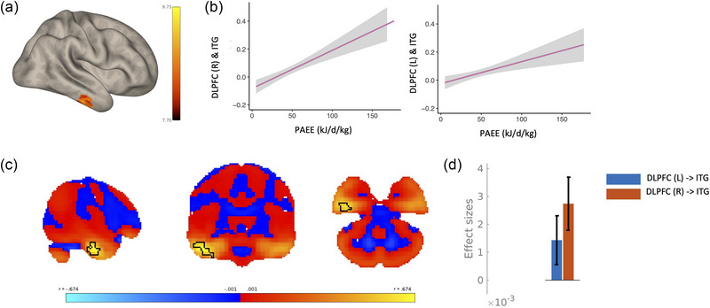
Methods: In a case-control study design, resting state function magnetic resonance imaging and physical activity data from a subset of participants (18-81 years old) from the Cambridge Centre for Ageing and Neuroscience (Cam-CAN) study was analyzed. Fifty-seven participants reported a prior head injury with loss of consciousness and 57 age and sex matched controls were selected. Seed-based functional connectivity analyses were performed using seeds in the dorsolateral prefrontal cortex and the inferior parietal lobule, to test for differences in functional connectivity between groups, associations between physical activity and functional connectivity within TBI as well as differential associations between physical activity and functional connectivity between TBI and controls.
Results: Seed-based connectivity analyses from the dorsolateral prefrontal cortex showed that those with a history of TBI had decreased positive connectivity between dorsolateral prefrontal cortex and intracalcarine cortex, lingual gyrus, and cerebellum, and increased positive connectivity between dorsolateral prefrontal cortex and cingulate gyrus and frontal pole in the TBI group. Results showed that higher physical activity was positively associated with increased connectivity between the dorsolateral prefrontal cortex and inferior temporal gyrus. Differential associations were observed between groups whereby the strength of the physical activity-functional connectivity association was different between the inferior parietal lobule and inferior temporal gyrus in TBI compared to controls.
Discussion: Individuals with a history of TBI show functional connectivity alterations of the FPCN. Moreover, engagement in physical activity is associated with functional network connectivity of the FPCN in those with a TBI. These findings are consistent with the evidence that physical activity affects FPCN connectivity in non-injured adults; however, this effect presents differently in those with a history of TBI.
Keywords: frontoparietal control network; physical activity; traumatic brain injury.
© 2024 The Author(s). Brain and Behavior published by Wiley Periodicals LLC.
Figures


References
-
- Ai, M. , Morris, T. P. , Zhang, J. , de la Colina, A. N. , Tremblay‐Mercier, J. , Villeneuve, S. , Whitfield‐Gabrieli, S. , Kramer, A. F. , & Geddes, M. R. (2023). Resting‐state MRI functional connectivity as a neural correlate of multidomain lifestyle adherence in older adults at risk for Alzheimer's disease. Scientific Reports, 13(1), Article 7487. 10.1038/s41598-023-32714-1 - DOI - PMC - PubMed
-
- Amir, J. , Nair, J. K. R. , Del Carpio‐O'Donovan, R. , Ptito, A. , Chen, J.‐K. , Chankowsky, J. , Tinawi, S. , Lunkova, E. , & Saluja, R. S. (2021). Atypical resting state functional connectivity in mild traumatic brain injury. Brain and Behavior, 11(8), e2261. 10.1002/brb3.2261 - DOI - PMC - PubMed
MeSH terms
Grants and funding
LinkOut - more resources
Full Text Sources
Medical
