Review
. 2024 May 7;16(Suppl 2):10008.
doi: 10.4081/dr.2024.10008.

When mycosis fungoides seems not to be within the spectrum of clinical and histopathological differential diagnoses

Affiliations
Review

When mycosis fungoides seems not to be within the spectrum of clinical and histopathological differential diagnoses

Monia Di Prete et al. Dermatol Reports. .

Abstract

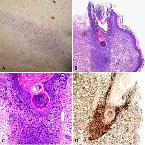
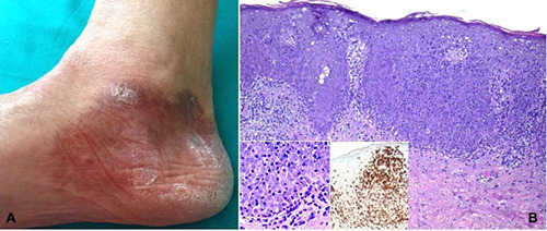
The most prevalent primary cutaneous T-cell lymphoma, mycosis fungoides (MF), is characterized by the development of plaques and nodules after an erythematous patchy phase that is non-specific. An infiltrate of atypical small- to medium-sized cerebriform lymphocytes in the superficial dermis, with variable epidermotropism, is the histopathological hallmark of the disease. In more advanced stages of the illness, large-cell transformation may be seen. Early diagnosis of MF can be very challenging based only on histopathologic or clinical findings, so it is critical to have a clinical-pathological correlation. Many atypical variants of MF that deviate from the classic Alibert-Bazin presentation of the disease have been described over the past 30 years, sometimes with different prognostic and therapeutic implications. Clinically or histopathologically, they can mimic a wide range of benign inflammatory skin disorders. To make a conclusive diagnosis in these cases, it is recommended to take multiple biopsies from various lesions and to carefully correlate the clinical and pathological findings. We have outlined the various facets of the illness in this review, positioning MF as a "great imitator", with an emphasis on the more recently identified variations, differential diagnosis, and its benign mimics.

Keywords: dermatopathology; mimickers; mycosis fungoides.


Conflict of interest statement

Conflict of interest: the authors declare no potential conflict of interest.

Figures

Figure 1.
Figure 1.
A) Oval patch with comedo-like papules on the flank; B) involvement of the infundibulum by lymphocytes with almost no epidermotropism; C) at the base of the infundibulum, a folliculotropic lymphocytic infiltrate is seen; D) immunohistochemical labeling with CD4 shows infiltration of the follicular epithelium by lymphocytes.
Figure 2.
Figure 2.
A) Erythematous plaque involving the forehead and eyebrows with alopecia of the medial brow; B) massive infiltration of the hair follicle with atypical T-cells and follicular mucinosis; C) immunohistochemical labeling with CD3 highlights the lymphocytic infiltration.
Figure 3.
Figure 3.
A) Solitary, minimally infiltrated plaque on the scalp; B, C) the clinical and dermoscopic hair loss pattern mimic androgenetic alopecia; D) Immunohistochemical labeling with CD4 shows infiltration of the follicular epithelium with lymphocytes.
Figure 4.
Figure 4.
A)Multiple skin-colored papules and plaques on the cheeks and forehead; no comedones visible; B-D) mucin within the folliculosebaceous units with a mild perifollicular and intraepithelial lymphocytic infiltrate.
Figure 5.
Figure 5.
A) Erythematous, scaly, distinct papules on the medial sole; B) band-like infiltrate of lymphocytesin the superficial dermis; C) dense mononuclear infiltration around deep dermal eccrine ducts and syringometaplasia.
Figure 6.
Figure 6.
A) Large, scaly, erythematous patch on the medial aspect of the heel; B) psoriasiform hyperplasia of the epidermis with overlying parakeratosis and dense intraepidermal lymphocytic infiltrate, characterized by a disproportion between the density of lymphocytes within the epidermis and that in the upper dermis, with only few perivascular cells in the dermis. In the inset, at higher power, the epidermotropic lymphocytes are medium to large with hyperchromatic and irregular nuclei, variably prominent nucleoli and minimal-to-abundant palestaining or eosinophilic cytoplasm. In the other inset, immunohistochemical labeling with CD8 shows epidermotropic medium-sized lymphocytes infiltrate.
Figure 7.
Figure 7.
A, B) Atrophic lax skin in the right axillary area; C, D) diffuse granulomatous dermal infiltrates composed of atypical lymphocytes, histiocytes and giant cells; E) multinucleated giant cell and adjacent atypical T lymphocytes; F) immunohistochemical labeling with CD4 highlights the atypical T-cells surrounding a multinucleated giant cell.
Figure 8.
Figure 8.
A, B) Diffuse erythematous, dusky discoloration involving the trunk, the limbs, and the neck; C, D) band-like lymphocytic infiltration of the papillary dermis with evident epidermotropism and a serum crust on the top.
Figure 9.
Figure 9.
A) Diffuse brownish discoloration of the skin; B-D) epidermotropic lymphocitic infiltrate, with scattered melanophages in the papillary dermis.
Figure 10.
Figure 10.
A-C) Diffuse erythematous papules on the trunk and arms; D) lymphocytic infiltrates in papillary dermis and small intraepidermal collection of lymphocytes.
Figure 11.
Figure 11.
A) Diffuse erythematous and scaly plaques on the back, mimicking psoriasis; B, C) psoriasiform epidermal hyperplasia in association with a dermal lymphocytic infiltrate, displaying epidermotropism.
Figure 12.
Figure 12.
A) Two, symmetric, erythematous patches on the medial aspect of the breast; B, C) band-like lymphocytic infiltration in the papillary dermis, with mild epidermotropism.
Figure 13.
Figure 13.
A) Diffuse maculo-papular scaly eruption on the trunk and extremities, including also hypopigmented lesions; B) in one biopsy, there is a lichenoid lymphocytic infiltrate in the papillary dermis, associated with a thick stratum corneum, alternating ortho- and parakeratosis; C) in another biopsy, the lichenoid infiltrate is more epidermotropic, with prominent vacuolar changes at the dermo-epidermal junction. Necrotic keratynocytes are absent and the lymphocytes are large and atypical, with hyperchromatic nuclei and clear perinuclear halo.
Figure 14.
Figure 14.
A) Erythematous, annular patches on the clavicular area; B) lichenoid lymphocytic infiltrate in the papillary dermis; C, D) the lymphocytes show moderate epidermotropism, associated with prominent vacuolar changes at the dermo-epidermal junction and necrotic keratinocytes at the tips of the rete ridges, which assumes a “squared off” aspect.

References

    1. Elder DE, Massi D, Scolyer RA WR. WHO Classification of Skin Tumours. 4th Edition. IARC Press; 2018.
    1. Muñoz-González H, Molina-Ruiz AM, Requena L. Clinicopathologic variants of mycosis fungoides. Actas Dermo-Sifiliográficas 2017;108:192-208. - PubMed
    1. Fauconneau A, Pham-Ledard A, Cappellen D, et al. Assessment of diagnostic criteria between primary cutaneous anaplastic large-cell lymphoma and CD30-rich transformed mycosis fungoides; A study of 66 cases. Br J Dermatol 2015;172:1547-54. - PubMed
    1. Massone C, Kodama K, Kerl H, Cerroni L. Histopathologic features of early (patch) lesions of mycosis fungoides: A morphologic study on 745 biopsy specimens from 427 patients. Am J Surg Pathol 2005;29:550-60. - PubMed
    1. Massone C, Crisman G, Kerl H, Cerroni L. The prognosis of early mycosis fungoides is not influenced by phenotype and T-cell clonality. Br J Dermatol 2008;159:881-6. - PubMed

LinkOut - more resources