. 2024 Sep 4:15:1439079.
doi: 10.3389/fphar.2024.1439079. eCollection 2024.

Paulownin elicits anti-tumor effects by enhancing NK cell cytotoxicity through JNK pathway activation

Affiliations

Paulownin elicits anti-tumor effects by enhancing NK cell cytotoxicity through JNK pathway activation

Eun Sun Park et al. Front Pharmacol. .

Abstract

Paulownin, a natural compound derived from Paulownia tomentosa wood, exhibits various physiological functions, including anti-bacterial and anti-fungal effects. However, the impact of paulownin on natural killer (NK) cell immune activity remains largely unknown. In this study, we investigated the effect of paulownin on NK cell activity both in vitro and in vivo, and explored its potential mechanisms. NK-92 cells were used for in vitro experiments and a BALB/c mouse model with B16F10 cells injected subcutaneously were used for in vivo anti-tumor analysis. We found that paulownin enhanced the cytolytic activity of NK-92 cells against leukemia, human colon, and human lung cancer cell lines. Paulownin treatment increased the expression of the degranulation marker protein CD107a and cytolytic granules, including granzyme B and perforin in NK-92 cells. Moreover, these enhancements of cytotoxicity and the expression of cytolytic granules induced by paulownin were also observed in human primary NK cells. Signaling studies showed that paulownin promoted the phosphorylation of JNK. The increased perforin expression and elevated cytotoxic activity induced by paulownin were effectively inhibited by pre-treatment with a JNK inhibitor. In vivo studies demonstrated that the administration of paulownin suppressed the growth of B16F10 melanoma cells allografted into mice. Paulownin administration promoted the activation of NK cells in the spleen of mice, resulting in enhanced cytotoxicity against YAC-1 cells. Moreover, the anti-tumor effects of paulownin were reduced upon the depletion of NK cells. Therefore, these results suggest that paulownin enhances NK cell cytotoxicity by activating the JNK signaling pathway and provide significant implications for developing new strategies for cancer immunotherapy.

Keywords: JNK; NK cells; immunotherapy; innate immunity; paulownin.


Conflict of interest statement

The authors declare that the research was conducted in the absence of any commercial or financial relationships that could be construed as a potential conflict of interest.

Figures

FIGURE 1
FIGURE 1
Paulownin enhances the cytotoxicity of NK-92 cells. (A) NK-92 cells were treated with 20 μM for 24 h or 48 h and then cytotoxicity assays were performed with K562 cells at a 5:1 ratio using a calcein-AM release assay. (B) NK-92 cells were treated with paulownin (0, 5, 10 and 20 μM) for 48 h. The cytotoxicity of NK-92 cells against K562 cells was analyzed by different E: T ratios (1.25:1, 2.5:1, and 5:1) for 2 h (C) NK-92 cells were treated with the indicated dose of paulownin and mixed with A549, PC-9, SW480, and HT29 cells. The cytolytic effects of NK-92 cells were evaluated by LDH release assay. The results represent the mean ± SD of three experiments (*p < 0.05, **p < 0.01, ***p < 0.001, ****p < 0.0001 by Student’s t-test, n = 3).
FIGURE 2
FIGURE 2
Paulownin stimulates cytolytic granule exocytosis in NK-92 cells. (A) NK-92 cells were treated with paulownin (0, 5, 10, and 20 μM) for 48 h. Subsequently, NK-92 cells were co-incubated with K562 cells at a 1:1 ratio for 2 h and stained with fluorescein isothiocyanate (FITC)-conjugated mouse anti-human CD107a antibody. Frequencies of CD56+ CD107a+ expression on NK cells were analyzed using Flow cytometry. Representative results are shown as dot plots and the average percentage of CD56+ CD107a+ expression is presented as statistical bar charts. NK-92 cells were treated with paulownin (0, 5, 10 and 20 μM) for 48 h. (B) The expression of mRNA level of perforin and granzyme B of NK-92 cells were evaluated by qPCR (C) Protein levels of perforin and granzyme B of NK-92 cells were determined by flow cytometry, with the numbers indicating the percentage of positive cells. (D) NK-92 cells were treated with or without 20 μM paulownin for 48 h. The cells were attached to PLL-coated slides. Then, slides were fixed, permeabilized, and stained with Alexa Flour 647-conjugated anti-perforin (Red) or Alexa Flour 647- conjugated anti- granzyme B (Red) along with 4′-6-Diamidino-2-phenylindole (DAPI, blue). Representative images are shown. The results represent the mean ± SD of three experiments (*p < 0.05, **p < 0.01, ***p < 0.001, ****p < 0.0001 by Student’s t-test, n = 3).
FIGURE 3
FIGURE 3
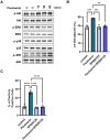
Paulownin activates NK cells through the JNK signaling pathway. NK-92 cells were stimulated with paulownin (20 μM) for 5, 15, 30, or 60 min. (A) Expression levels of total and phosphorylated Akt and MAPKs were analyzed by Western blotting. (B) NK-92 cells were treated with paulownin only, JNK inhibitor (SP00125) only, or both paulownin and SP00125. The cytotoxicity of NK-92 cells was assessed against K562 cells at a 5:1 ratio. (C) The expression of perforin was analyzed using FACS. The results represent the mean ± SD of three experiments (*p < 0.05, **p < 0.01, ***p < 0.001, ****p < 0.0001 by Student’s t-test, n = 3).
FIGURE 4
FIGURE 4
Paulownin enhances the cytotoxicity of primary NK cells. (A) pNK cells were treated with 20 μM paulownin for 24 h or 48 h The cytotoxicity of pNK cells toward K562 cells was evaluated using a calcein-AM release assay. K562 cells were used as target cells at an E: T ratio of 5:1 for 1 h (B) pNK cells were treated with paulownin for 48 h. The cytotoxicity of pNK cells against K562 cells was analyzed by different E: T (1.25:1, 2.5:1, and 5:1) ratios for 1 h. (C) Paulownin pretreated pNK cells were co-incubated with K562 cells at a 1:1 ratio for 1 h and stained with fluorescein isothiocyanate (FITC)-conjugated mouse anti-human CD107a antibody. Frequencies of CD107a expression on NK cells was analyzed using flow cytometry. Representative result shown as dot plot and average percentage of CD107a+ expression shown as statistical bar charts. pNK cells were treated with paulownin (0, 5, 10, and 20 μM). (D) The mRNA level of perforin and granzyme B of pNK cells were evaluated by qRT-PCR and (E) protein levels of perforin and granzyme B of pNK cells were determined by flow cytometry. Numbers indicate the percentage of positive target cells. The results represent the mean ± SD of three experiments (*p < 0.05, **p < 0.01, ***p < 0.001, ****p < 0.0001 by Student’s t-test, n = 3).
FIGURE 5
FIGURE 5
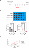
Paulownin inhibited the growth of B16F10 melanoma cancer allograft in Balb/c mice. (A) The experimental scheme to detect proper paulownin concentrations. B16F10 cells (5 × 10⁵) were subcutaneously injected into Balb/c mice. Paulownin was injected intraperitoneally every 2 or 3 days. The tumor volume and weight were measured after dissection on day 18 after injection of melanoma cells. (B) The tumor image and a summary graph of statistical bar showing each group of tumor volume and weight. (C) Splenocytes isolated form balb/c mice were co-cultured with YAC-1 target cell at 25:1, 50:1, or 100:1 for 24 h. The results represent the mean ± SD of three experiments (*p < 0.05, **p < 0.01, ***p < 0.001, ****p < 0.0001 by Student’s t-test, n = 5).
FIGURE 6
FIGURE 6
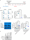
NK depletion abolished anticancer effect of paulownin in mice. (A) Overall scheme to investigate the NK activity-inducing effect of paulownin. Balb/c mice were subcutaneously injected with 5 × 10⁵ B16F10 cells and then received either paulownin or vehicle for 2 or 3 days with or without anti-asialoGM1 (100 μL) injection. (B) CD3- NKp46 + cells in the spleen of each group were measured with flow cytometry. The left panel shows as dot plot and the right panel shows as graph. (C) The tumor volume and weight were measured after dissection on day 18 after the injection of B16F10 cells. (D) The expression of Perforin and Granzyme B of spleenocytes was analyzed using Western blot. The results represent the mean ± SD of three experiments (*p < 0.05, **p < 0.01, ***p < 0.001, ****p < 0.0001 by Student’s t-test, n = 4).

References

    1. Aktas E., Kucuksezer U. C., Bilgic S., Erten G., Deniz G. (2009). Relationship between CD107a expression and cytotoxic activity. Cell Immunol. 254, 149–154. 10.1016/j.cellimm.2008.08.007 - DOI - PubMed
    1. Ali A. K., Nandagopal N., Lee S. H. (2015). IL-15-PI3K-AKT-mTOR: a critical pathway in the life journey of natural killer cells. Front. Immunol. 6, 355. 10.3389/fimmu.2015.00355 - DOI - PMC - PubMed
    1. Alter G., Malenfant J. M., Altfeld M. (2004). CD107a as a functional marker for the identification of natural killer cell activity. J. Immunol. Methods 294, 15–22. 10.1016/j.jim.2004.08.008 - DOI - PubMed
    1. Chang C. J., Chen Y. Y., Lu C. C., Lin C. S., Martel J., Tsai S. H., et al. (2014). Ganoderma lucidum stimulates NK cell cytotoxicity by inducing NKG2D/NCR activation and secretion of perforin and granulysin. Innate Immun. 20, 301–311. 10.1177/1753425913491789 - DOI - PubMed
    1. Chen X., Jin J., Hao F., Yang H., Sun H., Jiang C. (2021). Paulownia tomentosa flower polysaccharide as an effective immunopotentiator to enhance immune responses for Newcastle disease vaccine in mice. Italian J. Food Sci. 33, 11–20. 10.15586/ijfs.v33i4.2107 - DOI

LinkOut - more resources