. 2024 Sep 5;17(9):100963.
doi: 10.1016/j.waojou.2024.100963. eCollection 2024 Sep.

Sphk1 regulates HMGB1 via HDAC4 and mediates epithelial pyroptosis in allergic rhinitis

Affiliations

Sphk1 regulates HMGB1 via HDAC4 and mediates epithelial pyroptosis in allergic rhinitis

Wei Huang et al. World Allergy Organ J. .

Abstract

Background: Allergic rhinitis (AR) is a global health issue affecting millions of individuals worldwide. Pyroptosis has emerged as a major player in the development of AR, and targeting its inhibition with specific drugs holds promise for AR treatment. However, a comprehensive understanding of the precise mechanisms underlying pyroptosis in AR remains to be explored, warranting further investigation.

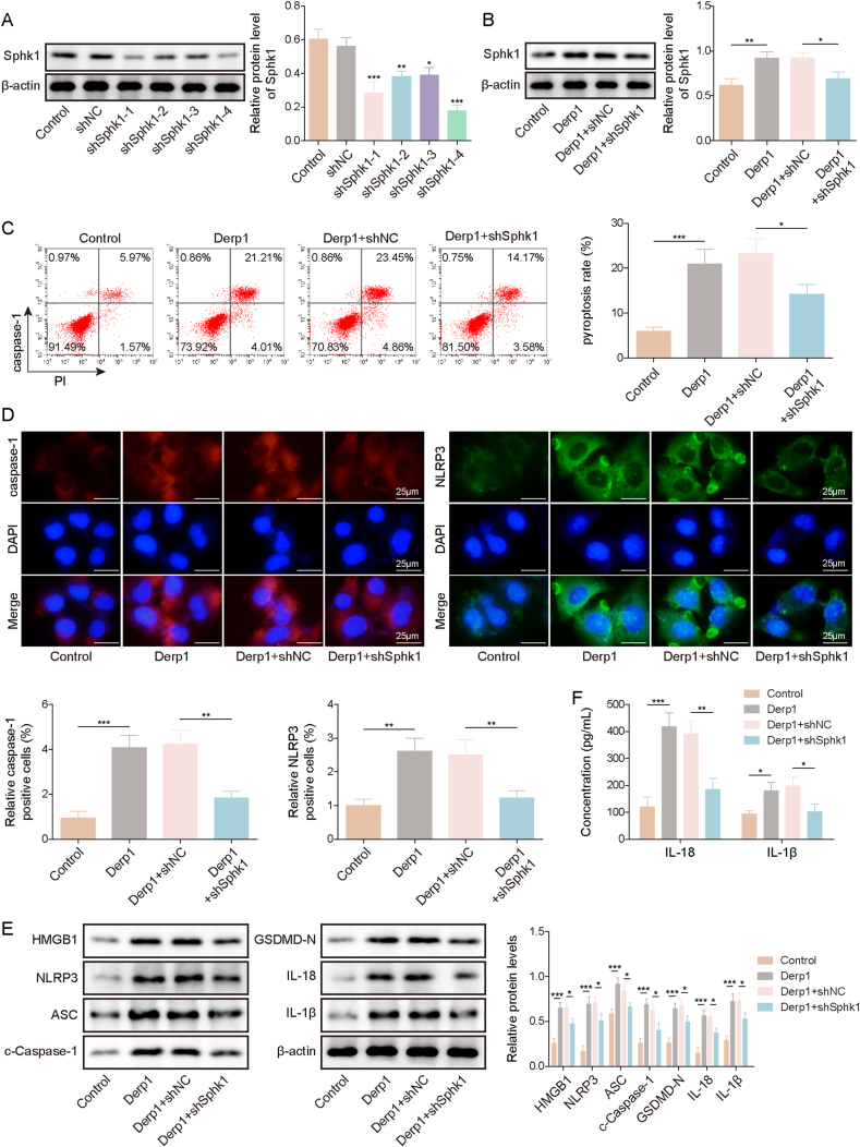
Objective: This study aims to elucidate the roles of HMGB1, Sphk1, and HDAC4 in regulating human nasal epithelial cell (hNEC) pyroptosis and AR.

Methods: An in vitro AR cell culture model and an in vivo AR mouse model were established. Western blot, ELISA, histological staining, and flow cytometry were utilized to confirm the gene and protein expression. The interactions among Sphk1, HDAC4, and HMGB1 were validated through ChIP, Co-IP, and Dual-luciferase assay.

Results and conclusion: We identified that the expression levels of Sphk1, HMGB1, and inflammasome components, including IL-18, and IL-1β were elevated in AR patients and mouse models. Knockdown of Sphk1 inhibited hNEC pyroptosis induced by dust mite allergen. Overexpression of HDAC4 suppressed HMGB1-mediated pyroptosis in hNECs. In addition, HDAC4 was found to mediate the transcriptional regulation of HMGB1 via MEF2C, a transcription factor. Additionally, Sphk1 was shown to interact with CaMKII-δ, promoting the phosphorylation of HDAC4 and inhibiting its cytoplasmic translocation. Knockdown of HDAC4 reversed the effect of Sphk1 knockdown on pyroptosis. These discoveries offer a glimpse into the molecular mechanisms underlying AR and suggest potential therapeutic targets for the treatment of this condition.

Keywords: Allergic; HDAC4; HMGB1; Pyroptosis; Rhinitis; Sphingosine kinase 1.


Conflict of interest statement

No conflicts of interest, financial or otherwise, are declared by the authors.

Figures

Fig. 1
Fig. 1
Sphk1, HMGB1, inflammasome, IL-18 and IL-1β expression was elevated in AR patients and mice. (A) The expression levels of IL-18 and IL-1β were measured in the nasal lavage fluid (NLF) of allergic rhinitis (AR) patients (n = 40) and normal healthy controls (n = 10) using ELISA. Next, an AR mouse model was established using ovalbumin (OVA) (N = 8 per group). (B) The number of scratches and sneezes was recorded in both the AR mouse group and the control group. (C) ELISA was performed to determine the expression levels of IgE, IL-4, HMGB1, IL-18, and IL-1β in the mouse serum and nasal mucosa tissue. (D) HE staining was conducted to evaluate the pathological changes in the mouse nasal mucosal tissue (scale bar = 100 μm). (E) IHC staining was employed to assess the expression levels of Sphk1, HMGB1, ASC, Caspase-1, and IL-1β in the mouse nasal mucosal tissue (scale bar = 100 μm). (F) Western Blot analysis was used to detect the expression levels of Sphk1, HMGB1, NLRP3, GSDMD-N, ASC, c-Caspase-1, IL-1β, and IL-18. ∗, P < 0.05; ∗∗, P < 0.01; ∗∗∗, P < 0.001
Fig. 2
Fig. 2
Knockdown of Sphk1 inhibited pyroptosis of Der p1-induced nasal epithelial cells. (A) Nasal epithelial cells (hNECs) were obtained from dust mite AR patients. Establish Sphk1 knockdown hNECs cells and assess the expression of Sphk1 using Western blot. An in vitro cell model of AR was established using dust mite protein Der p1 and then Sphk1 was knocked down in the AR cell model. (B) The expression of Sphk1 was determined using Western blot. (C) Flow cytometry detected the percentage of Caspase-1/PI-positive cells. (D) IF staining assessed the expression of Caspase-1 and NLRP3 (scale bar = 25 μm). (E) Western Blot measured the expression levels of HMGB1, NLRP3, GSDMD-N, ASC, c-Caspase-1, IL-1β, and IL-18. (F) ELISA examined the protein levels of IL-18, and IL-1β. All experiments were repeated at least three times. ∗, P < 0.05; ∗∗, P < 0.01; ∗∗∗, P < 0.001
Fig. 3
Fig. 3
HDAC4 overexpression suppressed Der p1-induced nasal mucosal epithelial cell pyroptosis through the regulation of HMGB1. (A) Nasal epithelial cells (hNECs) were obtained from dust mite AR patients. Established HDAC4 overexpressed hNECs and assessed the expression of HDAC4 using Western blot. The in vitro cell model of AR was established by treating the hNECs without or with dust mite protein Der p1. Afterward, HDAC4 was overexpressed in the AR cell model. (B) The expression of HDAC4 was determined using Western blot. (C) Flow cytometry detected the percentage of Caspase-1/PI-positive cells. (D) IF staining assessed the expression of Caspase-1 and NLRP3 (scale bar = 25 μm). (E) Western Blot measured the expression levels of HMGB1, NLRP3, GSDMD-N, ASC, c-Caspase-1, IL-1β, and IL-18. (F) ELISA examined the protein levels of IL-18, and IL-1β. All experiments were repeated at least three times. ∗, P < 0.05; ∗∗, P < 0.01; ∗∗∗, P < 0.001
Fig. 4
Fig. 4
HDAC4 mediated the transcription of HMGB1 via MEF2C. (A) Nasal epithelial cells (hNECs) were obtained from dust mite AR patients. Established HDAC4-depleted hNECs cells and assessed the expression of HDAC4 and HMGB1 using Western blot. (B) Bioinformatics analysis predicted the binding of MEF2C to the HMGB1 promoter. (C–D) ChIP and dual-luciferase reporter assay investigated the binding of MEF2C to the HMGB1 promoter. (E) Der p1 was used to treat hNECs to induce the in vitro AR model. Afterward, the dual-luciferase reporter assay investigated the binding of MEF2C to the HMGB1 promoter through HDAC4. All experiments were repeated at least three times. ∗, P < 0.05; ∗∗, P < 0.01; ∗∗∗, P < 0.001
Fig. 5
Fig. 5
Sphk1 directly interacted with CaMKII-δ to promote the phosphorylation of HDAC4 and inhibited the cytoplasmic translocation of HDAC4 to the nucleus in in vitro AR model. Nasal epithelial cells (hNECs) were obtained from dust mite AR patients. hNECs were treated without or with Der p1 or Der p1/Sphk1 inhibitor, SKI-5C. (A) Cytoplasm HDAC4 and nuclear HDAC4 expression were assessed via Western blot. (B) IF staining illustrated HDAC4 expression (scale bar = 25 μm). (C) Western blot detected phosphorylated HDAC4 and total HDAC4 expression levels. (D) CaMKII-δ activity was measured. (E) Western blot examined phosphorylated CaMKII-δ and total CaMKII-δ expression. (F) Co-IP assay investigated the interaction between Sphk1 and CaMKII-δ in hNECs. All experiments were repeated at least three times. ∗, P < 0.05; ∗∗, P < 0.01; ∗∗∗, P < 0.001
Fig. 6
Fig. 6
Downregulation of HDAC4 reversed the protective effect of Sphk1 knockdown on Der p1-induced nasal mucosal epithelial cell pyroptosis. Nasal epithelial cells (hNECs) were obtained from dust mite AR patients. hNECs were treated without or with Der p1. Afterward, Sphk1 and HDAC4 were knocked down by transfecting the Der p1-treated cells with shSphk1-4# and shHDAC4-2#, respectively. (A) The expression of Sphk1, HDAC4, and phosphorylated HDAC4 was determined using Western blot. (B) Flow cytometry detected the percentage of Caspase-1/PI-positive cells. (C) IF staining assessed the expression of Caspase-1 and NLRP3 (scale bar = 25 μm). (D) Western Blot measured the expression levels of HMGB1, NLRP3, GSDMD-N, ASC, c-Caspase-1, IL-1β, and IL-18. (E) ELISA examined the protein levels of HMGB1, IL-18, and IL-1β. All experiments were repeated at least three times. ∗, P < 0.05; ∗∗, P < 0.01; ∗∗∗, P < 0.001
Fig. 7
Fig. 7
Suppression of Sphk1 reduced HMGB1-regulated nasal mucosal epithelial cell pyroptosis in AR mouse models. Female BALB/c mice between seven and eight weeks were treated with ovalbumin (OVA) to establish the AR mouse model. Afterward, shSphk1 or negative control (shNC) was established and subcutaneously injected into mice. (A) The number of scratches and sneezes per minute was recorded in different mouse groups. (B) ELISA detected serum and nasal mucosa tissue protein levels of IgE, IL-4, HMGB1, IL-18 and IL-1β. (C) HE staining was performed to examine the pathological changes in the inferior turbinate mucosal tissue collected from different mouse groups (scale bar = 100 μm). (D) IHC staining was used to assess the expression levels of Sphk1, HMGB1, ASC, Caspase-1, and IL-1β in the inferior turbinate mucosal tissue collected from different mouse groups (scale bar = 100 μm). (E) Western Blot analysis was used to detect the expression levels of Sphk1, HMGB1, NLRP3, GSDMD-N, ASC, c-Caspase-1, IL-1β, and IL-18 in the inferior turbinate mucosal tissue collected from different mouse groups. N = 8 for per group. ∗, P < 0.05; ∗∗, P < 0.01; ∗∗∗, P < 0.001

Similar articles

References

    1. Cheng N., Wang Y., Gu Z. Understanding the role of NLRP3-mediated pyroptosis in allergic rhinitis: a review. Biomed Pharmacother. 2023;165 - PubMed
    1. Nur Husna S.M., Tan H.T., Md Shukri N., Mohd Ashari N.S., Wong K.K. Allergic rhinitis: a clinical and pathophysiological overview. Front Med (Lausanne) 2022;9:874114. - PMC - PubMed
    1. Bousquet J., Melén E., Haahtela T., et al. Rhinitis associated with asthma is distinct from rhinitis alone: the ARIA-MeDALL hypothesis. Allergy. 2023;78(5):1169–1203. - PubMed
    1. Yang Z., Liang C., Wang T., et al. NLRP3 inflammasome activation promotes the development of allergic rhinitis via epithelium pyroptosis. Biochem Biophys Res Commun. 2020;522(1):61–67. - PubMed
    1. Kelley N., Jeltema D., Duan Y., He Y. The NLRP3 inflammasome: an overview of mechanisms of activation and regulation. Int J Mol Sci. 2019;20(13):3328. - PMC - PubMed

LinkOut - more resources