. 2024 Aug 8;15(9):5098-5114.
doi: 10.1364/BOE.537268. eCollection 2024 Sep 1.

Chromatic cues for the sign of defocus in the peripheral retina

Affiliations

Chromatic cues for the sign of defocus in the peripheral retina

Len Zheleznyak et al. Biomed Opt Express. .

Abstract

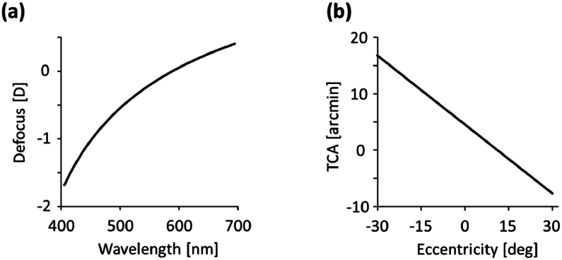
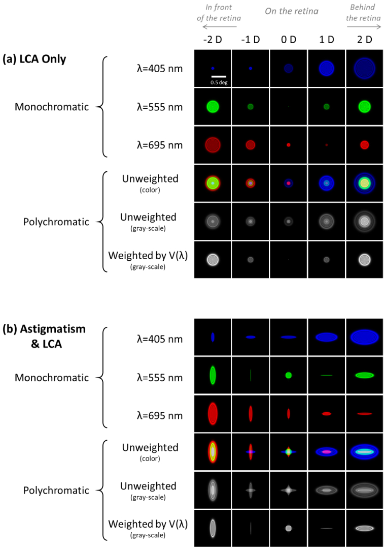
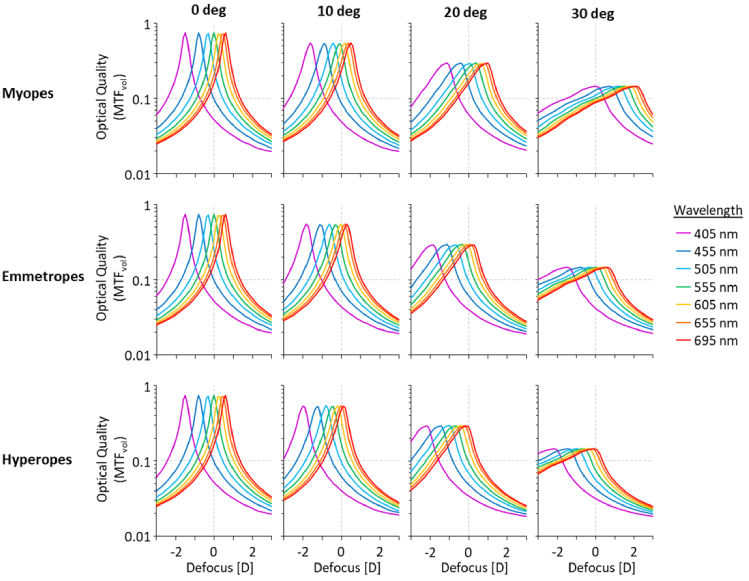
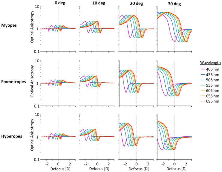
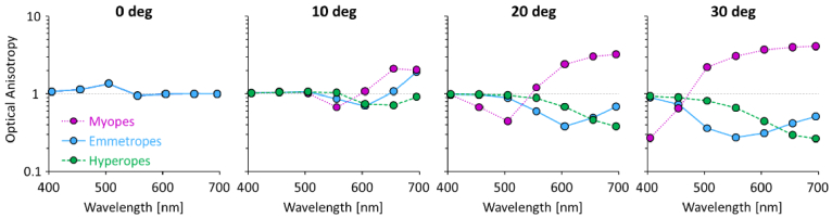
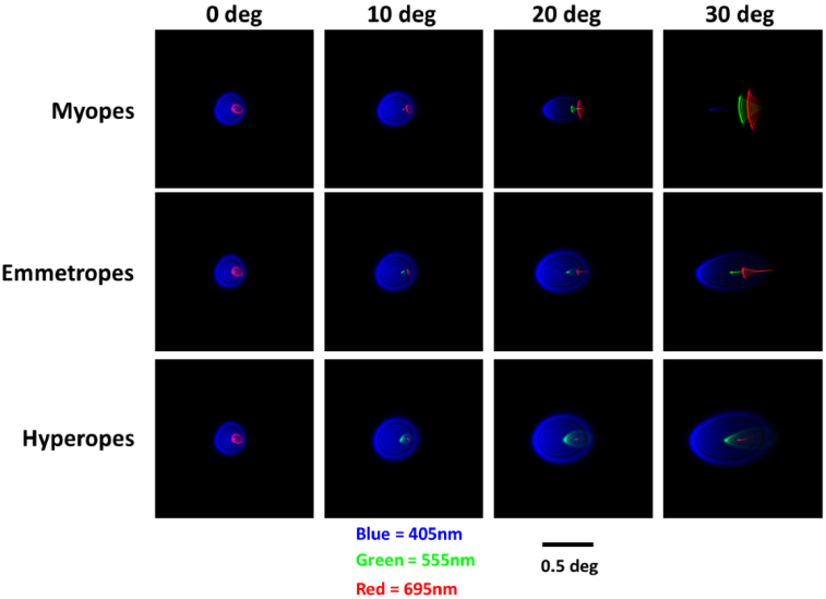
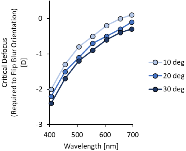
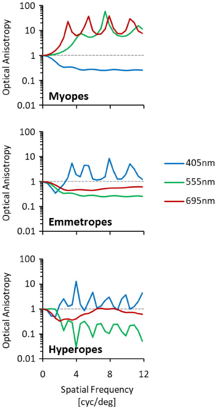
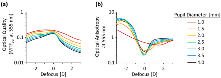
Detecting optical defocus at the retina is crucial for accurate accommodation and emmetropization. However, the optical characteristics of ocular defocus are not fully understood. To bridge this knowledge gap, we simulated polychromatic retinal image quality by considering both the monochromatic wavefront aberrations and chromatic aberrations of the eye, both in the fovea and the periphery (nasal visual field). Our study revealed two main findings: (1) chromatic and monochromatic aberrations interact to provide a signal to the retina (chromatic optical anisotropy) to discern positive from negative defocus and (2) that chromatic optical anisotropy exhibited notable differences among refractive error groups (myopes, emmetropes and hyperopes). These findings could enhance our understanding of the underlying mechanisms of defocus detection and their subsequent implications for myopia control therapies. Further research is needed to explore the retinal architecture's ability to utilize the optical signals identified in this study.


Conflict of interest statement

LZ: Clerio Vision (E, P), CL: None, SW: Clerio Vision (C)

Figures

Fig. 1.
Fig. 1.
(a) Longitudinal and (b) transverse chromatic aberration across the visible spectrum (405-695 nm).
Fig. 2.
Fig. 2.
Theoretical simulation of through-focus point spread functions for 2 conditions: (a) monochromatic aberration-free and (b) astigmatism of 2 D, both for a 4 mm pupil. PSFs are shown monochromatically at 405, 555 and 695 nm, and polychromatically in color and in grayscale (with and without V(λ) weighting [33,35]).
Fig. 3.
Fig. 3.
Through-focus monochromatic optical quality (MTFvol) for myopes, emmetropes and hyperopes across the visible spectrum and retinal eccentricities (nasal visual field) for a 4 mm pupil.
Fig. 4.
Fig. 4.
Through-focus monochromatic optical anisotropy (OA) for myopes, emmetropes and hyperopes across the visible spectrum and retinal eccentricities (nasal visual field) for a 4 mm pupil.
Fig. 5.
Fig. 5.
Optical anisotropy as a function of wavelength for hyperopes, emmetropes and myopes across the nasal visual field for a 4 mm pupil.
Fig. 6.
Fig. 6.
Point spread functions across the nasal visual field for foveally distance corrected eyes of hyperopes (top row), emmetropes (middle row) and myopes (bottom row). Within each frame, are superimposed monochromatic point spread functions of wavelengths 405 nm (in blue), 555 nm (in green) and 695 nm (in red) which are defocused and decentered from one another due to LCA and TCA, respectively.
Fig. 7.
Fig. 7.
Critical Defocus required to flip blur orientation from horizontal (OA < 1.0) to vertical (OA > 1.0) in the horizontal nasal visual field of the average emmetropic eye for a 4 mm pupil.
Fig. 8.
Fig. 8.
Monochromatic OA as a function of spatial frequency for refractive error groups at 405, 555 and 695 nm wavelengths. All data in this figure pertains to the 30 deg nasal visual field.
Fig. 9.
Fig. 9.
Through-focus (a) optical quality and (b) anisotropy for various pupil sizes at 30 deg in emmetropes at 555 nm.

Similar articles

Cited by

References

    1. Wallman J., Winawer J., “Homeostasis of eye growth and the question of myopia,” Neuron 43(4), 447–468 (2004).10.1016/j.neuron.2004.08.008 - DOI - PubMed
    1. Brown D. M., Mazade R., Clarkson-Townsend D., et al. , “Candidate pathways for retina to scleral signaling in refractive eye growth,” Exp. Eye Res. 219, 109071 (2022).10.1016/j.exer.2022.109071 - DOI - PMC - PubMed
    1. Zhang D.-Q., Wong K. Y., Sollars P. J., et al. , “Intraretinal signaling by ganglion cell photoreceptors to dopaminergic amacrine neurons,” Proc. Natl. Acad. Sci. 105(37), 14181–14186 (2008).10.1073/pnas.0803893105 - DOI - PMC - PubMed
    1. Summers J. A., Schaeffel F., Marcos S., et al. , “Functional integration of eye tissues and refractive eye development: Mechanisms and pathways,” Exp. Eye Res. 209, 108693 (2021).10.1016/j.exer.2021.108693 - DOI - PMC - PubMed
    1. Wolffsohn J. S., Kollbaum P. S., Berntsen D. A., et al. , “IMI-clinical myopia control trials and instrumentation report,” Invest. Ophthalmol. Visual Sci. 60(3), M132–M160 (2019).10.1167/iovs.18-25955 - DOI - PubMed

LinkOut - more resources