. 2024 Sep 4:11:1439181.
doi: 10.3389/fmed.2024.1439181. eCollection 2024.

SARS-CoV-2 replicates in the placenta after maternal infection during pregnancy

Affiliations

SARS-CoV-2 replicates in the placenta after maternal infection during pregnancy

Anda-Petronela Radan et al. Front Med (Lausanne). .

Abstract

Objectives: Pregnant women are at increased risk for severe SARS-CoV-2 infection and adverse neonatal outcome, primarily preterm birth and stillbirth. Our study aimed to investigate to which extent SARS-CoV-2 affects placental tissue and if viral replication within the placenta is evident, thus if there is a correlation between placental damage and adverse pregnancy outcome such as stillbirth.

Methods: We prospectively collected placentas from 61 SARS-CoV-2 infected pregnant women and 10 controls. Histopathological, immunohistochemical, and in situ hybridization studies were performed on all placentas with antibodies for SARS-CoV-2 proteins, ACE2, various immune cells, and inflammatory markers or probes for SARS-CoV-2 genes and an antisense strand.

Results: The measured scores of SARS-CoV-2 glycoprotein, nucleocapsid, and antisense strand indicating replication correlated with both the severity of maternal symptoms and presence of stillbirth. Specifically, 15/61 placentas exhibited replication, while the three cases with stillbirth had high or maximal replication scores. ACE2-H-score was significantly higher in COVID-19 patients, while the expression of various immune cells did not differ statistically. In multivariate analysis, presence of maternal comorbidities correlated with presence of severe COVID-19 infection.

Conclusion: We report evidence of active in vivo SARS-CoV-2 replication in the placenta after maternal infection in pregnancy in a case-control setting in a large population. Intensity of placental viral replication as well as viral levels were higher in women with severe or critical COVID-19 disease, supporting the rationale that severity of maternal SARS-CoV-2 infection could correlate with the severity of placentitis. Replication was maximal in cases of stillbirth, which suggests direct placental involvement in the pathophysiology of this dramatic outcome. Continuing to advocate for preventive measures against COVID-19 during pregnancy, including (re)vaccination, as well as appropriately counseling women with diagnosed infection, are of utter importance.

Keywords: COVID-19; SARS-CoV-2; SARS-CoV-2 replication; placenta; stillbirth.


Conflict of interest statement

The authors declare that the research was conducted in the absence of any commercial or financial relationships that could be construed as a potential conflict of interest.

Figures

Figure 1
Figure 1
Time point of infection. X axis shows weeks of gestation. Y axis shows number of cases.
Figure 2
Figure 2
Quantification of SARS-CoV-2 specific proreins and virus replication in mild/moderate and severe/critical COVID-19 cases. IRS score for (A) the spike glycoprotein (p = 0.0013) and (B) the nucleocapsid protein (p = 0.03) is significantly higher in severe or critical cases (pink) compared to mild or moderate cases (blue). (C) A significantly higher IRS score (p = 0.034) for the virus replication was detected in severe or critical cases. Data are represented as mean ± SD.
Figure 3
Figure 3
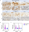
ACE2 in SARS-CoV-2 infected versus not infected placentas, with distribution between villi and decidua. (A) Case 1i images show COVID-19 miscarriage at 18 weeks of gestation. Case 2i images represent COVID-19 stillbirth at 30 weeks of gestation. Case 4i are images of a COVID-19 patient with severe course of the disease (pneumonia with decompensation) who was infected at 32 weeks of gestation and delivered at 33 1/7. The control cases images are placentas from COVID-19 negative women. Case 1c (control) are images of a placenta after stillbirth at 20 weeks, case 2c (control) images are from a term birth, and case 3c (control) was an IUGR case. Scale bar: 100 μm. (B) ACE2 H-Score quantification in the entire placenta. ACE2 H-score was significantly increased in COVID-19 cases (violet) compared to control cases (blue) (p = 0.0134). (C) ACE2 H-score was significantly increased in COVID-19 cases (violet) in the decidua compared to controls (blue) (p = 0.0149). No significant difference was observed in villi between COVID-19 (violet) and control cases (blue) (p = 0.07). Data are represented as mean ± SD.
Figure 4
Figure 4
Evidence of SARS-CoV-2 nucleocapsid and glycoprotein in SARS-CoV-2 infected placentas versus controls, as well as evidence of viral replication with RNAscope®. Cases are identical to Figure 3. (A–D) Representative images for the spike glycoprotein and (E–H) the nucleocapsid protein staining of cases and a control (negative). The red circles in case 3 point to a positive signal for spike glycoprotein (C), nucleocapsid protein (G). (I–P) Representative images of the RNAscope with magenta for SARS-CoV-2 S gene encoding the spike protein and cyan for SARS-CoV-2 Antisense strand of the orf1ab gene (virus replication). (K) Red circles point to a positive signal for virus replication. (M) Representative overview and (N) representative high magnification image after RNAscope staining of case 1i show high viral replication (cyan). (O) Representative overview and (P) representative high magnification image after RNAscope staining of case 2i show high viral replication (cyan).
Figure 5
Figure 5
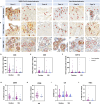
Quantification of immune cells in COVID-19 and control placentas. Cases are identical to Figure 3. Representative images of COVID-19 cases and images of control cases with (A) CD8+ staining in the decidua, with (B) CD3+ staining in the decidua and with (C) CD68+ staining in the villous stroma. (A,B) Arrowheads point to some positive CD3 and CD8 cells. (C) Red circles indicate hotspots of CD68 positive cells. (D–F) Quantification of CD3-, CD8-, and CD11b positive cell number in hotspots in the decidua and villi. (G,H) Quantification of positive area of CD68 and CD86 staining in hotspots in the decidua and villi. (I) IRS-score of IL6 and (J) positive area of TNFa were additionally analyzed. In all cases, no significant difference was detected between COVID-19 and control cases (see Table 2, data represented as mean ± SD).
Figure 6
Figure 6
Evidence of marked chronic histiocytic intervillositis. Representative images of case 2i with stillbirth at 30 weeks of gestation.
Figure 7
Figure 7
Mechanism of placental damage in SARS-CoV-2 infection during pregnancy.

References

    1. Di Mascio D, Buca D, Berghella V, Khalil A, Rizzo G, Odibo A, et al. Counseling in maternal-fetal medicine: SARS-CoV-2 infection in pregnancy. Ultrasound Obstet Gynecol. (2021) 57:687–97. doi: 10.1002/uog.23628, PMID: - DOI - PMC - PubMed
    1. Gurol-Urganci I, Jardine JE, Carroll F, Draycott T, Dunn G, Fremeaux A, et al. Maternal and perinatal outcomes of pregnant women with SARS-CoV-2 infection at the time of birth in England: national cohort study. Am J Obstet Gynecol. (2021) 225:522.e1–522.e11. doi: 10.1016/j.ajog.2021.05.016, PMID: - DOI - PMC - PubMed
    1. Roberts DJ, Edlow AG, Romero RJ, Coyne CB, Ting DT, Hornick JL, et al. A standardized definition of placental infection by SARS-CoV-2, a consensus statement from the National Institutes of Health/Eunice Kennedy Shriver National Institute of Child Health and Human Development SARS-CoV-2 placental infection workshop. Am J Obstet Gynecol. (2021) 225:593.e1–9. doi: 10.1016/j.ajog.2021.07.029, PMID: - DOI - PMC - PubMed
    1. Stenton S, McPartland J, Shukla R, Turner K, Marton T, Hargitai B, et al. SARS-COV2 placentitis and pregnancy outcome: a multicentre experience during the alpha and early Delta waves of coronavirus pandemic in England. EClinicalMedicine. (2022) 47:101389. doi: 10.1016/j.eclinm.2022.101389, PMID: - DOI - PMC - PubMed
    1. Marton T, Hargitai B, Hunter K, Pugh M, Murray P. Massive Perivillous fibrin deposition and chronic histiocytic Intervillositis a complication of SARS-CoV-2 infection. Pediatr Dev Pathol. (2021) 24:450–4. doi: 10.1177/10935266211020723, PMID: - DOI - PubMed