. 2024 Sep 4:12:1442198.
doi: 10.3389/fcell.2024.1442198. eCollection 2024.

Golgi clustering by the deficiency of COPI-SNARE in Drosophila photoreceptors

Affiliations

Golgi clustering by the deficiency of COPI-SNARE in Drosophila photoreceptors

Tatsuya Tago et al. Front Cell Dev Biol. .

Abstract

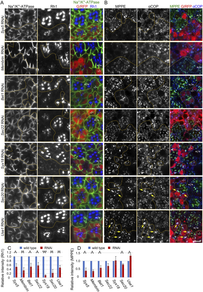
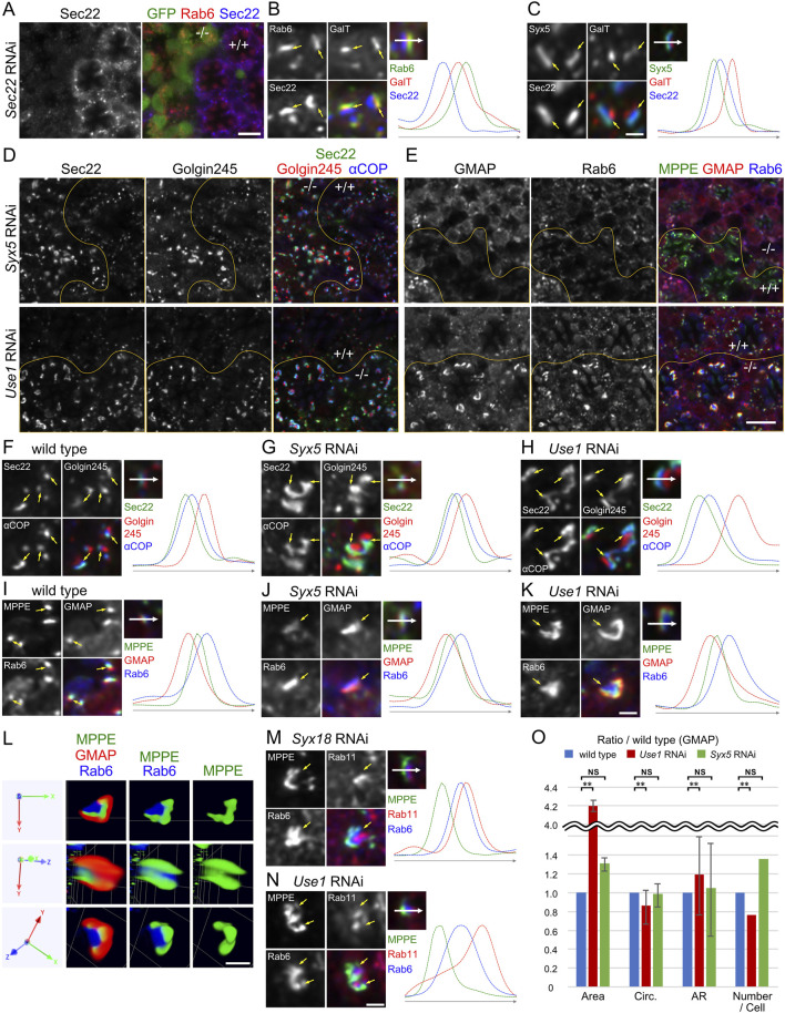
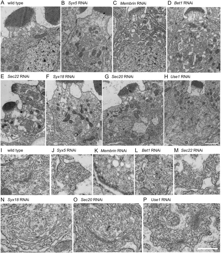
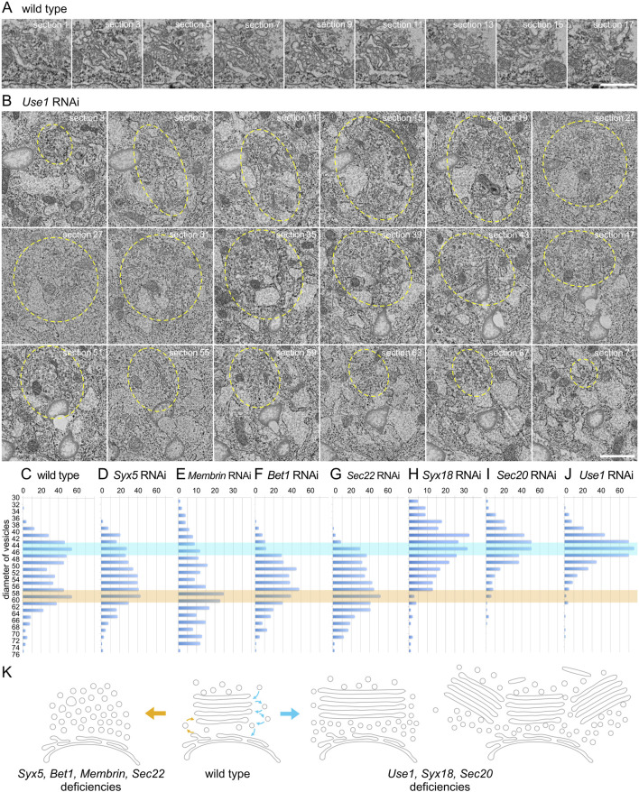
A comprehensive study of soluble N-ethylmaleimide-sensitive factor attachment protein receptors (SNAREs) in the fly genome by RNAi in Drosophila photoreceptors indicated that knockdown of any of the COPI-SNAREs, Syx18, Sec20, and Use1, resulted in the same characteristic phenotypes: Golgi stacks gathering on their trans-side, laterally expanded Golgi cisternae, and a reduced number of discrete Golgi stacks. These Golgi stacks are reminiscent of mammalian Golgi ribbons and Brefeldin A (BFA)-bodies in Drosophila S2 cells. As previously reported, BFA suppresses trans-Golgi network (TGN) fission and Golgi stack separation to form a BFA-body, which is a cluster of Golgi stacks cored by recycling endosomes. We found that the impairing each of COPI-SNAREs results in clustered Golgi stacks similar to BFA-bodies, indicating that COPI-SNAREs have a role to separate clustered Golgi stacks. These results further support the idea that the movement of Golgi stacks and the balance of fusion and fission of the TGN determine the level of clustering and ribbon formation of Golgi stacks within cells.

Keywords: BFA-body; Drosophila; Golgi stacks; photoreceptors; recycling endosomes.


Conflict of interest statement

The authors declare that the research was conducted in the absence of any commercial or financial relationships that could be construed as a potential conflict of interest.

Figures

FIGURE 1
FIGURE 1
Reduction of Rh1 in the rhabdomeres by knockdown of SNAREs for COPI and COPII fusion. (A) Immunostaining of SNARE RNAi construct-expressing retina by eyeless-CoinFLP-longGMR-Gal4 (Syx5, Bet1, Syx18, and Sec20) or eyeless-CoinFLP-Act5C-Gal4 (Membrin, Sec22, and Use1) using anti-Na+/K+-ATPase-α (green) and anti-Rh1 (blue) antibodies. RFP/GFP (red) represents the cells expressing RNAi constructs. (B) Immunostaining of SNARE RNAi construct-expressing retina by eyeless-CoinFLP-longGMR-Gal4 (Syx5, Bet1, Syx18, and Sec20) or eyeless-CoinFLP-Act5C-Gal4 (Membrin, Sec22, and Use1) using anti-MPPE (green) and anti-αCOP (blue) antibodies. RFP/GFP (red) represents the cells expressing RNAi constructs. The anti-MPPE antibody stains medial Golgi and also rhabdomere tips, the latter is likely representing cross-reactivity. (C, D) The ratio of integrated fluorescence density for Rh1 (C), and MPPE (D) staining of the cytoplasm compared to that of whole cells was plotted. Blue bars indicate wild-type cells and red bars indicate cells expressing RNAi constructs. Error bars indicate the SD of three retinas. Significance according to two-tailed unpaired Student’s t-test: **p < 0.01 and *p < 0.05. Scale bar: 5 μm (A, B).
FIGURE 2
FIGURE 2
Golgi stacks are enlarged by knockdown of SNAREs for COPI fusion. (A) Immunostaining of Sec22 RNAi construct-expressing retina by eyeless-CoinFLP-Act5C-Gal4 using anti-Rab6 (red) and anti-Sec22 (blue) antibodies. GFP (green) represents the cells expressing Sec22 RNAi construct. (B) Left: wild-type Golgi stacks expressing GalT::CFP (red) immunostained by anti-Rab6 (green) and anti-Sec22 (blue) antibodies. Arrows indicate the relative position of staining. Right: the plot of signal intensities along the 1.5 μm from the top image. (C) Left: wild-type Golgi stacks expressing GalT::CFP (red) and Syx5::Myc immunostained by anti-Myc (green) and anti-Sec22 (blue) antibodies. Arrows indicate the relative position of staining. Right: the plot of signal intensities along the 1.5 μm from the top image. (D) Immunostaining of Syx5 (upper) or Use1 (lower) RNAi construct-expressing retina by eyeless-CoinFLP-longGMR-Gal4 (upper) or eyeless-CoinFLP-Act5C-Gal4 (lower) using anti-Sec22 (green), anti-Golgin245 (red) and anti-αCOPI (blue) antibodies. (E) Immunostaining of Syx5 (upper) or Use1 (lower) RNAi construct-expressing retina by eyeless-CoinFLP-longGMR-Gal4 (upper) or eyeless-CoinFLP-Act5C-Gal4 (lower) using anti-MPPE (green), anti-GMAP (red) and anti-Rab6 (blue) antibodies. (F–H) Left: wild-type (F), Syx5 knockdown (G) and Use1 knockdown (H) Golgi stacks immunostained by anti-Sec22 (green), anti-Golgin245 (red) and anti-αCOPI (blue) antibodies. Arrows indicate the relative positions of staining. Right: the plot of signal intensities along the 1.5 μm from the top image. (I–K) Left: wild-type (I), Syx5 knockdown (J) and Use1 knockdown (K) Golgi stacks immunostained with anti-MPPE (green), anti-GMAP (red), and anti-Rab6 (blue) antibodies. Arrows indicate the relative positions of staining. Right: the plot of signal intensities along the 1.5 μm from the top image. (L) Volumetrically rendered images of a Use1 knockdown Golgi stack immunostained with anti-MPPE (green), anti-GMAP (red), and anti-Rab6 (blue) antibodies presented from three different angles. (M, N) Left: Golgi stacks with Syx18 knockdown (M) and Use1 knockdown (N) immunostained with anti-MPPE (green), anti-Rab11 (red), and anti-Rab6 (blue) antibodies. Arrows indicate the relative positions of staining. Right: the plot of signal intensities along the 1.5 μm from the top image.(O) Plots of the ratio of area, number, circularity (Circ), and aspect ratio (AR: major axis/minor axis) of Golgi stacks in Use1 RNAi- or Syx5 RNAi-expressing photoreceptors compared to those in wild-type photoreceptors. Blue, red, and green bars indicate wild-type, Use1 RNAi, or Syx5 RNAi-expressing photoreceptors, respectively. Error bars indicate standard SD of four retinas. Significance according to two-tailed unpaired Student’s t-test: **p < 0.01.Scale bars: 5 μm (A,D, E) and 1 μm (B, C, F–K, L, M, N).
FIGURE 3
FIGURE 3
Morphologies of Golgi stacks in COPI- or COPII- SNARE knockdown photoreceptors. (A–H) Electron micrographs of SNARE RNAi construct-expressing photoreceptors by eyeless-CoinFLP-longGMR-Gal4 (Syx5, Bet1, Syx18, and Sec20) or eyeless-CoinFLP-Act5C-Gal4 (Membrin, Sec22 and Use1) at late pupae. (I–P) Golgi stack or vesicle clusters in COPI- or COPII-SNARE knockdown photoreceptor. Scale bars: 2 μm (A–H) and 500 nm (I–P).
FIGURE 4
FIGURE 4
Enlarged Golgi stack in Use1 knockdown photoreceptor. (A) Serial sections of a Golgi stack at 50-nm intervals in the wild-type photoreceptor, numbered as indicated. (B) Serial sections of a cluster of Golgi stacks at 50-nm intervals in the Use1 RNAi construct expressing photoreceptor by eyeless-CoinFLP-longGMR-Gal4, numbered as indicated. A cluster of Golgi stacks circled with yellow line. (C–J) Plots of the number of vesicles with indicated diameters found near Golgi stacks or vesicle clusters in the wild-type (C) or SNARE RNAi construct-expressing photoreceptors by eyeless-CoinFLP-longGMR-Gal4 (Syx5 (D), Bet1 (F), Syx18 (I), and Sec20 (J) or eyeless-CoinFLP-Act5C-Gal4 (Membrin (E), Sec22 (G) and Use1 (H)) at late pupae. (K) Model of structural changes in Golgi stacks in COPI- and COPII-SNARE knockdown photoreceptor cells. COPII-SNARE knockdown transformed Golgi stacks into vesicle clusters with the same diameter as COPII vesicles (left). In contrast, with COPI-SNARE knockdown, Golgi stacks expanded or assembled around TGNs (right). These expanded or clustered Golgi stacks were accompanied by vesicles with COPI vesicle diameters. Scale bars: 500 nm (A, B).

Similar articles

Cited by

References

    1. Belgareh-Touzé N., Corral-Debrinski M., Launhardt H., Galan J. M., Munder T., Le Panse S., et al. (2003). Yeast functional analysis: identification of two essential genes involved in ER to Golgi trafficking. Traffic 4, 607–617. 10.1034/j.1600-0854.2003.00116.x - DOI - PubMed
    1. Benvenuto G., Leone S., Astoricchio E., Bormke S., Jasek S., D'Aniello E., et al. (2024). Evolution of the ribbon-like organization of the Golgi apparatus in animal cells. Cell. Rep. 43, 113791. 10.1016/j.celrep.2024.113791 - DOI - PubMed
    1. Bosch J. A., Tran N. H., Hariharan I. K. (2015). CoinFLP: a system for efficient mosaic screening and for visualizing clonal boundaries in Drosophila. Development 142, 597–606. 10.1242/dev.114603 - DOI - PMC - PubMed
    1. Cao J., Li Y., Xia W., Reddig K., Hu W., Xie W., et al. (2011). A Drosophila metallophosphoesterase mediates deglycosylation of rhodopsin. EMBO J. 30, 3701–3713. 10.1038/emboj.2011.254 - DOI - PMC - PubMed
    1. Caracci M. O., Fuentealba L. M., Marzolo M. P. (2019). Golgi complex dynamics and its implication in prevalent neurological disorders. Front. Cell. Dev. Biol. 7, 75. 10.3389/fcell.2019.00075 - DOI - PMC - PubMed

LinkOut - more resources