

A simple transconjunctival technique for the management of intraconal orbital hydatid cyst
- PMID: 39297476
- PMCID: PMC11991541
- DOI: 10.4103/IJO.IJO_756_24
A simple transconjunctival technique for the management of intraconal orbital hydatid cyst
Abstract
Purpose: To present a simple transconjunctival technique for the excision of intraconal orbital hydatid cysts.
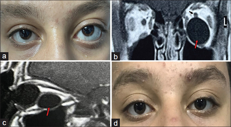
Methods: This retrospective, non-comparative, clinical intervention case study was conducted between April 2018 and October 2023. The study included five patients presented to the Orbital unit of Assiut University Hospital with an intraconal orbital cyst, which histologically proved to be a hydatid cyst. In all cases, a conjunctival incision near the fornix was made depending on the cyst location as revealed by computed tomography (CT) or magnetic resonance imaging (MRI). A traction suture was applied to the two relevant recti muscles to guide the globe toward the desired direction. Blunt orbital dissection was made toward the cyst until exposing its anterior surface. A 20-gauge needle was introduced into the cyst and followed by aspiration of its content. The collapsed cyst was then removed by non-toothed forceps and followed by copious irrigation of the field. The follow-up period ranged from 11 to 58 months.
Results: The age of patients ranged from 11 to 44 years. Three were males and two were females. The cyst was iso-dense to the vitreous on CT and iso-intense to the vitreous on MRI. In all cases after aspiration of the content, the collapsed cyst was easily removed. None of the five patients developed recurrence during the follow-up period.
Conclusion: The removal of the collapsed orbital hydatid cyst in the intraconal space after the aspiration of its content via transconjunctival anterior orbitotomy is a simple, fast technique with early recovery and maximum cosmesis.
Copyright © 2024 Indian Journal of Ophthalmology.
Conflict of interest statement
There are no conflicts of interest.
Figures


References
-
- Anandpara KM, Aswani Y, Hira P, Sathe PA. Isolated primary orbital hydatid disease presenting as multiple cystic lesions: A rare cause of proptosis. Ann Parasitol. 2015;61:193–5. - PubMed
-
- Hammoud M, Benzagmout M, Lakhdar F, Chakour K, Chaoui M. Fronto orbital approach for primary orbital hydatid cyst: Case report. J Neurol Stroke. 2020;10:53–6.
-
- Kahveci R, Sanli AM, Gurer B, Sekerci Z. Orbital hydatid cyst. J Neurosurg Pediatr. 2012;9:42–4. - PubMed
-
- Awad Mohammad Ael N, Ray CJ, Karcioglu ZA. Echinococcus cysts of the orbit and substernum. Am J Ophthalmol. 1994;118:676–8. - PubMed
MeSH terms
LinkOut - more resources
Full Text Sources
Miscellaneous
