. 2024 Oct 1;27(10):pyae041.
doi: 10.1093/ijnp/pyae041.

Ketamine Prevents Inflammation-Induced Reduction of Human Hippocampal Neurogenesis via Inhibiting the Production of Neurotoxic Metabolites of the Kynurenine Pathway

Affiliations

Ketamine Prevents Inflammation-Induced Reduction of Human Hippocampal Neurogenesis via Inhibiting the Production of Neurotoxic Metabolites of the Kynurenine Pathway

Gargi Mandal et al. Int J Neuropsychopharmacol. .

Abstract

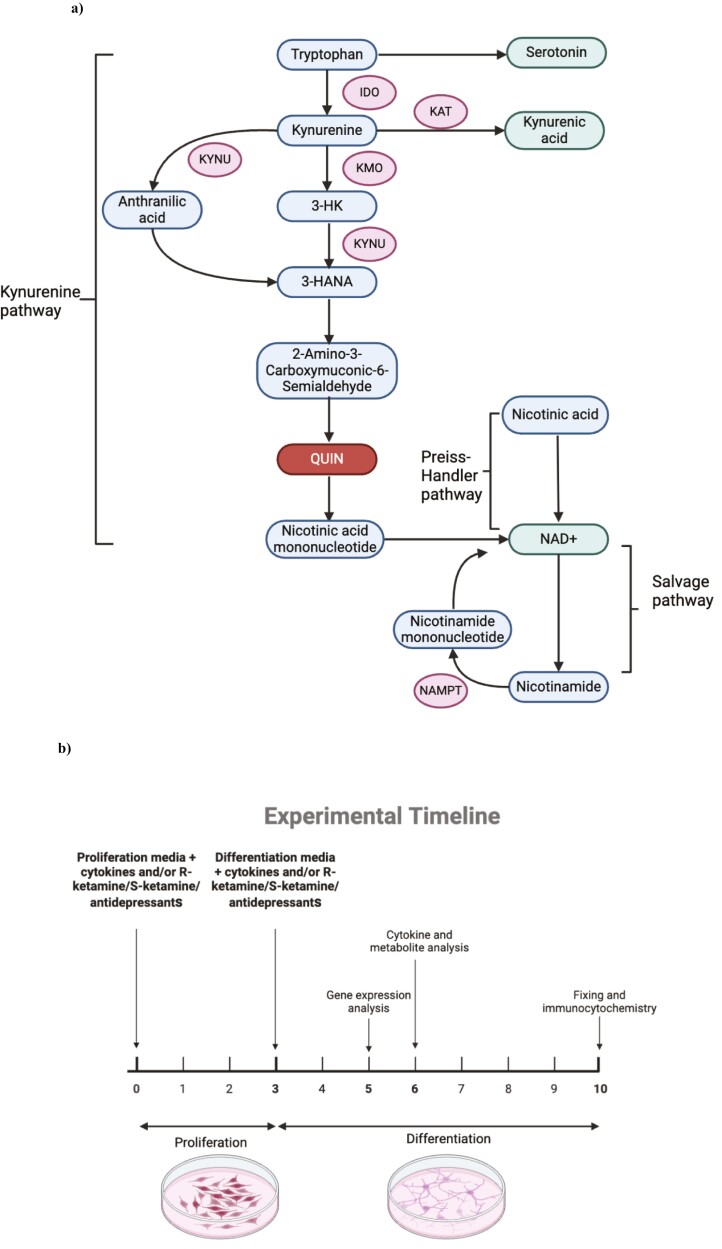
Background: Understanding the precise mechanisms of ketamine is crucial for replicating its rapid antidepressant effects without inducing psychomimetic changes. Here, we explore whether the antidepressant-like effects of ketamine enantiomers are underscored by protection against cytokine-induced reductions in hippocampal neurogenesis and activation of the neurotoxic kynurenine pathway in our well-established in vitro model of depression in a dish.

Methods: We used the fetal hippocampal progenitor cell line (HPC0A07/03C) to investigate ketamine's impact on cytokine-induced reductions in neurogenesis in vitro. Cells were treated with interleukin- 1beta (IL-1b) (10 ng/mL) or IL-6 (50 pg/mL), alone or in combination with ketamine enantiomers arketamine (R-ketamine, 400 nM) or esketamine (S-ketamine, 400 nM) or antidepressants sertraline (1 mM) or venlafaxine (1 mM).

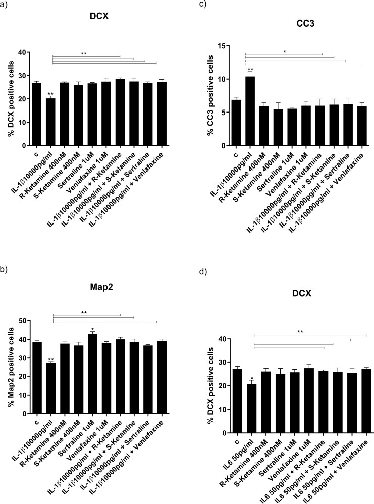
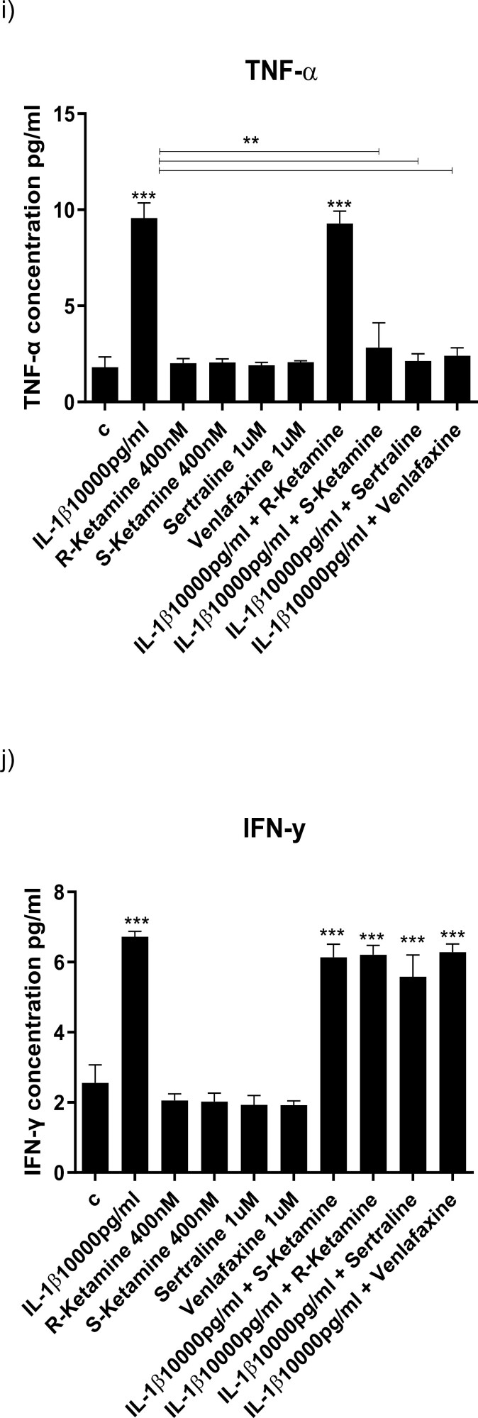
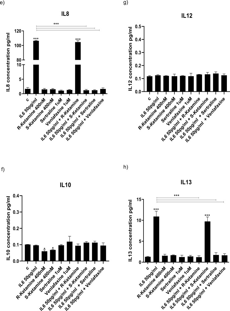
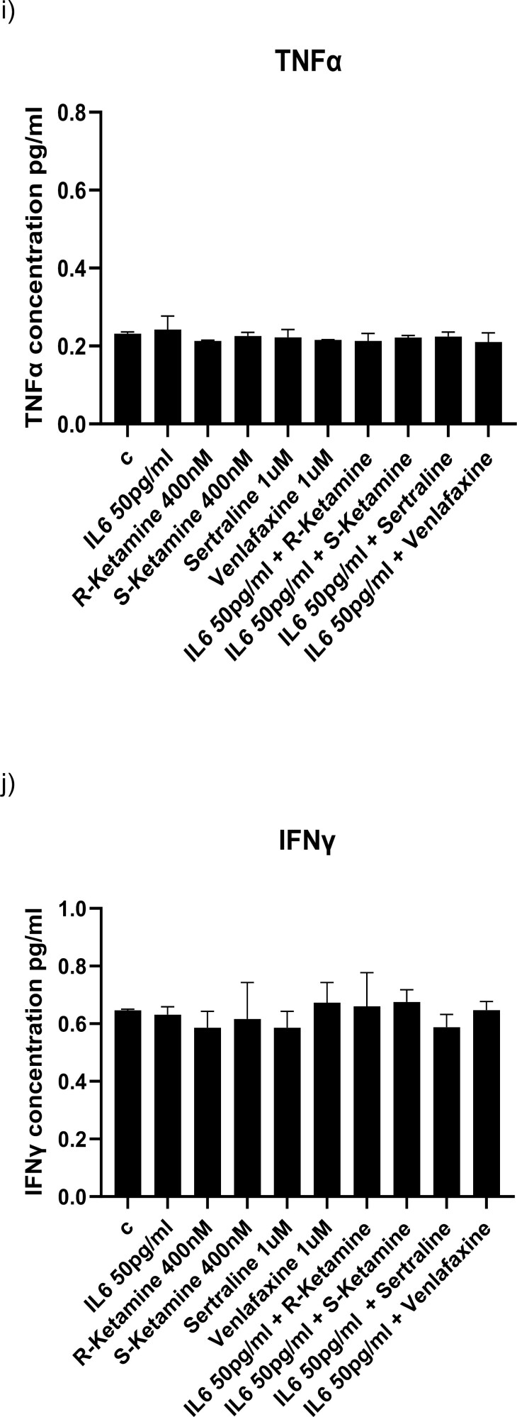
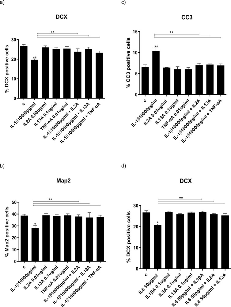
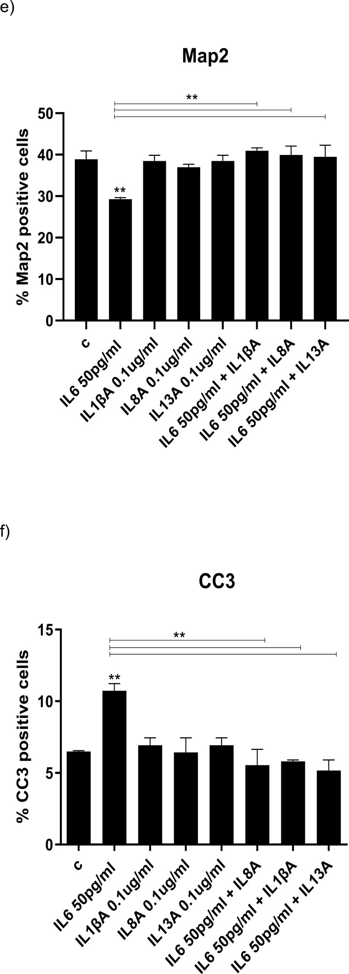
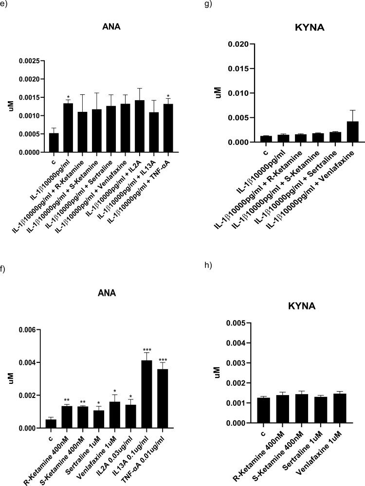
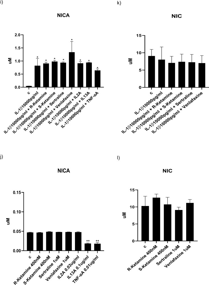
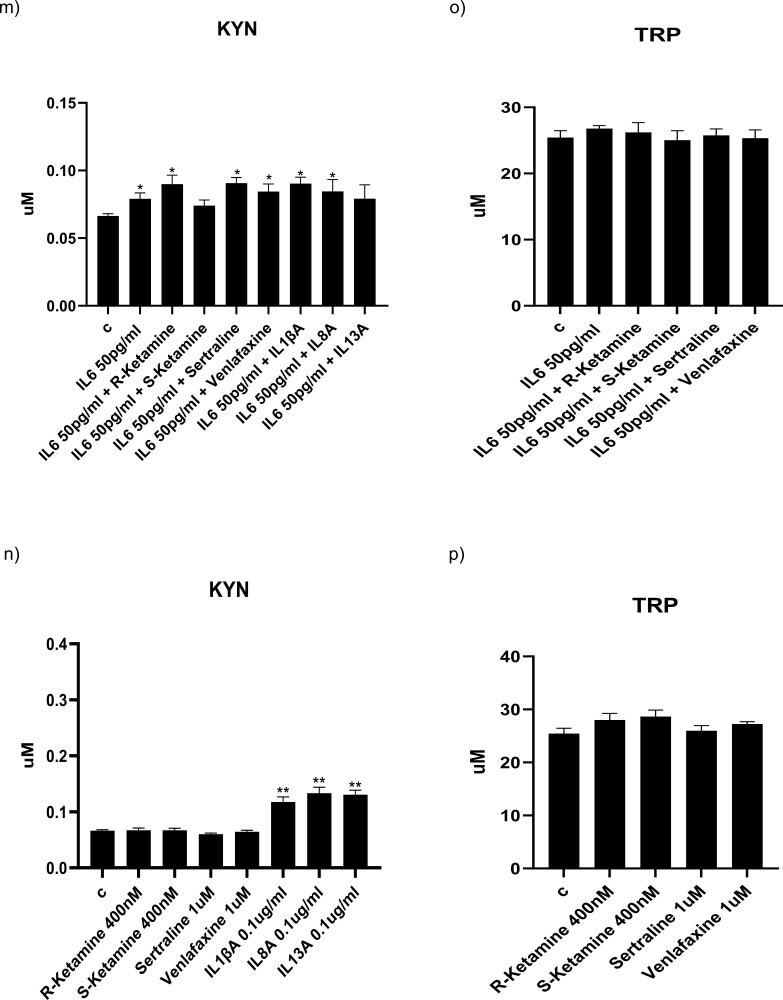
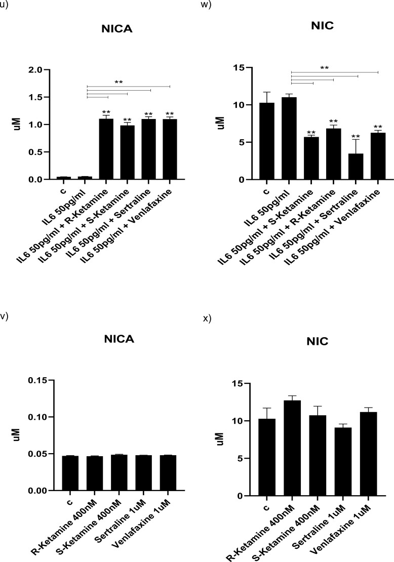
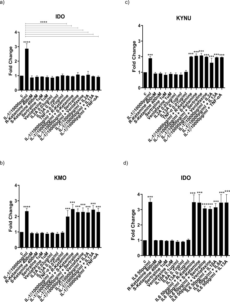
Results: Resembling the effect of antidepressants, both ketamine enantiomers prevented IL-1b- and IL-6-induced reduction in neurogenesis and increase in apoptosis. This was mediated by inhibition of IL-1b-induced production of IL-2 and IL-13 by R-ketamine and of IL-1b-induced tumor necrosis factor-alpha by S-ketamine. Likewise, R-ketamine inhibited IL-6-induced production of IL-13, whereas S-ketamine inhibited IL-6-induced IL-1b and IL-8. Moreover, both R- and S-ketamine prevented IL-1b-induced increases in indoleamine 2,3-dioxygenase expression as well as kynurenine production, which in turn was shown to mediate the detrimental effects of IL-1b on neurogenesis and apoptosis. In contrast, neither R- nor S-ketamine prevented IL-6-induced kynurenine pathway activation.

Conclusions: Results suggest that R- and S-ketamine have pro-neurogenic and anti-inflammatory properties; however, this is mediated by inhibition of the kynurenine pathway only in the context of IL-1b. Overall, this study enhances our understanding of the mechanisms underlying ketamine's antidepressant effects in the context of different inflammatory phenotypes, ultimately leading to the development of more effective, personalized therapeutic approaches for patients suffering from depression.

Keywords: Cytokines; apoptosis; hippocampal neurogenesis; kynurenine pathway.


Figures

Figure 1.
Figure 1.
The kynurenine pathway of tryptophan metabolism (A) and timeline of cellular experiments (B). Simplified kynurenine pathway of tryptophan metabolism leading to production of either neurotoxic or neuroprotective metabolites (A); the experimental timeline used in this study: cells were first treated for 3 days with reduced modified media with growth factors (proliferation media) with or without IL-1β or IL-6 and/or R-ketamine, S-ketamine, sertraline or venlafaxine. After 3 days, media was removed, and cells were given media without growth factors (differentiation media) and treatment with all compounds continued. For analysis of gene expression, RNA was isolated after 2 days in differentiation. For analysis of cytokines and kynurenine pathway metabolites, supernatant was collected after 3 days in differentiation. For analysis of differentiation and apoptosis, cells were fixed after 7 days of differentiation (10 days total treatment) and immunocytochemistry was performed (B). Abbreviations: 3-HANA, 3-hydroxyanthranilic acid; 3-HK, 3-Hydorxykynurenine; IDO, indolamine-2,3,dioxygenase; KAT, kynurenine aminotransferase; KMO, kynurenine-3-monooxygenase; KYNU, kynureninase; NAD+, nicotinamide adenine dinucleotide; NAMPT, nicotinamide phosphoribosyl transferase; QUIN, quinolinic acid.
Figure 2.
Figure 2.
Ketamine and antidepressants reverse the IL-1b- and IL-6–induced reductions in neurogenesis and increases in apoptosis in human hippocampal progenitor cells. Treatment with IL-1b (10 000 pg/mL) decreases the number of DCX-positive (A) and MAP2-positive (B) neurons and increases the number of CC3 positive neurons (C). Treatment with R-ketamine, S-ketamine, sertraline, or venlafaxine reverse the IL-1b-induced decrease in DXC-positive cells (A) and MAP2 positive cells (B), and the increase in CC3 positive cells (C). Treatment with IL-6 (50 pg/mL) decreases the number of DCX-positive (D) and MAP2-positive (E) neurons and increases the number of CC3 positive neurons (F). Treatment with R-ketamine, S-ketamine, sertraline, or venlafaxine reverse the IL-6–induced decrease in DXC-positive cells (D) and MAP2 positive cells (E), and the increase in CC3 positive cells (F). Two-way ANOVA was performed. Data are shown as mean; *P < .05, **P < .01, ***P < .001 comparisons as indicated.
Figure 2.
Figure 2.
Ketamine and antidepressants reverse the IL-1b- and IL-6–induced reductions in neurogenesis and increases in apoptosis in human hippocampal progenitor cells. Treatment with IL-1b (10 000 pg/mL) decreases the number of DCX-positive (A) and MAP2-positive (B) neurons and increases the number of CC3 positive neurons (C). Treatment with R-ketamine, S-ketamine, sertraline, or venlafaxine reverse the IL-1b-induced decrease in DXC-positive cells (A) and MAP2 positive cells (B), and the increase in CC3 positive cells (C). Treatment with IL-6 (50 pg/mL) decreases the number of DCX-positive (D) and MAP2-positive (E) neurons and increases the number of CC3 positive neurons (F). Treatment with R-ketamine, S-ketamine, sertraline, or venlafaxine reverse the IL-6–induced decrease in DXC-positive cells (D) and MAP2 positive cells (E), and the increase in CC3 positive cells (F). Two-way ANOVA was performed. Data are shown as mean; *P < .05, **P < .01, ***P < .001 comparisons as indicated.
Figure 3.
Figure 3.
Cytokine release in the supernatant of cells exposed to IL-1b alone or in co-treatment with ketamine or antidepressants. Concentrations of cytokines in the supernatant of cells treated for 3 days during proliferation followed by 3 days during differentiation with IL-1b alone or in combination with R-ketamine, S-ketamine, sertraline, or venlafaxine. Treatment with IL-1b alone increased the concentration of all cytokines in the supernatant (A–J). Treatment with antidepressants alone increased the concentration of IL-2 (B) and decreased the concentration of IL-6 (D), while treatment with ketamine alone decreased the concentration of IL-4 (C), IL-6 (D), and IL-10 (F). R-ketamine, sertraline, and venlafaxine prevented the IL-1b–induced increase in IL-2 (B) and IL-13 (H). S-ketamine, sertraline, and venlafaxine prevented the IL-1b–induced increased production of TNF-a (I). Two-way ANOVA was performed. Data are shown as mean: *P < .05, **P < .01, ***P < .001 comparisons as indicated.
Figure 3.
Figure 3.
Cytokine release in the supernatant of cells exposed to IL-1b alone or in co-treatment with ketamine or antidepressants. Concentrations of cytokines in the supernatant of cells treated for 3 days during proliferation followed by 3 days during differentiation with IL-1b alone or in combination with R-ketamine, S-ketamine, sertraline, or venlafaxine. Treatment with IL-1b alone increased the concentration of all cytokines in the supernatant (A–J). Treatment with antidepressants alone increased the concentration of IL-2 (B) and decreased the concentration of IL-6 (D), while treatment with ketamine alone decreased the concentration of IL-4 (C), IL-6 (D), and IL-10 (F). R-ketamine, sertraline, and venlafaxine prevented the IL-1b–induced increase in IL-2 (B) and IL-13 (H). S-ketamine, sertraline, and venlafaxine prevented the IL-1b–induced increased production of TNF-a (I). Two-way ANOVA was performed. Data are shown as mean: *P < .05, **P < .01, ***P < .001 comparisons as indicated.
Figure 4.
Figure 4.
Production of cytokines in the supernatant of cells exposed to IL-6 alone or in co-treatment with ketamine or antidepressants. Concentrations of cytokines in the supernatant of cells treated for 3 days during proliferation followed by 3 days during differentiation with IL-6 alone or in combination with R-ketamine, S-ketamine, sertraline, or venlafaxine (A-J). Treatment with IL-6 alone increased the concentration of IL-1b (A), IL-6 (D), IL-8 (E), and IL-13 (H) in the supernatant. Treatment with antidepressants alone increased the concentration of IL-2 (B) and decreased the concentration of IL-6 (D), while treatment with ketamine alone decreased the concentration of IL-4 (C), IL-6 (D), and IL-10 (F). S-ketamine, sertraline, and venlafaxine prevented the IL-6–induced increase in IL-1b (A) and IL-8 (E). R-ketamine, sertraline, and venlafaxine prevented the IL-6–induced increased production of IL-13 (H). Two-way ANOVA was performed. Data are shown as mean: *P < .05, **P < .01, ***P < .001 comparisons as indicated.
Figure 4.
Figure 4.
Production of cytokines in the supernatant of cells exposed to IL-6 alone or in co-treatment with ketamine or antidepressants. Concentrations of cytokines in the supernatant of cells treated for 3 days during proliferation followed by 3 days during differentiation with IL-6 alone or in combination with R-ketamine, S-ketamine, sertraline, or venlafaxine (A-J). Treatment with IL-6 alone increased the concentration of IL-1b (A), IL-6 (D), IL-8 (E), and IL-13 (H) in the supernatant. Treatment with antidepressants alone increased the concentration of IL-2 (B) and decreased the concentration of IL-6 (D), while treatment with ketamine alone decreased the concentration of IL-4 (C), IL-6 (D), and IL-10 (F). S-ketamine, sertraline, and venlafaxine prevented the IL-6–induced increase in IL-1b (A) and IL-8 (E). R-ketamine, sertraline, and venlafaxine prevented the IL-6–induced increased production of IL-13 (H). Two-way ANOVA was performed. Data are shown as mean: *P < .05, **P < .01, ***P < .001 comparisons as indicated.
Figure 5.
Figure 5.
Antibodies against IL-2, IL-13, and TNF-a, and IL-1b, IL-8 and IL-13 prevent, respectively, IL-1b– and IL-6–induced reductions in neurogenesis and increases in apoptosis. Treatment with IL-2A (0.03 mg/mL), IL-13A (0.1 mg/mL), and TNF-aA (0.01 mg/mL) prevents the decrease in DCX- and MAP2-positive cells and the increase in CC3-positive cells induced by IL-1b (A–C). Treatment with IL-1bA (0.1 mg/mL), IL-8A (0.1 µg/mL), and IL-13A (0.1 mg/mL) prevents the decrease in DCX- and MAP2-positive cells and the increase in CC3-positive cells induced by IL-6 (D–F). Two-way ANOVA was performed. Data are shown as mean; *P < .05, **P < .01 comparisons as indicated.
Figure 5.
Figure 5.
Antibodies against IL-2, IL-13, and TNF-a, and IL-1b, IL-8 and IL-13 prevent, respectively, IL-1b– and IL-6–induced reductions in neurogenesis and increases in apoptosis. Treatment with IL-2A (0.03 mg/mL), IL-13A (0.1 mg/mL), and TNF-aA (0.01 mg/mL) prevents the decrease in DCX- and MAP2-positive cells and the increase in CC3-positive cells induced by IL-1b (A–C). Treatment with IL-1bA (0.1 mg/mL), IL-8A (0.1 µg/mL), and IL-13A (0.1 mg/mL) prevents the decrease in DCX- and MAP2-positive cells and the increase in CC3-positive cells induced by IL-6 (D–F). Two-way ANOVA was performed. Data are shown as mean; *P < .05, **P < .01 comparisons as indicated.
Figure 6.
Figure 6.
Ketamine and antidepressants prevent IL-1b– but not IL-6–induced activation of the kynurenine pathway via inhibiting IL-2, IL-13, and TNFα. Concentrations of kynurenine pathway metabolites in the supernatant of cells treated for 3 days during proliferation followed by 3 days during differentiation with IL-1b or IL-6 alone or in combination with R-ketamine, S-ketamine, sertraline, or venlafaxine (A–X). Treatment with IL-1b alone increased the concentration of kynurenine (KYN) (A), anthranilic acid (E), and nicotinic acid (NICA) (I). Co-treatment with R-ketamine, S-ketamine, sertraline, venlafaxine, IL-2A, IL-13A, or TNF-aA prevents the IL-1b–induced increased production of KYN (A). Treatment with R-ketamine, S-ketamine, sertraline, venlafaxine, IL-2A, IL-13A, or TNF-a alone increases the concentration of anthranilic acid (F). Treatment with IL-13A or TNF-a alone increases the concentration of KYN (A) and decreases the concentration of NICA (J). Treatment with IL-6 increased production of KYN (M), ANA (Q), and KYNA (S), none of which were prevented by co-treatment with R-ketamine, S-Ketamine, sertraline, venlafaxine, IL-1bA, IL-8A, or IL-13A. Treatment with IL-1bA, IL-8A, or IL-13A alone increased the concentration of KYN (N), while R-ketamine, S-ketamine, sertraline, venlafaxine, IL-1bA, IL-8A, or IL-13A alone increased the concentration of ANA (R) and NICA (U) and decreased the concentration of nicotinamide (NIC) (W). Two-way ANOVA (A, C, E, G, I, K, M, O, Q, S, U, W) and 2-way ANOVA (B, D, F, H, J, L, N, P, R, T, V, X) were performed. Data are shown as mean: *P < .05, **P < .01, ***P < .001 comparisons as indicated.
Figure 6.
Figure 6.
Ketamine and antidepressants prevent IL-1b– but not IL-6–induced activation of the kynurenine pathway via inhibiting IL-2, IL-13, and TNFα. Concentrations of kynurenine pathway metabolites in the supernatant of cells treated for 3 days during proliferation followed by 3 days during differentiation with IL-1b or IL-6 alone or in combination with R-ketamine, S-ketamine, sertraline, or venlafaxine (A–X). Treatment with IL-1b alone increased the concentration of kynurenine (KYN) (A), anthranilic acid (E), and nicotinic acid (NICA) (I). Co-treatment with R-ketamine, S-ketamine, sertraline, venlafaxine, IL-2A, IL-13A, or TNF-aA prevents the IL-1b–induced increased production of KYN (A). Treatment with R-ketamine, S-ketamine, sertraline, venlafaxine, IL-2A, IL-13A, or TNF-a alone increases the concentration of anthranilic acid (F). Treatment with IL-13A or TNF-a alone increases the concentration of KYN (A) and decreases the concentration of NICA (J). Treatment with IL-6 increased production of KYN (M), ANA (Q), and KYNA (S), none of which were prevented by co-treatment with R-ketamine, S-Ketamine, sertraline, venlafaxine, IL-1bA, IL-8A, or IL-13A. Treatment with IL-1bA, IL-8A, or IL-13A alone increased the concentration of KYN (N), while R-ketamine, S-ketamine, sertraline, venlafaxine, IL-1bA, IL-8A, or IL-13A alone increased the concentration of ANA (R) and NICA (U) and decreased the concentration of nicotinamide (NIC) (W). Two-way ANOVA (A, C, E, G, I, K, M, O, Q, S, U, W) and 2-way ANOVA (B, D, F, H, J, L, N, P, R, T, V, X) were performed. Data are shown as mean: *P < .05, **P < .01, ***P < .001 comparisons as indicated.
Figure 7.
Figure 7.
Expression of genes involved in the activation of the kynurenine pathway, and the effect on cells of IL-1b– and IL-6–induced kynurenine metabolites. Gene expression changes in cells treated for 3 days during proliferation followed by 2 days during differentiation with IL-1b or IL-6 alone or in combination with R-ketamine, S-ketamine, sertraline, or venlafaxine. Treatment with IL-1b increased expression of IDO, KMO, and KYNU (A–C). Co-treatment with R-ketamine, S-ketamine, sertraline, venlafaxine, and IL-2A, IL-13A, and TNF-aA prevented the IL-1b–induced increased expression of IDO (A). Treatment with IL-6 alone increased the concentrations of IDO, KMO, and KYNU (D–F); however, none of these increases were prevented by co-treatment with R-ketamine, S-ketamine, sertraline, venlafaxine, or antibodies for IL-1b, IL-8, and IL-13. Additionally, cells were treated between day 3 and 7 of differentiation directly to the same concentrations of KYN, ANA, NICA, or KYNA previously identified upon treatment with IL-1b or IL-6 alone. Treatment of cells with KYN (1 mM), ANA (0.0015 mM), and NICA (1 mM) decreased the percentage of DCX- and MAP2-positive neurons and increased the percentage of CC3-positive neurons to the same degree as IL-1b treatment (G–I). Similarly, KYN (0.08 mM) and ANA (0.002 mM) decreased the percentage of DCX- and MAP2-positive neurons and increased the percentage of CC3-positive neurons to the same degree as IL-6 treatment (J–L). Conversely, treatment with KYNA (0.002 mM) increased the percentage of DCX- and MAP2-positive neurons and decreased the percentage of CC3-positive neurons compared with control (J–L). Two-way ANOVA (A–F), and 2-way ANOVA (G–L) were performed. Data are shown as mean; *P < .05, **P < .01, ***P < .001, ****P < .0001 comparisons as indicated.
Figure 7.
Figure 7.
Expression of genes involved in the activation of the kynurenine pathway, and the effect on cells of IL-1b– and IL-6–induced kynurenine metabolites. Gene expression changes in cells treated for 3 days during proliferation followed by 2 days during differentiation with IL-1b or IL-6 alone or in combination with R-ketamine, S-ketamine, sertraline, or venlafaxine. Treatment with IL-1b increased expression of IDO, KMO, and KYNU (A–C). Co-treatment with R-ketamine, S-ketamine, sertraline, venlafaxine, and IL-2A, IL-13A, and TNF-aA prevented the IL-1b–induced increased expression of IDO (A). Treatment with IL-6 alone increased the concentrations of IDO, KMO, and KYNU (D–F); however, none of these increases were prevented by co-treatment with R-ketamine, S-ketamine, sertraline, venlafaxine, or antibodies for IL-1b, IL-8, and IL-13. Additionally, cells were treated between day 3 and 7 of differentiation directly to the same concentrations of KYN, ANA, NICA, or KYNA previously identified upon treatment with IL-1b or IL-6 alone. Treatment of cells with KYN (1 mM), ANA (0.0015 mM), and NICA (1 mM) decreased the percentage of DCX- and MAP2-positive neurons and increased the percentage of CC3-positive neurons to the same degree as IL-1b treatment (G–I). Similarly, KYN (0.08 mM) and ANA (0.002 mM) decreased the percentage of DCX- and MAP2-positive neurons and increased the percentage of CC3-positive neurons to the same degree as IL-6 treatment (J–L). Conversely, treatment with KYNA (0.002 mM) increased the percentage of DCX- and MAP2-positive neurons and decreased the percentage of CC3-positive neurons compared with control (J–L). Two-way ANOVA (A–F), and 2-way ANOVA (G–L) were performed. Data are shown as mean; *P < .05, **P < .01, ***P < .001, ****P < .0001 comparisons as indicated.

References

    1. Anacker C, Cattaneo A, Luoni A, Musaelyan K, Zunszain PA, Milanesi E, Rybka J, Berry A, Cirulli F, Thuret S, Price J, Riva MA, Gennarelli M, Pariante CM (2013a) Glucocorticoid-related molecular signaling pathways regulating hippocampal neurogenesis. Neuropsychopharmacology 38:872–883. - PMC - PubMed
    1. Anacker C, Cattaneo A, Musaelyan K, Zunszain PA, Horowitz M, Molteni R, Luoni A, Calabrese F, Tansey K, Gennarelli M, Thuret S, Price J, Uher R, Riva MA, Pariante CM (2013b) Role for the kinase SGK1 in stress, depression, and glucocorticoid effects on hippocampal neurogenesis. Proc Natl Acad Sci USA 110:8708–8713. - PMC - PubMed
    1. Autry AE, Adachi M, Nosyreva E, Na ES, Los MF, Cheng PF, Kavalali ET, Monteggia LM (2011) NMDA receptor blockade at rest triggers rapid behavioural antidepressant responses. Nature 475:91–95. - PMC - PubMed
    1. Beilin B, Rusabrov Y, Shapira Y, Roytblat L, Greemberg L, Yardeni IZ, Bessler H (2007) Low-dose ketamine affects immune responses in humans during the early postoperative period. Br J Anaesth 99:522–527. - PubMed
    1. Benson CA, Powell HR, Liput M, Dinham S, Freedman DA, Ignatowski TA, Stachowiak EK, Stachowiak MK (2020) Immune factor, TNFα, disrupts human brain organoid development similar to schizophrenia-schizophrenia increases developmental vulnerability to TNFα. Front Cell Neurosci 14:233. - PMC - PubMed

Publication types