. 2024 Oct 1;18(39):27077-27089.
doi: 10.1021/acsnano.4c11652. Epub 2024 Sep 19.

Blood Distribution of SARS-CoV-2 Lipid Nanoparticle mRNA Vaccine in Humans

Affiliations

Blood Distribution of SARS-CoV-2 Lipid Nanoparticle mRNA Vaccine in Humans

Stephen J Kent et al. ACS Nano. .

Abstract

Lipid nanoparticle mRNA vaccines are an exciting but emerging technology used in humans. There is limited understanding of the factors that influence their biodistribution and immunogenicity. Antibodies to poly(ethylene glycol) (PEG), which is on the surface of the lipid nanoparticle, are detectable in humans and boosted by human mRNA vaccination. We hypothesized that PEG-specific antibodies could increase the clearance of mRNA vaccines. To test this, we developed methods to quantify both the vaccine mRNA and ionizable lipid in frequent serial blood samples from 19 subjects receiving Moderna SPIKEVAX mRNA booster immunization. Both the vaccine mRNA and ionizable lipid peaked in blood 1-2 days post vaccination (median peak level 0.19 and 3.22 ng mL-1, respectively). The vaccine mRNA was detectable and quantifiable up to 14-15 days postvaccination in 37% of subjects. We measured the proportion of vaccine mRNA that was relatively intact in blood over time and found that the decay kinetics of the intact mRNA and ionizable lipid were identical, suggesting the intact lipid nanoparticle recirculates in blood. However, the decay rates of mRNA and ionizable lipids did not correlate with baseline levels of PEG-specific antibodies. Interestingly, the magnitude of mRNA and ionizable lipid detected in blood did correlate with the boost in the level of PEG antibodies. Furthermore, the ability of a subject's monocytes to phagocytose lipid nanoparticles was inversely related to the rise in PEG antibodies. This suggests that the circulation of mRNA lipid nanoparticles into the blood and their clearance by phagocytes influence the PEG immunogenicity of the mRNA vaccines. Overall, this work defines the pharmacokinetics of lipid nanoparticle mRNA vaccine components in human blood after intramuscular injection and the factors that influence these processes. These insights should be valuable in improving the future safety and efficacy of lipid nanoparticle mRNA vaccines and therapeutics.

Keywords: COVID-19; PEGylated lipid nanoparticle; biomolecular coronas; immunoglobulins; kinetics of mRNA; particle–immune cell interactions.


Conflict of interest statement

The authors declare no competing financial interest.

Figures
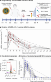
Figure 1
Figure 1
In vivo kinetics of mRNA and ionizable lipid from the SPIKEVAX SARS-CoV-2 mRNA vaccine in human blood. (a) Schematic illustration of the project design to track the kinetics of mRNA vaccine in human blood of 19 healthy subjects who received one dose of Moderna SPIKEVAX COVID-19 bivalent mRNA vaccine (see Table S1 for subject information). Plasma samples were collected at prevaccination (day 0) and at a median of 8 (range 4–14) other time points between 4 h and 28 days postvaccination. Created with BioRender.com. (b) Longitudinal vaccine mRNA concentrations in the plasma of the 19 subjects. To improve readability, the detailed mRNA kinetics within the first 24 h of vaccination is shown in Figure S2. (c) Representative image showing the peak intensity of SM-102 signals in a set of plasma samples from a subject at day 0–6 postvaccination determined by liquid chromatograph mass spectrometry. (d) Longitudinal SM-102 ionizable lipid concentrations in the plasma of the 19 subjects. The concentrations of vaccine mRNA and SM-102 ionizable lipid were calculated based on the linear standard curves and raw data in Figure S1.
Figure 2
Figure 2
Integrity of vaccine mRNA in plasma after vaccination with SPIKEVAX SARS-CoV-2 mRNA vaccine. (a) Schematic illustration of a duplex ddPCR assay using a two-primer set targeting two regions (mRNA1273-1 nt 876–1010 and mRNA1273-2 nt 2370–2474) of the mRNA1273 sequence. (b) Representative dot plot profiles of FAM-labeled mRNA1273-1 primer and probe (channel 1, amplitude) and HEX-labeled mRNA1273-2 primer and probe (channel 2, amplitude) at day 0 (left panel), 1 (middle panel), and 7 (right panel) postvaccination. Droplets emitting 2D signals were separated into four groups (gray: double negative for mRNA1273-1 and mRNA1273-2; blue: positive for mRNA1273-1, negative for mRNA1273-2; green: positive for mRNA1273-2, negative for mRNA1273-1; and orange: double positive for both mRNA1273-1 and mRNA1273-2). (c) Vaccine mRNA integrity in plasma of 19 subjects postvaccination. Vaccine mRNA integrity was assessed by mRNA linkage (%), which was expressed as the estimated percent of linked molecules (correcting for the frequency of random association of probes). The number of droplets in each single or double positive group was derived by QX Manager software. (d) Longitudinal intact and nonintact mRNA levels in the plasma of the 19 subjects before and after vaccination. The intact mRNA levels were calculated by multiplying the mRNA linkage (%) by the total mRNA levels detected in plasma. The LLOQ (shown as a dashed line) is determined based on the linear standard curves of vaccine mRNA at 0.001 ng mL–1 (Figure S1a). To improve readability, the detailed mRNA integrity kinetics within the first 24 h of vaccination is shown in Figure S4.
Figure 3
Figure 3
Dynamics of vaccine mRNA and SM-102 lipids in the plasma. (a) Comparison of decay rate between total mRNA, intact mRNA, and SM-102 lipid. Statistics assessed by the likelihood ratio test. (b) Best-fit decay slops of SM-102 lipids, total mRNA, intact mRNA, nonintact mRNA, and the rate of degradation of intact mRNA. The responses at the first time point (the peak time) for each parameter are set to 100%, and the change (%) over time and half-life are shown. As the decay slopes of SM-102 lipid and intact mRNA overlap, the curve of the SM-102 lipid slope was plotted with higher thickness than that of the intact mRNA slope to improve readability.
Figure 4
Figure 4
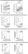
Kinetics of anti-PEG antibody and their correlation with vaccine mRNA kinetics. (a) Longitudinal anti-PEG IgG and IgM titers in the plasma before and after IM inoculation of SPIKEVAX SARS-CoV-2 mRNA vaccine. Statistics assessed by nonparametric Friedman’s test with Dunn’s multiple comparisons test (n = 17 as 17 subjects have all five time points). (b) No significant correlation between pre-existing anti-PEG antibody titers and total mRNA decay rate across the 19 subjects. (c) Significant positive correlation between the peak levels of total mRNA in plasma and anti-PEG IgG expansion (growth rate/fold change) across the 19 subjects. (d) Significant positive correlation between the peak levels of ionizable lipid (SM-102) and anti-PEG IgM expansion (growth rate or fold change) across the 19 subjects. Statistics in (a–c) were assessed by Spearman correlation analysis (n = 19).
Figure 5
Figure 5
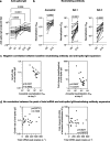
Kinetics of antispike IgG and neutralizing antibody. (a) Longitudinal antispike IgG titers in the plasma of the Moderna bivalent mRNA vaccinee cohort (Table S1). Statistics assessed by nonparametric Friedman’s test with Dunn’s multiple comparisons test (n = 17 as 17 subjects have all five time points). (b) Comparing live SARS-COV-2 neutralization titer (inhibitory concentration 50, IC50) before vaccination (day 0) and postvaccination at day 14 of the Moderna bivalent mRNA vaccinee cohort. The limit of detection at a titer of 1:20 is shown in the dashed line. Statistics assessed by nonparametric Wilcoxon’s matched-pairs signed rank test (n = 19). (c) Significant negative correlation between baseline neutralizing antibodies and antispike IgG expansion (growth rate or fold change). (d) The peak levels of total vaccine mRNA in the plasma do not influence the expansion of antispike IgG and neutralizing antibody. Statistics in (c,d) were assessed by Spearman correlation analysis.
Figure 6
Figure 6
Human blood assay to assess lipid nanoparticle–immune cell interactions. (a) Schematic illustration of the in vitro assay to assess the person-specific cellular interactions of primary immune cells with lipid nanoparticles. PBMCs (collected from the 19 subjects before receiving the Moderna SPIKEVAX bivalent vaccination) were washed by centrifugation with serum-free media multiple times to completely remove plasma. Lipid nanoparticles were preincubated with human plasma from 1 subject and then incubated with PBMCs from the 19 subjects in serum-free media for 1 h at 37 °C, followed by phenotyping cells with antibody cocktails and analysis by flow cytometry. Created with BioRender.com. (b) Flow cytometry histograms represent the monocyte phagocytosis of lipid nanoparticles after incubating with PBMCs of 3 different subjects. Cell-only control groups show the respective cell populations without particles in the incubation media. (c) Significant negative correlation between monocyte phagocytosis of lipid nanoparticles (median fluorescence intensity, MFI) and anti-PEG IgG expansion (growth rate or fold change). Statistics were assessed by Spearman correlation analysis.

References

    1. Ye Z.; Harmon J.; Ni W.; Li Y.; Wich D.; Xu Q. The mRNA Vaccine Revolution: COVID-19 Has Launched the Future of Vaccinology. ACS Nano 2023, 17, 15231–15253. 10.1021/acsnano.2c12584. - DOI - PubMed
    1. Fertig T. E.; Chitoiu L.; Marta D. S.; Ionescu V.-S.; Cismasiu V. B.; Radu E.; Angheluta G.; Dobre M.; Serbanescu A.; Hinescu M. E.; Gherghiceanu M. Vaccine mRNA Can Be Detected in Blood at 15 Days Post-Vaccination. Biomedicines 2022, 10, 1538.10.3390/biomedicines10071538. - DOI - PMC - PubMed
    1. Castruita J. A. S.; Schneider U. V.; Mollerup S.; Leineweber T. D.; Weis N.; Bukh J.; Pedersen M. S.; Westh H. SARS-CoV-2 Spike mRNA Vaccine Sequences Circulate in Blood up to 28 Days after COVID-19 Vaccination. APMIS 2023, 131, 128–132. 10.1111/apm.13294. - DOI - PMC - PubMed
    1. Hanna N.; De Mejia C. M.; Heffes-Doon A.; Lin X.; Botros B.; Gurzenda E.; Clauss-Pascarelli C.; Nayak A. Biodistribution of mRNA COVID-19 Vaccines in Human Breast Milk. EBioMedicine 2023, 96, 104800.10.1016/j.ebiom.2023.104800. - DOI - PMC - PubMed
    1. Hanna N.; Heffes-Doon A.; Lin X.; Manzano De Mejia C.; Botros B.; Gurzenda E.; Nayak A. Detection of Messenger RNA Covid-19 Vaccines in Human Breast Milk. JAMA Pediatr. 2022, 176, 1268–1270. 10.1001/jamapediatrics.2022.3581. - DOI - PMC - PubMed

Publication types