

AI-based lumbar central canal stenosis classification on sagittal MR images is comparable to experienced radiologists using axial images
- PMID: 39299953
- PMCID: PMC11913898
- DOI: 10.1007/s00330-024-11080-0
AI-based lumbar central canal stenosis classification on sagittal MR images is comparable to experienced radiologists using axial images
Abstract
Objectives: The assessment of lumbar central canal stenosis (LCCS) is crucial for diagnosing and planning treatment for patients with low back pain and neurogenic pain. However, manual assessment methods are time-consuming, variable, and require axial MRIs. The aim of this study is to develop and validate an AI-based model that automatically classifies LCCS using sagittal T2-weighted MRIs.
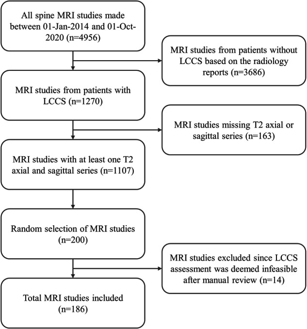
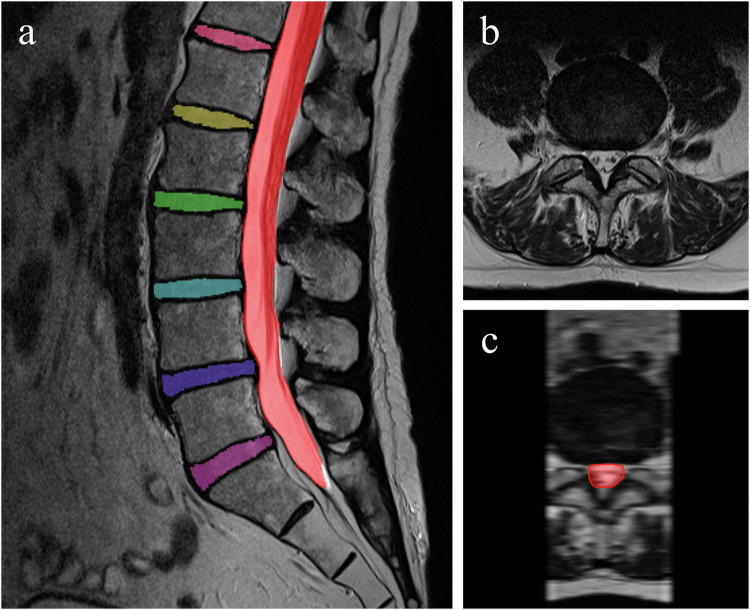
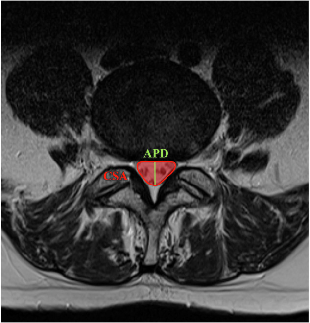
Methods: A pre-existing 3D AI algorithm was utilized to segment the spinal canal and intervertebral discs (IVDs), enabling quantitative measurements at each IVD level. Four musculoskeletal radiologists graded 683 IVD levels from 186 LCCS patients using the 4-class Lee grading system. A second consensus reading was conducted by readers 1 and 2, which, along with automatic measurements, formed the training dataset for a multiclass (grade 0-3) and binary (grade 0-1 vs. 2-3) random forest classifier with tenfold cross-validation.
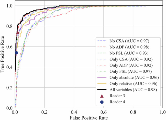
Results: The multiclass model achieved a Cohen's weighted kappa of 0.86 (95% CI: 0.82-0.90), comparable to readers 3 and 4 with 0.85 (95% CI: 0.80-0.89) and 0.73 (95% CI: 0.68-0.79) respectively. The binary model demonstrated an AUC of 0.98 (95% CI: 0.97-0.99), sensitivity of 93% (95% CI: 91-96%), and specificity of 91% (95% CI: 87-95%). In comparison, readers 3 and 4 achieved a specificity of 98 and 99% and sensitivity of 74 and 54%, respectively.
Conclusion: Both the multiclass and binary models, while only using sagittal MR images, perform on par with experienced radiologists who also had access to axial sequences. This underscores the potential of this novel algorithm in enhancing diagnostic accuracy and efficiency in medical imaging.
Key points: Question How can the classification of lumbar central canal stenosis (LCCS) be made more efficient? Findings Multiclass and binary AI models, using only sagittal MR images, performed on par with experienced radiologists who also had access to axial sequences. Clinical relevance Our AI algorithm accurately classifies LCCS from sagittal MRI, matching experienced radiologists. This study offers a promising tool for automated LCCS assessment from sagittal T2 MRI, potentially reducing the reliance on additional axial imaging.
Keywords: Deep learning; Lumbar central canal stenosis; MRI; Machine learning; Spine.
© 2024. The Author(s).
Conflict of interest statement
Compliance with ethical standards. Guarantor: The scientific guarantor of this publication is M.d.K. Conflict of interest: The authors of this manuscript declare relationships with the following companies: B.v.G. is CSO of Thirona; N.L. is employed at Stryker. The remaining authors declare no conflicts of interest. Statistics and biometry: One of the authors has significant statistical expertise. Informed consent: Written informed consent was not required for this study because it was exempted, given the use of retrospective anonymized MRI examinations. This retrospective study was approved by the institutional review board at Radboud University Medical Center (IRB 2016-2275). Ethical approval: Institutional Review Board approval was obtained. Study subjects or cohorts overlap: No study subjects or cohorts have been previously reported. Methodology: Retrospective Experimental Performed at one institution
Figures


References
-
- Ota Y, Connolly M, Srinivasan A et al (2020) Mechanisms and origins of spinal pain: from molecules to anatomy, with diagnostic clues and imaging findings. Radiographics 40:1163–1181. 10.1148/rg.2020190185 - PubMed
-
- Van Der Graaf JW, Kroeze RJ, Buckens CFM et al (2023) MRI image features with an evident relation to low back pain: a narrative review. Eur Spine J 32:1830–1841. 10.1007/s00586-023-07602-x - PubMed
-
- Jensen RK, Jensen TS, Koes B, Hartvigsen J (2020) Prevalence of lumbar spinal stenosis in general and clinical populations: a systematic review and meta-analysis. Eur Spine J 29:2143–2163. 10.1007/s00586-020-06339-1 - PubMed
Publication types
MeSH terms
LinkOut - more resources
Full Text Sources
Medical
