. 2024 Sep 19;15(1):8226.
doi: 10.1038/s41467-024-52537-6.

MFSD7C protects hemolysis-induced lung impairments by inhibiting ferroptosis

Affiliations

MFSD7C protects hemolysis-induced lung impairments by inhibiting ferroptosis

Huirui Wang et al. Nat Commun. .

Abstract

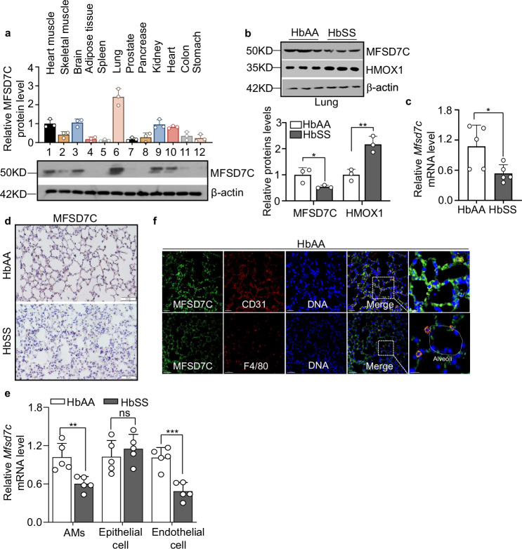
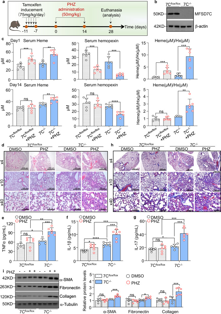
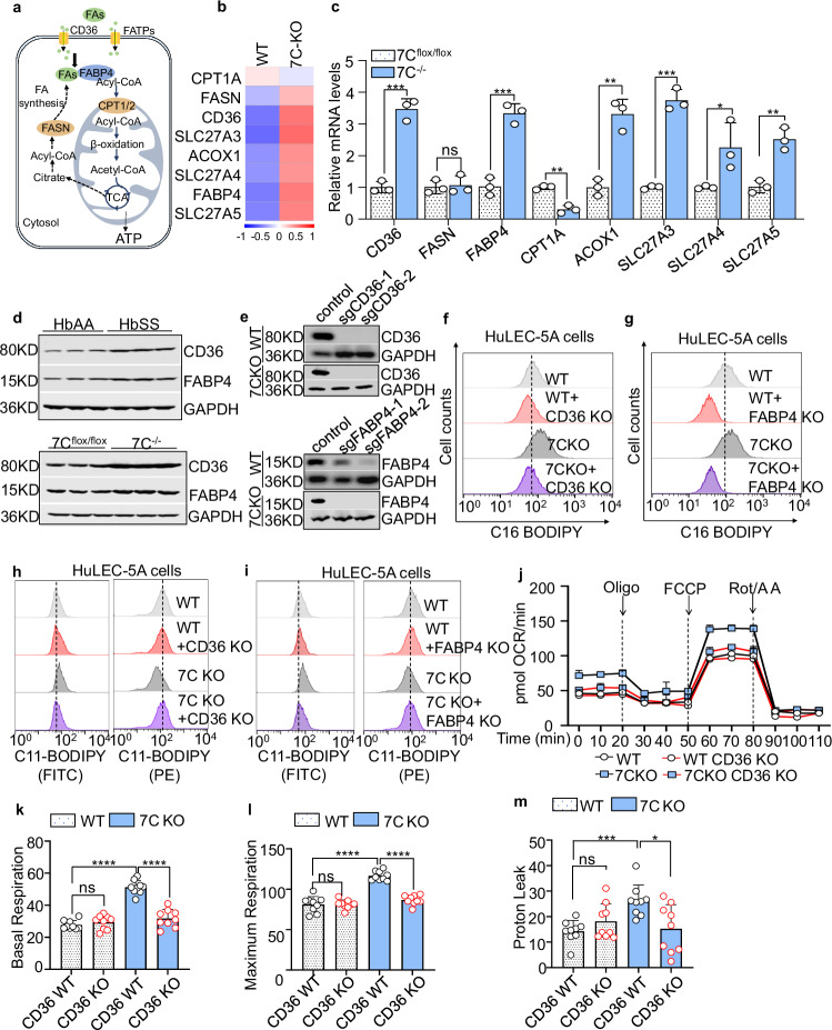
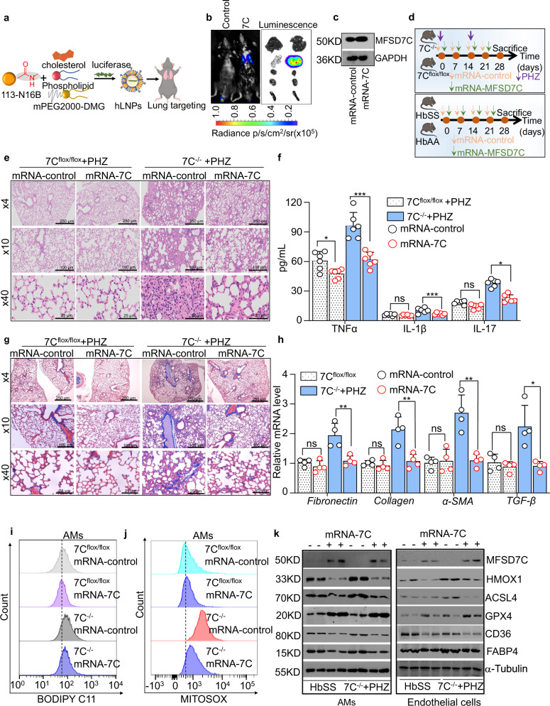
Hemolysis drives susceptibility to lung injury and predicts poor outcomes in diseases, such as malaria and sickle cell disease (SCD). However, the underlying pathological mechanism remains elusive. Here, we report that major facilitator superfamily domain containing 7 C (MFSD7C) protects the lung from hemolytic-induced damage by preventing ferroptosis. Mechanistically, MFSD7C deficiency in HuLEC-5A cells leads to mitochondrial dysfunction, lipid remodeling and dysregulation of ACSL4 and GPX4, thereby enhancing lipid peroxidation and promoting ferroptosis. Furthermore, systemic administration of MFSD7C mRNA-loaded nanoparticles effectively prevents lung injury in hemolytic mice, such as HbSS-Townes mice and PHZ-challenged 7 C-/- mice. These findings present the detailed link between hemolytic complications and ferroptosis, providing potential therapeutic targets for patients with hemolytic disorders.


Conflict of interest statement

The authors declare no competing interests.

Figures

Fig. 1
Fig. 1. Hemolytic-related lung damage was observed in SCD mice.
af HbAA- and HbSS-Townes mice were analyzed at 12 months. a Serum heme and hemopexin (Hx) levels were measured by ELISA. n = 6. b Representative micrographs on lung sections (H&E, x4, x10, x40) and inflammation score were shown. n = 6. c Cytokines levels, including TNFα, IL-1β, and IL-17 in murine BALF were measured by ELISA. n = 4. d Representative micrographs on lung sections (Masson’s trichrome, x4, x10, x40) and fibrosis score were shown. n = 6. Representative western blot (e) and quantitative analysis (f) of α-SMA, Fibronectin, and Collagen expression in lung tissue. Four biological replicates were performed for each sample. Data are presented as mean + standard deviation (SD). *p < 0.05; **p < 0.01; ***p < 0.001; ****p < 0.0001.
Fig. 2
Fig. 2. The MFSD7C expression level was downregulated in alveolar macrophages and endothelial cells of hemolytic lung.
a Relative MFSD7C protein levels in various tissues of 8 weeks old HbAA-Townes mice. n = 3. bd 12-month old HbAA- and HbSS-Townes mice were analyzed. b Expression levels of MFSD7C and HMOX1 in lung tissues were analyzed using western blot. Three biological replicates were performed for each sample. c Changes in the mRNA expression of MFSD7C of lung tissues from hemolytic mouse model were evaluated by RT-qPCR. n = 5. d IHC staining of MFSD7C in lung samples. e Relative MFSD7C mRNA level in AMs, epithelial cells, and endothelial cells isolated from lung tissues. n = 5. f Representative immunofluorescence images of lung tissues in HbAA mice (MFSD7C in green, CD31 and F4/80 in red, and nuclei in blue, photomicrograph bar = 5 µm). Data are presented as mean + standard deviation (SD). *p < 0.05; **p < 0.01; ***p < 0.001.
Fig. 3
Fig. 3. Deficiency of MFSD7C aggravates pulmonary damage in PHZ-induced hemolytic diseases.
a Experimental scheme of tamoxifen inducement and PHZ administration. b Representative western blot of MFSD7C expression in lung tissues from 7Cflox/flox and 7C−/− mice 7 days after Tamoxifen treatment. Four biological replicates were performed for each sample. c Serum heme and hemopexin (Hx) levels were measured at day 1 and day 14 (just before 2nd PHZ administration) by ELISA. n = 6. di 7Cflox/flox and 7C−/− mice treated with DMSO or PHZ were analyzed at day 28. d Representative lung H&E staining sections (x4, x10, x40). TNFα (e), IL-1β (f), and IL-17 (g) levels in BALF were measured by ELISA. n = 6. h Representative lung Masson’s trichrome staining (x4, x10, x40). i Expression levels of α-SMA, Fibronectin, and Collagen expression in lung tissues were analyzed using western blot. n = 6. Data are presented as mean + standard deviation (SD). *p < 0.05; **p < 0.01; ***p < 0.001; ****p < 0.0001.
Fig. 4
Fig. 4. MFSD7C regulates lipid remodeling by causing mitochondria dysfunction.
a KEGG pathway analysis of differentially expressed genes (DEGs) (log2(fold change) > 1.2; P < 0.05) in MFSD7C-KO HuLEC-5A cells versus control. Seahorse analysis of cellular oxygen consumption rate (OCR) (b), ATP production (c) in AMs isolated from the lung of 7Cflox/flox, 7C−/−, and hemolytic mice. n = 3. d Lipid ROS production in AMs were determined by flow cytometry using C11-Bodipy. n = 3. e Mitochondrial ROS measured by MitoSOX green in AMs. f ATP luminescent levels of WT, 7C-KO1, 7C-KO2 and 7C-Rescued HuLEC-5A cells. Right: Western blot analysis of MFSD7C knockout and rescue efficiency. n = 3. g Immunofluorescence analysis of JC-1-stained WT, MFSD7C-KO, and rescued HuLEC-5A cells for the detection of mitochondrial membrane potential changes. Monomer in green, Aggregate in red, and nuclei in blue, photomicrograph bar = 5 µm. Right: quantification of fluorescence intensity. n = 3. h Uptake of long chain FAs (Bodipy c16) in HuLEC-5A cells was determined by FACS analysis. n = 3. i Volcano plot showing changes in lipid profile in AMs from 7C−/− mice versus 7Cflox/flox mice. Right: indicated lipid screened by the variable importance in the projection VIP > 1. j Volcano plots showing the changes in TG, PE, PE(P) and PE(O). Changes were grouped as PUFA (red fill) and SFA/MUFA (white fill). Data are presented as mean + standard deviation (SD). *p < 0.05; **p < 0.01; ***p < 0.001; ****p < 0.0001.
Fig. 5
Fig. 5. MFSD7C deficiency increased lipid flux by inducing CD36 and FABP4 expression.
a Schematic diagram for fatty acid transportation and oxidation. b Heatmaps of relative gene expression in MFSD7C-KO HuLEC-5A cells versus control. c RT-qPCR analysis of CD36, FASN, FABP4, CPT1A, SLC27A3, SLC27A4, SLC27A5 and ACOX1 relative mRNA levels in AMs from 7Cflox/flox and 7C−/− mice. n = 3. d Representative western blot of CD36 and FABP4 expression in AMs. Three biological replicates were performed for each sample. e Western blot analysis of CD36 and FABP4 knockout efficiency in MFSD7C-WT and KO HuLEC-5A cells. Three biological replicates were performed for each sample. Uptake of long chain FAs (Bodipy C16) in CD36 KO (f) or FKBP4 KO (g) HuLEC-5A cells was determined by FACS analysis. hi Lipid ROS production in HuLEC-5A cells were determined by flow cytometry using BODIPY C11. Seahorse analysis of cellular oxygen consumption rate (OCR) (j), basal respiration (k), maximum respiration (l), and proton leak (m) in CD36 KO HuLEC-5A cells. n = 3. Data are presented as mean + standard deviation (SD). *p < 0.05; **p < 0.01; ***p < 0.001; ****p < 0.0001.
Fig. 6
Fig. 6. Lipid peroxidation contributes the ferroptosis sensitivity caused by MFSD7C deficiency.
a Volcano plot illustrating DEGs in MFSD7C-KO HuLEC-5A cells versus control, highlighting ferroptosis related genes. b Representative western blot of GPX4, ACSL4, HMOX1 and MFSD7C expression in AMs and endothelia cells from lung tissues of 12-month-old HbAA and HbSS mice. Four biological replicates were performed for each sample. c MDA levels in lung tissues were assessed by TBARS assay. n = 6. d Cell viability of AMs treated with an increasing RSL3 concentration (0.01–5 μM) for 24 h. n = 3. e DMSO or CD36 inhibitor (SSO) (20 µM, 12 h) pretreated WT and MFSD7C-KO HuLEC-5A cells were treated with RSL3 (1 μM), Heme (10 μM) and DMSO (control) for 24 h and cell viability was analyzed. n = 4. f Representative electron microscope images of WT and MFSD7C-KO HuLEC-5A cells treated with RSL3 (1 μM) for 8 h, with arrows indicating mitochondria. Three biological replicates were performed for each sample. g Cytokine levels, including TNFα, IL-1β, and IL-17 in murine BALF were measured by ELISA with DMSO or Fer-1(1 μM) pretreatment for 12 h. n = 4. h Conditioned medium (CM) of RSL3 (0.5 μM) treated MFSD7C-KO and WT HuLEC-5A cells were collected after 72 h and transferred to recipient nHLFs for 72 h. Cells were pretreated with DMSO or Fer-1 (1 μM) for 1 h before RSL3 treatment. i ECM deposition assay in nHLFs exposed to MFSD7C-KO or WT HuLEC-5A cells derived CM. n = 3. j H&E staining and Masson’s trichrome staining (x4, x10, x40) of lung tissues were shown. Three biological replicates were performed for each sample. Data are presented as mean + standard deviation (SD). *p < 0.05; **p < 0.01; ***p < 0.001; ****p < 0.0001.
Fig. 7
Fig. 7. Administration of MFSD7C mRNA improves lung damage in 7C−/− mice.
a Schematic illustration of lung targetability of hybrid 113-N16B lipid nanoparticles. hLNP was formulated by 113-N16B: cholesterol: phospholipids: DMG-PEG2000 (50:38.5:10:1.5). b Representative whole-body and organs bioluminescence images of mice by IVIS imaging system. Mice were intravenously administered with Control mRNA-loaded hLNPs or Luc+7C mRNA-loaded hLNPs at a single dose of 0.5 mg/kg. Images were taken at 6 h post injection. c Western blot analysis of MFSD7C expression in lung of 7Cflox/flox mice treated with 7C mRNA-loaded hLNPs at 6 h post injection. Three biological replicates were performed for each sample. d Treatment design for hemolytic mice and SCD mice. 12-month old HbAA- and HbSS-Townes mice and 8-week old 7Cflox/flox and 7C−/− mice (n = 6) received MFSD7C or control mRNA-loaded nanoparticles weekly for four weeks and were sacrificed for analysis at one week after last treatment. 7C−/− and 7Cflox/flox mice were additionally injected with 50 mg/kg of PHZ on day 0 and day 7 to induce hemolysis. e H&E staining of lung tissues (x4, x10, x40). f Cytokine levels, including TNFα, IL-1β, and IL-17 in murine BALF were measured by ELISA. n = 4. g Masson’s trichrome staining of lung tissues (x4, x10, x40). h Changes in the mRNA expression of Fibronectin, Collagen, α-SMA, and TGF-β were evaluated by RT-qPCR. n = 3. i Lipid ROS production in AMs were determined by flow cytometry using BODIPY C11. j Mitochondrial ROS measured by MitoSOX green in AMs. k Representative western blot of MFSD7C, HMOX1, ACSL4, GPX4, CD36 and FABP4 expression in AMs and endothelial cells isolated from HbSS-Townes mice and PHZ-challenged 7C−/− mice treated with MFSD7C mRNA or control mRNA. Data are presented as mean + standard deviation (SD). *p < 0.05; **p < 0.01; ***p < 0.001; ****p < 0.0001.
Fig. 8
Fig. 8. MFSD7C protects hemolysis-induced lung impairments by inhibiting ferroptosis.
Elevated levels of free heme in serum due to hemolysis led to a reduction in MFSD7C expression in AMs and endothelial cells within lung tissue of mice. The decrease of MFSD7C promotes the uptake of fatty acid by CD36. Meanwhile, the downregulation of MFSD7C causes mitochondria dysfunction and dysregulation of ACSL4 and GPX4, which promotes lipid peroxidation and triggers ferroptosis, resulting in hemolytic lung damage. Intravenous administration of MFSD7C mRNA-loaded nanoparticles can restore MFSD7C protein levels, mitigating ferroptosis, and thereby alleviating hemolytic-induced lung damage.

References

    1. Kato, G. J., Steinberg, M. H. & Gladwin, M. T. Intravascular hemolysis and the pathophysiology of sickle cell disease. J. Clin. Investig.127, 750–760 (2017). - PMC - PubMed
    1. Lamikanra, A. A. et al. Malarial anemia: of mice and men. Blood110, 18–28 (2007). - PubMed
    1. Hill, A. & Hill, Q. A. Autoimmune hemolytic anemia. Hematol. Am. Soc. Hematol. Educ. Program2018, 382–389 (2018). - PMC - PubMed
    1. Colden, M. A., Kumar, S., Munkhbileg, B. & Babushok, D. V. Insights into the emergence of paroxysmal nocturnal hemoglobinuria. Front. Immunol.12, 830172 (2021). - PMC - PubMed
    1. Chiabrando, D., Vinchi, F., Fiorito, V., Mercurio, S. & Tolosano, E. Heme in pathophysiology: a matter of scavenging, metabolism and trafficking across cell membranes. Front. Pharm.5, 61 (2014). - PMC - PubMed

Publication types

MeSH terms

Substances

Associated data