. 2024 Sep 19;7(1):1179.
doi: 10.1038/s42003-024-06803-4.

Targeted protein degradation using chimeric human E2 ubiquitin-conjugating enzymes

Affiliations

Targeted protein degradation using chimeric human E2 ubiquitin-conjugating enzymes

Jonathan D Taylor et al. Commun Biol. .

Abstract

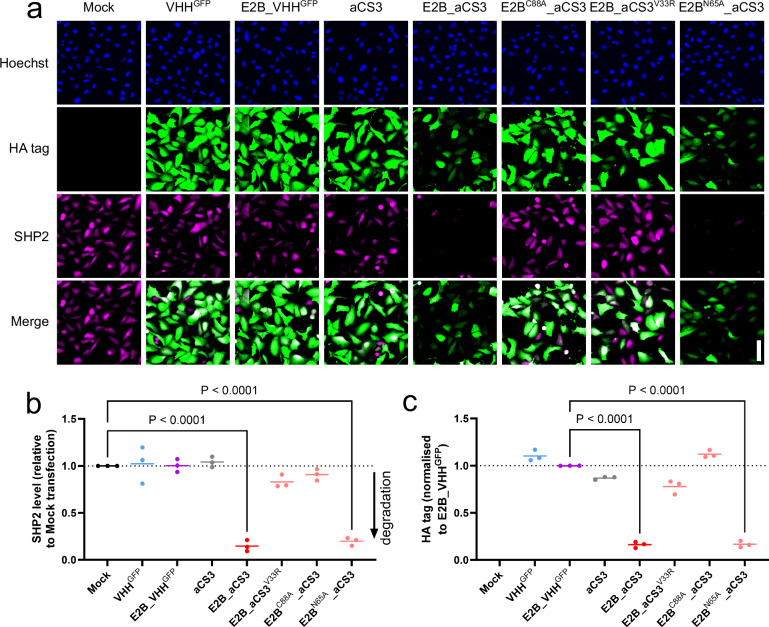
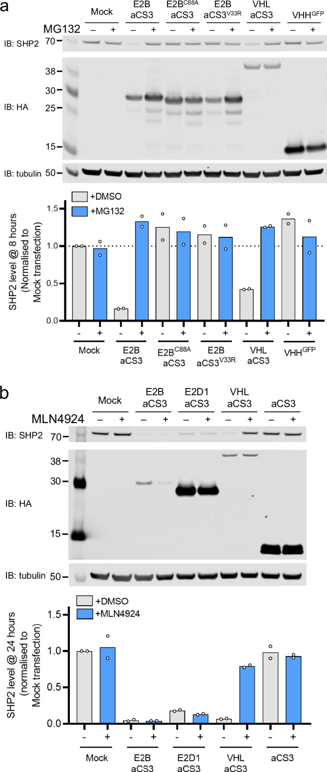
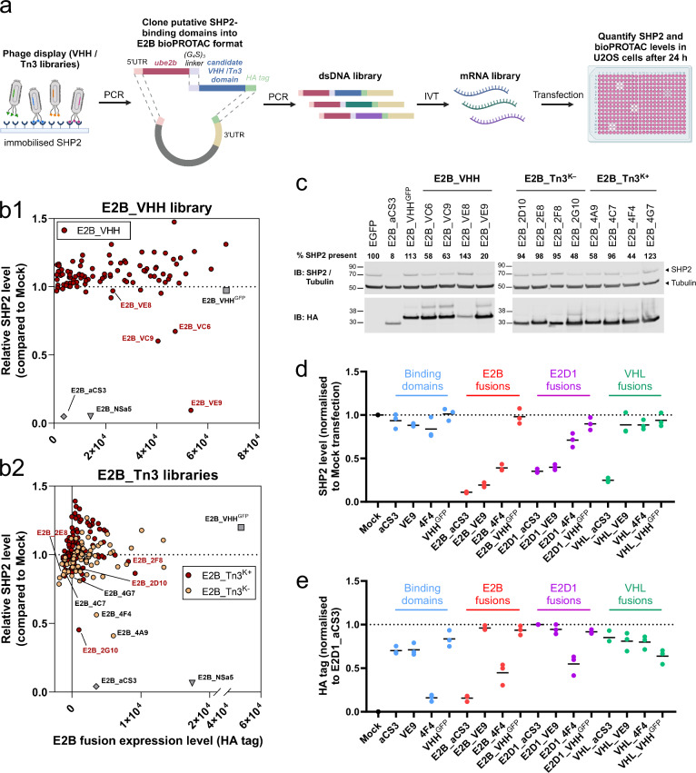
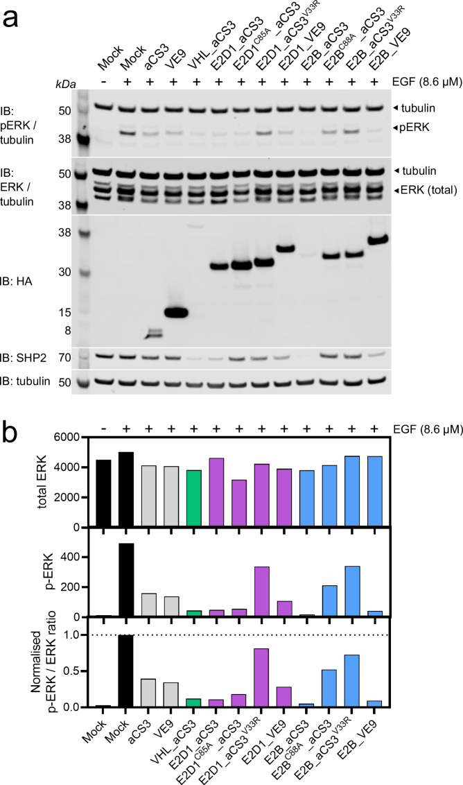
Proteins can be targeted for degradation by engineering biomolecules that direct them to the eukaryotic ubiquitination machinery. For instance, the fusion of an E3 ubiquitin ligase to a suitable target binding domain creates a 'biological Proteolysis-Targeting Chimera' (bioPROTAC). Here we employ an analogous approach where the target protein is recruited directly to a human E2 ubiquitin-conjugating enzyme via an attached target binding domain. Through rational design and screening we develop E2 bioPROTACs that induce the degradation of the human intracellular proteins SHP2 and KRAS. Using global proteomics, we characterise the target-specific and wider effects of E2 vs. VHL-based fusions. Taking SHP2 as a model target, we also employ a route to bioPROTAC discovery based on protein display libraries, yielding a degrader with comparatively weak affinity capable of suppressing SHP2-mediated signalling.


Conflict of interest statement

All authors are present, or former employees of AstraZeneca. J.D.T., T.M.E., S.M.C., A.R., C.B., N.B., J.H., R.M., M.B., F.P., R.C., D.G.R., A.Z., and J.D. hold AstraZeneca stock. R.M. and S.L. are employees of Alchemab Therapeutics, Cambridge, UK. K.C. is an employee of C4 Therapeutics, Watertown, MA, USA. This work is related to patent WO2022106869A1.

Figures

Fig. 1
Fig. 1. Degradation of SHP2 by E2D1-based bioPROTACs.
a Proposed intracellular biological mechanism for E2 bioPROTAC activity. Ubiquitin is first captured by the E2 enzymatic domain (step 1). The target protein is recruited via the attached binding domain [BD] and subsequently ubiquitinated (step 2). Re-loading of the E2 bioPROTAC (step 3) enables further target ubiquitination, ultimately leading to its degradation in the proteasome (step 4). Created with BioRender.com. b Immunofluorescence (IF) images of U2OS cells fixed 24 h after transfection with bioPROTAC or isotype control mRNAs. Scale bar (lower right corner image) = 100 µm. c, d Dot plots showing mean cellular SHP2 levels and expressed HA-tagged protein levels from IF images in (b). Data from three biological replicates. e Representative western blot of U2OS cells transfected with SHP2-targeted bioPROTAC or isotype control mRNAs and harvested after 24 h. f Mean SHP2 densitometry data from (e) (n = 3 biological replicates). Statistical significance for data in (c, d, f) was calculated using a one-way ANOVA.
Fig. 2
Fig. 2. Enhanced SHP2 degradation induced by E2B_aCS3.
a Representative immunofluorescence images of U2OS cells 24 h post-transfection with E2B_aCS3 mRNA, or isotype controls. Scale bar (lower right corner image) = 100 µm. b, c Dot plots showing mean cellular SHP2 levels and expressed HA-tagged protein levels from IF images in (a). Data from three biological replicates. Statistical significance was calculated using a one-way ANOVA.
Fig. 3
Fig. 3. E2 bioPROTACs induce proteasomal degradation independently of CRL E3 ligases.
Western blots of U2OS cells transfected with mRNA encoding bioPROTACs, inactive variants or domains, and incubated in the absence (−) or presence (+) of a 5 µM MG132 for 8 h or b 1 µM MLN4294 for 24 h. Representative blots are shown from two biological replicates. Bar graphs display the mean SHP2 band densitometry relative to mock-transfected control cells (individual data points are shown for each replicate). Related to Supplementary Fig. 1.
Fig. 4
Fig. 4. Global proteomics analysis of E2 and VHL-based bioPROTAC expression.
Volcano plots showing data for 7657 proteins from U2OS cells transfected with either ag bioPROTAC mRNA (wild type or mutant, as indicated) or h an siRNA pair targeting shp2 transcripts. The degradation domain (e.g. E2D1) and SHP2 are highlighted on each plot. Dashed lines indicate significance thresholds (log2FC beyond ±0.58; adjusted p value < 0.05). Data obtained from four biological replicates. i Bar graph representing the numbers and distribution of proteomic hits for the active bioPROTACs. j Circos plot for the wild type bioPROTACs. Purple lines connect common hits present in ≥2 datasets. k Heatmap displaying functional enrichment analyses for the active bioPROTACs. Datasets were individually analysed within Metascape and curated to highlight the significant changes to biological pathways for each bioPROTAC. The individual cells are coloured according to the functional enrichment score (−10logP), with darker shading indicating greater statistical significance. Related to Supplementary Figs. 5–9 and Supplementary Data 1–3.
Fig. 5
Fig. 5. Identification of novel SHP2-degrading E2 bioPROTACs from display library fusions.
a E2 bioPROTAC discovery workflow. The VHH or Tn3 domain encoded in each selected phage is PCR-amplified and ligated 3’ to a common E2B-linker dsDNA fragment. The library of plasmids containing unique domains serves as a template for downstream in vitro transcription (IVT) followed by transfection and screening. Created with BioRender.com. b Immunofluorescence data obtained from U2OS cells expressing E2B fusion mRNAs created from randomly selected sequence-unique phage display output clones. x-axis = HA-tagged fusion level; y-axis = SHP2 level relative to Mock cells. Two types of fusion were tested: b1 E2B_VHH (n = 94) and b2 E2B_Tn3 (i.e. Tenascin Fibronectin-type III fusions using either Tn3K+ (n = 93) or Tn3K− (n = 73) domains i.e. with or without lysine, respectively, in sequence variable regions). Data shown are representative of two biological replicates. Domains taken forward for analysis are individually labelled with their library identifier (V = VHH, 2 = Tn3K+ and 4 = Tn3K−) and source well position. c Selected E2B fusions from (b) were re-analysed by western blotting. The % SHP2 remaining compared to EGFP-transfected control cells was quantified by densitometry and shown numerically for each fusion. Data shown are representative to two biological replicates. Dot plots indicating mean d SHP2 or e HA-tagged protein levels as determined by immunofluorescence. U2OS cells were transfected with mRNA encoding either single domains (VE9, 4F4, aCS3, or VHHGFP) or the same domains fused E2D1, E2B, or VHL. Data from three biological replicates. Related to Supplementary Figs. 10 and 11.
Fig. 6
Fig. 6. Attenuation of ERK phosphorylation by SHP2-targeted E2 bioPROTACs.
a Western blots of serum-starved MDA-MB-468 cells transfected with mRNA encoding bioPROTACs (or controls) after 24 h, followed by a 15 min exposure to DMSO (−) or Epidermal Growth Factor (EGF, +). Blots were probed using antibodies specific for phosphorylated ERK (pERK), total ERK (tERK), HA-tag, SHP2, or tubulin (loading control) as indicated. Data shown is representative from two biological replicates. b Bar graphs displaying densitometry data from (a) for pERK, tERK, and the pERK/tERK ratio, normalised to that of untransfected control cells. Related to Supplementary Fig. 12.

References

    1. Békés, M., Langley, D. R. & Crews, C. M. PROTAC targeted protein degraders: the past is prologue. Nat. Rev. Drug Discov.21, 181–200 (2022). - PMC - PubMed
    1. Sasso, J. M. et al. Molecular glues: the adhesive connecting targeted protein degradation to the clinic. Biochemistry. 10.1021/acs.biochem.2c00245 (2022). - PMC - PubMed
    1. Dikic, I. & Schulman, B. A. An expanded lexicon for the ubiquitin code. Nat. Rev. Mol. Cell Biol. 1–15 10.1038/s41580-022-00543-1 (2022). - PMC - PubMed
    1. Guenette, R. G., Yang, S. W., Min, J., Pei, B. & Potts, P. R. Target and tissue selectivity of PROTAC degraders. Chem. Soc. Rev.51, 5740–5756 (2022). - PubMed
    1. Paiva, S.-L. & Crews, C. M. Targeted protein degradation: elements of PROTAC design. Curr. Opin. Chem. Biol.50, 111 (2019). - PMC - PubMed

Publication types

MeSH terms

Substances

LinkOut - more resources