. 2024 Sep 19;14(1):21915.
doi: 10.1038/s41598-024-72255-9.

Treatment resistance to melanoma therapeutics on a single cell level

Affiliations

Treatment resistance to melanoma therapeutics on a single cell level

Lijun Yao et al. Sci Rep. .

Abstract

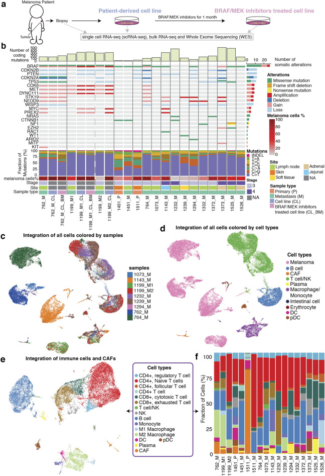
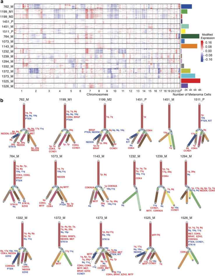
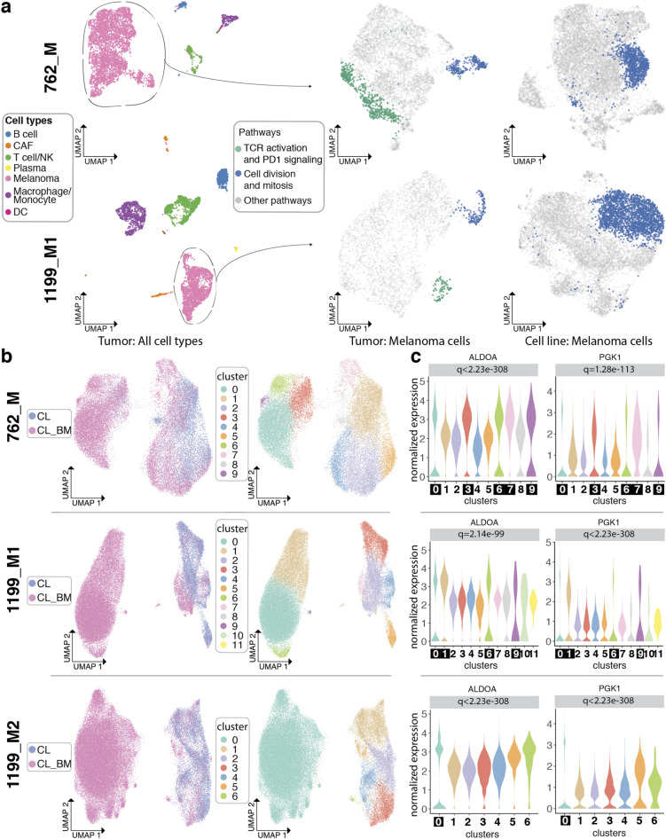
Therapy targeting the BRAF-MEK cascade created a treatment revolution for patients with BRAF mutant advanced melanoma. Unfortunately, 80% patients treated will progress by 5 years follow-up. Thus, it is imperative we study mechanisms of melanoma progression and therapeutic resistance. We created a scRNA (single cell RNA) atlas of 128,230 cells from 18 tumors across the treatment spectrum, discovering melanoma cells clustered strongly by transcriptome profiles of patients of origins. Our cell-level investigation revealed gains of 1q and 7q as likely early clonal events in metastatic melanomas. By comparing patient tumors and their derivative cell lines, we observed that PD1 responsive tumor fraction disappears when cells are propagated in vitro. We further established three anti-BRAF-MEK treatment resistant cell lines using three BRAF mutant tumors. ALDOA and PGK1 were found to be highly expressed in treatment resistant cell populations and metformin was effective in targeting the resistant cells. Our study suggests that the investigation of patient tumors and their derivative lines is essential for understanding disease progression, treatment response and resistance.


Conflict of interest statement

The authors declare no competing interests.

Figures

Fig. 1
Fig. 1
Study design, datasets, genomic landscape and cell populations revealed by scRNA-seq. (a) Establishment of melanoma patient-derived cell lines and BRAF-MEK inhibitor resistant cell lines. (b) Landscape of driver mutations, copy number variations (CNV), mutation signatures, melanoma cell percentage, clinical information and data availability across 15 patients, with vertical black lines separating patients and dashed lines separating 2 metastatic samples taken from the same patient. Copy number amplification/gain and copy number deletion/loss are shown in red/light red, blue/light blue respectively. NA = not applicable, P = primary, M = metastasis, CL = cell line, CL_BM = BRAF/MEK inhibitors treated cell line, scRNA-seq = single-cell RNA sequencing, WES = Whole Exome Sequencing. (c) UMAP plot shows integrated tumor and its microenvironment cells from 8 samples. (d) UMAP plot shows integrated tumor and its microenvironment cells from 8 samples as in panel c, colored by main cell types. CAF = cancer associated fibroblasts, DC = dendritic cells, pDC = plasmacytoid dendritic cells, and NK = natural killer cells. (e) UMAP plot shows integrated immune cells and CAFs from 8 samples (as in panel c and d, colored by cell subtypes. Melanoma cells, erythrocytes and gastrointestinal cells have been removed. (f) Bar chart shows the fraction of immune cell subsets for each sample with color indicating cell types.
Fig. 2
Fig. 2
Single-cell copy number variation analysis of cutaneous melanomas. (a) Single-cell CNV landscape from 17 samples with hierarchical clustering of CNV profiles of melanoma cells within every tumor. Bar plot on the right indicates the number of melanoma cells in each tumor. (b) Clonality trees of driver CNVs for each sample. The length of branches corresponds to the number of cells in the calculated subclone. Copy number gain is labelled in red and copy number loss is labelled in blue.
Fig. 3
Fig. 3
Transcriptional changes of melanoma cells from patient tumors, to patient tumor-derived cell lines, to BRAF/MEK inhibitors resistant cell lines. (a) UMAPs show all cell populations in 762_M and 1199_M1 tumors (left), melanoma cells in 762_M and 1199_M1 with cells colored by related pathways (middle), and melanoma cells in tumor-derived cell lines (762_M_CL and 1199_M1_CL) with cells colored by related pathways (right). (b) UMAPs show integrated melanoma cells from tumor-derived cell lines (762_M_CL, 1199_M1_CL, and 1199_M2_CL) and BRAF/MEK inhibitor resistant cell lines (762_M_CL_BM, 1199_M1_CL_BM and 1199_M2_CL_BM), with cells colored by samples (left) or clusters (right). c. Violin plots show normalized expression of ALDOA and PGK1 in each cluster for sample 762_M (top), 1199_M1 (middle) and 1199_M2 (bottom). Clusters mainly composed of cells from BRAF/MEK inhibitors treated cell lines are highlighted in black boxes.
Fig. 4
Fig. 4
Transcriptional changes of BRAF/MEK resistant cells before and after metformin treatment. (a) Bar graphs show metformin related cell death on initial lines (762_M_CL and 1199_M1_CL) and BRAF/MEK inhibitors treated cell lines (762_M_CL_BM and 1199_M1_CL_BM). 762_M is shown at the top and 1199_M at the bottom. CL_BM_MF = metformin treated cell lines. mM = millimolar, ** = p < 0.01, *** = p < 0.001. (b) UMAPs show integrated melanoma cells from tumor-derived cell lines (762_M_CL, 1199_M1_CL), BRAF/MEK inhibitors resistant cell lines (762_M_CL_BM, 1199_M1_CL_BM) and metformin treated cell lines (762_M_CL_BM_MF, 1199_M1_CL_BM_MF), with cells colored by samples (left) or clusters (right). (c) Violin plots show normalized expression of ALDOA and PGK1 in each cluster for sample 762_M (left), 1199_M1 (right). Clusters mainly composed of cells from BRAF/MEK inhibitors treated cell lines and/or metformin treated cell lines are highlighted in black boxes. (d) Violin plots show normalized expression of ALDOA and PGK1 of cells from 762 and 762_M_CL_BM_MF in boxed clusters of panel c. * = p < 0.05, ** = p < 0.01, *** = p < 0.001 (e) Violin plots show normalized expression of ALDOA and PGK1 of cells from 1199_M1_CL_BM and 1199_M1_CL_BM_MF in boxed clusters of panel c. * = p < 0.05, ** = p < 0.01, *** = p < 0.001 (f) Violin plots show normalized expression of representative DEGs for cell populations disappearing post metformin treatment, such as cluster 9 in 762_M and cluster 1 in 1199_M1, highlighted in black boxes. (g) Violin plots show normalized expression of representative DEGs for cell population emerging post metformin treatment, such as cluster 10 in 762_M, highlighted in black boxes.

References

    1. Larkin, J. et al. Combined vemurafenib and cobimetinib in BRAF-mutated melanoma. N. Engl. J. Med.371, 1867–1876 (2014). - PubMed
    1. Long, G. V. et al. Adjuvant Dabrafenib plus Trametinib in stage III BRAF-mutated melanoma. N. Engl. J. Med.377, 1813–1823 (2017). - PubMed
    1. Robert, C. et al. Five-year outcomes with Dabrafenib plus Trametinib in metastatic melanoma. N. Engl. J. Med.381, 626–636 (2019). - PubMed
    1. Ojha, R. et al. ER translocation of the MAPK pathway drives therapy resistance in BRAF-mutant melanoma. Cancer Discov.9, 396–415 (2019). - PMC - PubMed
    1. Johannessen, C. M. et al. A melanocyte lineage program confers resistance to MAP kinase pathway inhibition. Nature504, 138–142 (2013). - PMC - PubMed

MeSH terms