. 2024 Sep 16;7(1):1155.
doi: 10.1038/s42003-024-06864-5.

SPRR1B+ keratinocytes prime oral mucosa for rapid wound healing via STAT3 activation

Affiliations

SPRR1B+ keratinocytes prime oral mucosa for rapid wound healing via STAT3 activation

Xinyang Xuanyuan et al. Commun Biol. .

Abstract

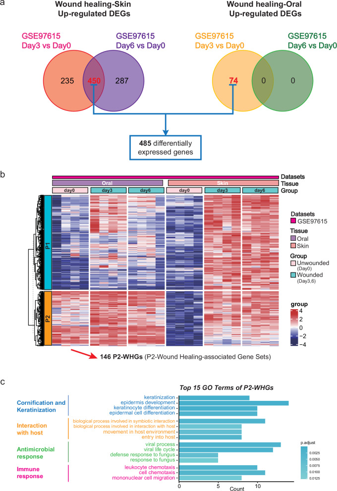
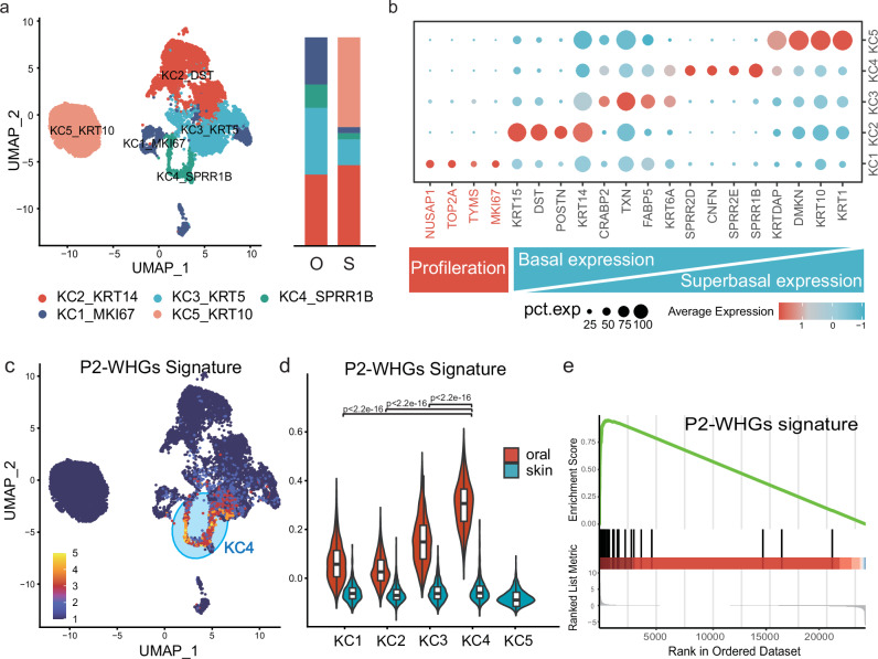
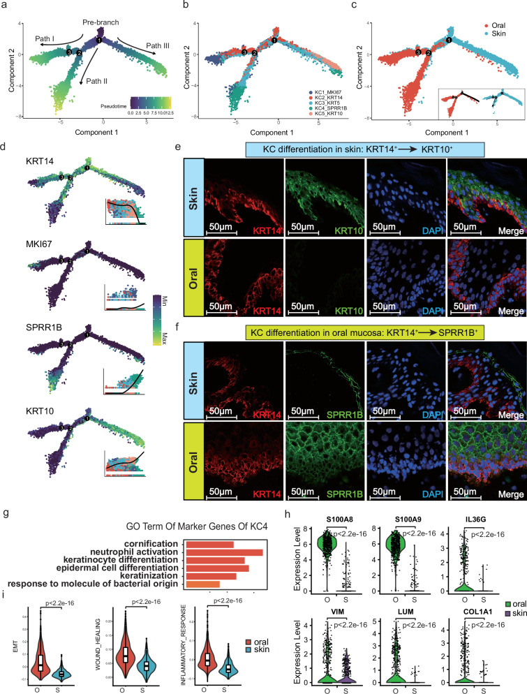
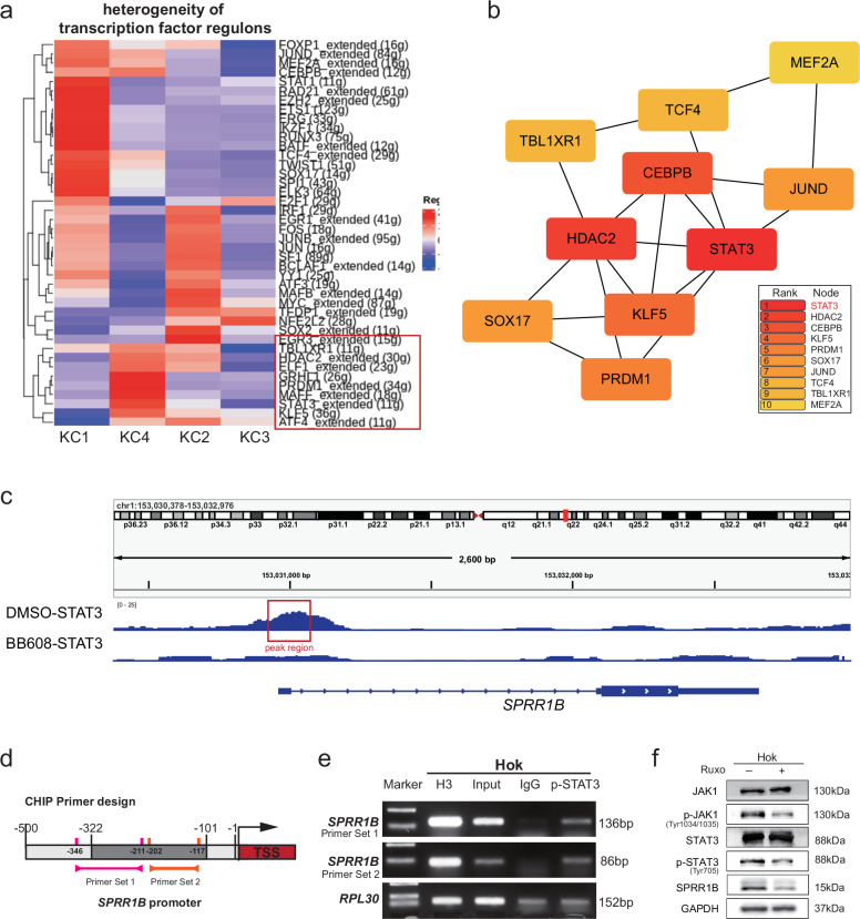
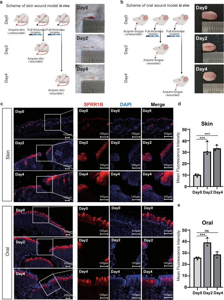
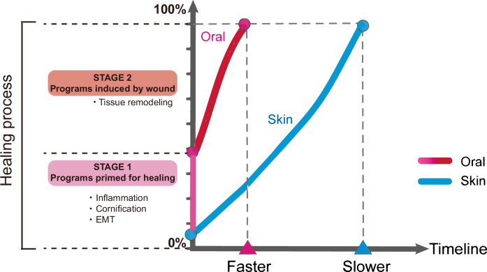
Oral mucosal wounds exhibit accelerated healing with reduced scarring compared to cutaneous wounds, representing an optimal wound healing paradigm. However, the specific cellular subtypes orchestrating the efficient healing of mucosal tissues remain elusive. Through a comprehensive analysis integrating bulk-mRNA and single-cell sequencing data during the wound healing process in oral mucosa and skin, we have delineated a distinct set of genes markedly upregulated during tissue repair. This collection of wound healing-associated genesets was highly enriched in a specific keratinocyte subpopulation identified as STAT3-activated SPRR1B+ keratinocytes. Notably, despite the inherent rapidity of oral mucosal healing, the induction of SPRR1B+ keratinocytes is evident in both skin and mucosal wound healing processes in murine model. Intriguingly, these wound healing-promoting SPRR1B+ keratinocytes, which are induced via STAT3 activation, inherently abundant in unwounded normal mucosa but absent in normal skin. SPRR1B knockdown significantly inhibits mucosal keratinocyte migration, a critical attribute for effective wound healing. In summary, through analysis of human oral and skin wound healing processes at single-cell resolution, coupled with validation in murine model, suggests STAT3-activated SPRR1B+ keratinocytes are associated with the rapid mucosal repair process. This discovery underscores the potential application of SPRR1B+ keratinocytes in the therapeutic management of chronic or non-healing wounds.


Conflict of interest statement

The authors declare no competing interests.

Figures

Fig. 1
Fig. 1. Identification of P2-Wound Healing-associated Gene Sets (P2-WHGs) during oral mucosa and skin wound healing.
a Differentially expressed genes (DEGs) between injured skin (left) or injured oral (right) vs uninjured counterpart were selected for further analysis. (|logFC| > 1 and adjusted p < 0.05). b Heatmap showing expression of selected DEGs between injured and uninjured group in each GSE dataset. c Gene ontology (GO) enrichment analysis revealed the top 15 GO Terms of P2-WHGs.
Fig. 2
Fig. 2. Keratinocytes are the major contributing cells for P2-WHGs signature, identified by Single-cell RNA-seq analysis of all cells in human normal skin and oral mucosa.
a Unbiased clustering of 103,758 cells based on the single cell RNA-seq data from normal skin and oral mucosa reveals 9 cellular clusters. Clusters are distinguished by different colors. The identity of each cell cluster is shown at the top (Fig. 2a; left). The proportion of cell lineages in oral mucosa and skin (Fig. 2a; right). (O: oral mucosa; S: skin). b Violin plot analysis of cluster-specific genes for each cell type. c Heatmap of differentially expressed genes. For each cluster, the top 10 genes and their relative expression levels in all sequenced cells are shown (Fig. 2c; left). Bubble plot showing representative GO term enrichment of biological processes according to cell types (Fig. 2c: right). d UMAP plots based on the P2-WHGs score of all cells, and high P2-WHGs score cell clusters are highlighted. e Violin plots based on the P2-WHGs score of all cells. p values are from two-sided Wilcoxon rank-sum tests, ****p < 2.2e-16.
Fig. 3
Fig. 3. SPRR1B+ keratinocyte cluster (KC4) shows the highest P2-WHGs signature, and is more enriched in normal oral mucosa than skin.
a Identification of 5 distinct keratinocyte subtypes based on the single cell sequencing data (Fig. 3a; left). The proportion of each keratinocyte subtypes in mucosa and skin (Fig. 3a; right). (O: oral mucosa; S: skin). b Dot plot analysis of cluster-specific genes for each subtype of keratinocyte. c UMAP plots based on the P2-WHGs score of keratinocytes. High P2-WHGs score cell clusters are highlighted. d Violin plots based on the P2-WHGs score of each keratinocyte subclusters in oral mucosa and skin. Two-sided Wilcoxon rank-sum tests were performed. ****p < 2.2e-16. e GSEA enrichment plots for P2-WHGs signaling upregulated in oral KC4 compared to skin KC4.
Fig. 4
Fig. 4. Define the characteristics and trajectory of SPRR1B+ keratinocyte cluster (KC4).
ac Reconstruction of a trajectory with keratinous subpopulation; Results of pseudotime analysis of keratinocytes, colored by subclusters (Fig. 4a), pseudotime (Fig. 4b) and samples (Fig. 4c). d Expression levels for each cell are colored and overlaid onto pseudotime. Genes were selected as specifically expressed in celltypes. Bright yellow indicates maximum relative expression, and dark blue indicates low or no expression of this gene, each were accompanied by color-coded pseudotime feature plots. e, f Representative immunofluorescent images of normal skin and oral mucosa stained with the basal cell marker keratin 14 (K14, red), differentiation markers keratin 10 (K10, green), and Small Proline-Rich Protein IB (SPRR1B, green). Scale bar 50 µm. g Gene Ontology analysis of marker genes of the KC4 keratinocyte cluster. h The violin plot represents the gene expression levels in different group. i Gene scoring analysis of KC4 between oral and skin using the indicated molecular signatures. Two-sided Wilcoxon rank-sum tests were performed (h, i). ****p < 2.2e-16.
Fig. 5
Fig. 5. STAT3 regulates wound healing gene expression in SPRR1B+ keratinocytes.
a Heatmaps showing the transcriptional factors (TFs) which were differently expressed in each keratinocyte clusters. b The 10 TF genes with the highest scores in the Protein–Protein Interaction (PPI) network obtained by using the MCC algorithm of the CytoHubba plugin. c The predicted binding sites of STAT3 at the promoter region of SPRR1B gene. d Schematic of SPRR1B transcriptional start site (TSS) (red) and 0 to 500 bp upstream sequence. Design of PCR products for chromatin immunoprecipitation (ChIP) experiments (orange and red vertical lines) and predicted STAT3 binding sites (gray band) are indicated. e ChIP-PCR detection of STAT3 binding to the SPRR1B promoter region. f Western blot of STAT3, p-STAT3, JAK1, p-JAK1 and SPRR1B in HOK cells with JAK1/2 inhibitor ruxolitinib (Ruxo).
Fig. 6
Fig. 6. In vivo model confirms that SPRR1B+ keratinocytes are constitutively enriched in oral mucosa and are increased during both oral mucosa and skin wound healing.
a, b Schematic diagram of the experimental design in mice wound healing model. (Fig. 6a: skin; Fig. 6b: oral. Created with BioRender.com). The skin and tongue tissue of mice during the wound healing process are shown on the right (skin: n = 3, oral: n = 3). c Immunofluorescence images of oral and skin samples on different days during wound healing. Scale bar: 100 µm. d, e Quantification of the mean fluorescence intensity of SPRR1B over a 500-pixel area on either side of the skin and oral wound (n = 3). Error bars are shown as mean ± SEM from n = 3. One-way analysis of variance (ANOVA) were performed (d, e). *p < 0.05, **p < 0.01, ***p < 0.001. ns not significant.
Fig. 7
Fig. 7. SPRR1B regulates the migration ability of keratinocytes.
a Western blot of SPRR1B and internal control (GAPDH) between HOK and HaCat cells. b Western blot of SPRR1B in HOK cells expressing control shRNA or either of three independent shRNAs targeting SPRR1B. c Migrating HOK cells transduced with shRNAs of SPRR1B and shNC (control) by lentiviral delivery. Images were taken every 3 h from 0 to 12 h after scratching. d Line plot representing relative change in wound area size of HOK cells as a function of time (n = 3). e Western blot to show overexpression of SPRR1B after transfection with lentiviruses in HaCat cells. f Migrating HaCat cells transduced with SPRR1B and GFP (control) by lentiviral delivery. Images were taken every 3 h from 0 to 21 h after scratching. g Line plot representing relative change in wound area size of HaCat cells as a function of time (n = 3). Error bars are shown as mean ± SEM from n = 3. Two-way analysis of variance (ANOVA) were performed (d, g). *p < 0.05, **p < 0.01, ***p < 0.001. ns not significant.
Fig. 8
Fig. 8. Schematic diagram of the wound healing model of skin and oral mucosa.
The wound healing process model of skin and oral mucosa. The oral mucosa is primed for the wound healing stage, resulting in a faster healing rate compared to skin. (Oral: Pink and red lines; Skin: Blue lines). EMT, epithelial-mesenchymal transition.

References

    1. Martin, R. F. Wound healing. Surg. Clin. North Am.100, ix–xi (2020). - PMC - PubMed
    1. Choi, M. et al. The case : a nonhealing skin ulcer in a patient 5 years after successful transplantation. Kidney Int.100, 1357–1358 (2021). - PubMed
    1. Kuntz, T., Koushk-Jalali, B. & Kreuter, A. Sunitinib-induced pyoderma gangrenosum-like skin ulcer. Cmaj192, E552 (2020). - PMC - PubMed
    1. Pugliese, E., Coentro, J. Q., Raghunath, M. & Zeugolis, D. I. Wound healing and scar wars. Adv. Drug Deliv. Rev.129, 1–3 (2018). - PubMed
    1. Waasdorp, M. et al. The bigger picture: why oral mucosa heals better than skin. Biomolecules11, 1165 (2021). - PMC - PubMed

Publication types

Associated data

LinkOut - more resources