. 2024 Sep 19;22(1):578.
doi: 10.1186/s12951-024-02822-9.

Macrophage membrane-camouflaged biomimetic nanoparticles for rheumatoid arthritis treatment via modulating macrophage polarization

Affiliations

Macrophage membrane-camouflaged biomimetic nanoparticles for rheumatoid arthritis treatment via modulating macrophage polarization

Renpeng Zhou et al. J Nanobiotechnology. .

Abstract

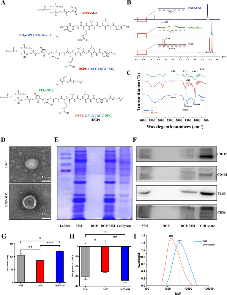
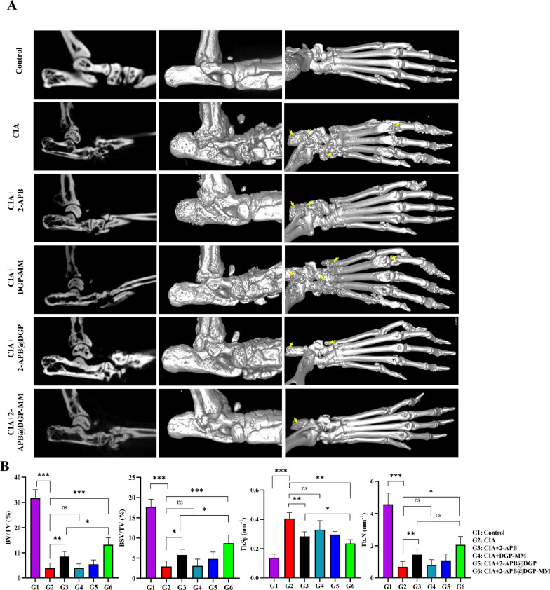
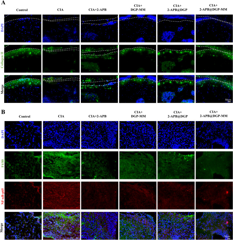
Rheumatoid arthritis (RA) is a debilitating autoimmune disease characterized by chronic joint inflammation and cartilage damage. Current therapeutic strategies often result in side effects, necessitating the development of targeted and safer treatment options. This study introduces a novel nanotherapeutic system, 2-APB@DGP-MM, which utilizes macrophage membrane (MM)-encapsulated nanoparticles (NPs) for the targeted delivery of 2-Aminoethyl diphenylborinate (2-APB) to inflamed joints more effectively. The NPs are designed with a matrix metalloproteinase (MMP)-cleavable peptide, allowing for MMP-responsive drug release within RA microenvironment. Comprehensive in vitro and in vivo assays confirmed the successful synthesis and loading of 2-APB into the DSPE-GPLGVRGC-PEG (DGP) NPs, as well as their ability to repolarize macrophages from a pro-inflammatory M1 to an anti-inflammatory M2 phenotype. The NPs demonstrated high biocompatibility, low cytotoxicity, and enhanced cellular uptake. In a collagen-induced arthritis (CIA) mouse model, intra-articular injection of 2-APB@DGP-MM significantly reduced synovial inflammation and cartilage destruction. Histological analysis corroborated these findings, demonstrating marked improvements in joint structure and delayed disease progression. Above all, the 2-APB@DGP-MM nanotherapeutic system offers a promising and safe approach for RA treatment by modulating macrophage polarization and delivering effective agents to inflamed joints.

Keywords: 2-APB; Inflammation; Macrophage polarization; Membrane-encapsulated nanoparticles; Rheumatoid arthritis.


Conflict of interest statement

The authors declare no competing interests.

Figures

Scheme 1
Scheme 1
Construction of 2-APB@DGP-MM and their inhibition mechanisms of synovial inflammation in rheumatoid arthritis
Fig. 1
Fig. 1
2-APB promotes the phenotypic transformation of macrophages from M1 to M2. A, B The effect of 2-APB on the expression of polarization marker proteins iNOS and Arg-1 in RAW264.7 cells after LPS/IL-4 treatment. C, D Effect of 2-APB on the expression of iNOS and Arg-1 on RAW264.7 via confocal fluorescence (scale bar: 50 μm). E Quantitative analysis of the iNOS and Arg-1 fluorescence intensity (n = 3). F Effect of 2-APB on the expression of CD206 and Arg-1 mRNA in RAW264.7. ns = no significance; *P < 0.05; **P < 0.01
Fig. 2
Fig. 2
Synthesis and characterization of DGP-MM and 2-APB@DGP. A The synthesis process of DSPE-GPLGVRGC-PEG (DGP). B The characteristic peak of DSPE-PEG, GPLGVRGC and DGP in 1H-NMR assay. C The characteristic wavelength numbers in the FT-IR assay. D TEM images of DGP and DGP-MM (scale bar: 50 nm). E Coomassie blue fast staining assay for the MM, DGP and DGP-MM. F Western blot detection for the macrophage markers, including CD11b, CD49d, F4/80, and CD86. G, H The mean particle size and mean zeta potential of MM, DGP, and DGP-MM. I The GPC detection of DGP and DGP plus MMP2 (30 ng/mL). ns = no significance; *P < 0.05; **P < 0.01; ***P < 0.001
Fig. 3
Fig. 3
2-APB@DGP-MM promotes macrophage polarization from M1 to M2 via enhancing cellular uptake and drug release. A The effects of DGP-MM and 2-APB@DGP-MM on cell viability at different time points and concentrations. B The mean diameter alteration of different NPs at different pH conditions after incubation for various time, and the release percentage of 2-APB from different groups at different times. C Hematotoxicity of DGP-MM or 2-APB@DGP-MM on the fresh red blood cell from mice at different concentrations of DGP-MM NPs or 2-APB@DGP-MM NPs (0, 12.5, 25, 50 and 100 μg/mL), 1% Triton-X 100 (positive control) and sterile PBS (negative control) for 1 h. D The uptake efficiency of Cy5-loaded 2-APB@DGP or Cy5-loaded 2-APB@DGP-MM in BMDMs (scale bar: 40 μm). E The protein expression of iNOS and Arg-1 after different treatments for 4 days. FI The relative mRNA expression of M1 polarization markers (iNOS and IL-2b) and M2 polarization markers (Arg-1 and CD206) after corresponding treatments. *p < 0.05; **P < 0.01; ***P < 0.001; ****P < 0.0001
Fig. 4
Fig. 4
2-APB@DGP-MM inhibits CIA progression. A Schematic diagram of treatment schedule. B Representative photographs of mice ankle joints after 2-APB@DGP-MM treatment. C H&E and D S/F staining of ankle joints or synovium from mice after different treatments. E The arthritis scores were calculated for the inflammatory joints with different treatments. F The paw thickness of the inflamed joints for different treatments. G Statistical analysis for the synovitis score and cartilage thickness after different treatments. *P < 0.05; **P < 0.01; ***P < 0.001
Fig. 5
Fig. 5
2-APB@DGP-MM alleviates joints destruction in CIA mice. A Micro-CT images of ankle joints of CIA mice after 2-APB@DGP-MM, 2-APB@DGP, DGP-MM or 2-APB treatment. B Quantitative analysis of the BV/TV, BS/TV, Tb.Sp and Tb.N. ns = no significance; *P < 0.05; **P < 0.01; ***P < 0.001
Fig. 6
Fig. 6
2-APB@DGP-MM restores the level of collagen II and reduces the level of NF-κB-p65 in the joints of CIA mice. Representative fluorescence images of Collagen II (A) and NF-κB-p65 (B) in the joint tissues after different treatments (scale bar: 50 μm)
Fig. 7
Fig. 7
2-APB@DGP-MM modulates macrophage polarization in CIA mice joint. Representative fluorescence images of the co-localization of iNOS (A) and CD206 (B) with macrophage, respectively, in the joint after different treatment (scale bar: 50 μm)

References

    1. Smolen JS, Aletaha D, McInnes IB. Rheumatoid arthritis. Lancet. 2016;388:2023–38. - PubMed
    1. Diseases GBD, Injuries C. Global burden of 369 diseases and injuries in 204 countries and territories, 1990–2019: a systematic analysis for the Global Burden of Disease Study 2019. Lancet. 2020;396:1204–22. - PMC - PubMed
    1. Fan XX, Xu MZ, Leung EL, Jun C, Yuan Z, Liu L. ROS-responsive berberine polymeric micelles effectively suppressed the inflammation of rheumatoid arthritis by targeting mitochondria. Nanomicro Lett. 2020;12:76. - PMC - PubMed
    1. Fan Z, Li J, Liu J, Jiao H, Liu B. Anti-inflammation and joint lubrication dual effects of a novel hyaluronic acid/curcumin nanomicelle improve the efficacy of rheumatoid arthritis therapy. ACS Appl Mater Interfaces. 2018;10:23595–604. - PubMed
    1. Asquith DL, Ballantine LE, Nijjar JS, Makdasy MK, Patel S, Wright PB, Reilly JH, Kerr S, Kurowska-Stolarska M, Gracie JA, McInnes IB. The liver X receptor pathway is highly upregulated in rheumatoid arthritis synovial macrophages and potentiates TLR-driven cytokine release. Ann Rheum Dis. 2013;72:2024–31. - PubMed

Substances

LinkOut - more resources