

Combining optical genome mapping and RNA-seq for structural variants detection and interpretation in unsolved neurodevelopmental disorders
- PMID: 39300495
- PMCID: PMC11414247
- DOI: 10.1186/s13073-024-01382-9
Combining optical genome mapping and RNA-seq for structural variants detection and interpretation in unsolved neurodevelopmental disorders
Abstract
Background: Structural variations (SVs) are key genetic contributors to neurodevelopmental disorders (NDDs). Exome sequencing (ES), the current first-line tool for genetic testing of NDDs, falls short in SVs detection. This diagnostic gap is being actively addressed by new methods such as optical genome mapping (OGM).
Methods: This study evaluated the utility of combining OGM and RNA-seq in the detection and interpretation of SVs in ES-negative NDDs. OGM was performed in 43 patients with NDDs with inconclusive ES results. Candidate SVs were selected based on disease association and pathogenicity evaluation, and further validated or reconstructed by alternative methods, including long-read sequencing for a complex rearrangement event. RNA-Seq was performed on blood samples from patients with candidate SVs to facilitate interpretation of pathogenicity.
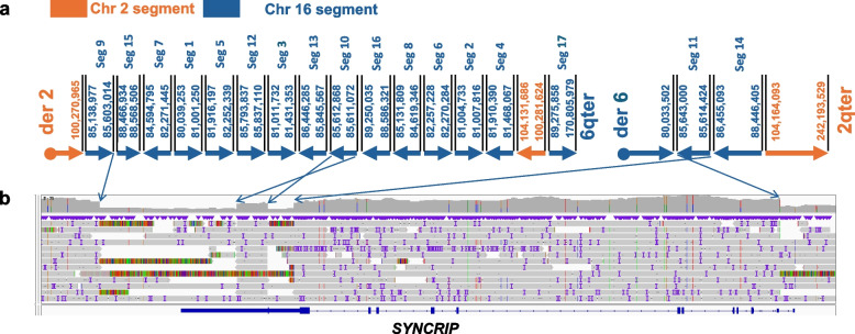
Results: OGM detected four candidate SVs, and RNA-seq confirmed the pathogenicity of three SVs in the patient cohort. This combined approach solved three cases-two cases with de novo SVs in genes associated with autosomal dominant NDDs, including a deletion encompassing the promoter and 5'UTR of MBD5 and an intragenic duplication of PAFAH1B1, and a third case possessing an intragenic duplication in trans with a pathogenic single-nucleotide variant of PLA2G6, associated with autosomal recessive NDDs. The expression alteration of the affected genes and the tandem positioning of two intragenic duplications were confirmed by RNA-seq. In the fourth case, OGM detected a complex rearrangement involving chromosomes 2 and 6, much more complex than the de novo t(2:6)(q13;q15) indicated by conventional cytogenetic analysis. Reconstruction showed that 17 segments of 6q15 spanning 9.3 Mb were disarranged and joined 2q11.2, with four breakpoints detected in the 5' and 3' non-coding region of the NDD-associated gene SYNCRIP. RNA-seq revealed largely preserved SYNCRIP expression, leaving the pathogenicity of this complex rearrangement event uncertain.
Conclusions: SVs in ES-negative NDDs can be identified by OGM, which is particularly useful for SVs in non-coding regions not covered by ES. OGM helps to construct complex SVs and provides information on the location and orientation of duplications, which is crucial for pathogenicity interpretation. The integration of RNA-seq facilitates the interpretation of the functional consequences of SVs at the transcriptional level. These findings demonstrate the utility and feasibility of combining OGM and RNA-seq in ES-negative cases with NDDs.
Keywords: Neurodevelopmental disorders; Optical genome mapping; RNA-seq; Structural variants.
© 2024. The Author(s).
Conflict of interest statement
The authors declare no competing interests.
Figures


References
-
- Smith AC, Neveling K, Kanagal-Shamanna R. Optical genome mapping for structural variation analysis in hematologic malignancies. Am J Hematol. 2022;97(7):975–82. - PubMed
MeSH terms
LinkOut - more resources
Full Text Sources
Miscellaneous
