. 2024 Sep 19;21(1):38.
doi: 10.1186/s12989-024-00600-x.

The chemical composition of secondary organic aerosols regulates transcriptomic and metabolomic signaling in an epithelial-endothelial in vitro coculture

Affiliations

The chemical composition of secondary organic aerosols regulates transcriptomic and metabolomic signaling in an epithelial-endothelial in vitro coculture

Svenja Offer et al. Part Fibre Toxicol. .

Abstract

Background: The formation of secondary organic aerosols (SOA) by atmospheric oxidation reactions substantially contributes to the burden of fine particulate matter (PM2.5), which has been associated with adverse health effects (e.g., cardiovascular diseases). However, the molecular and cellular effects of atmospheric aging on aerosol toxicity have not been fully elucidated, especially in model systems that enable cell-to-cell signaling.

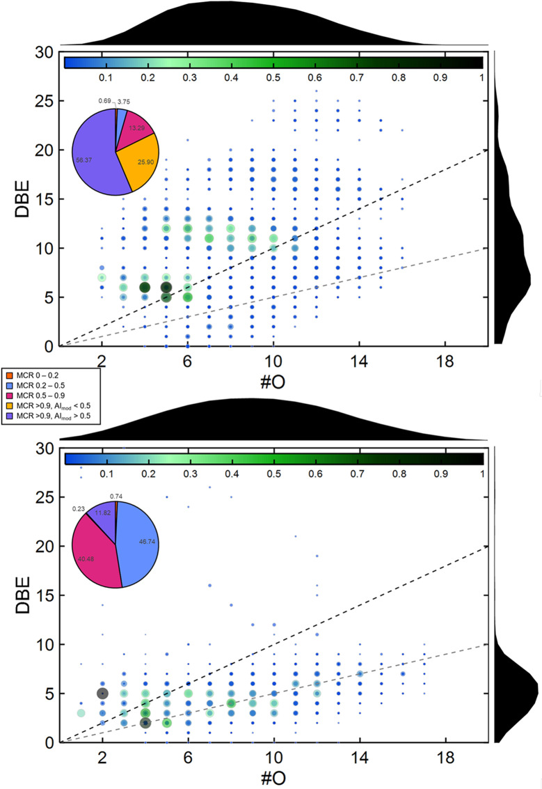
Methods: In this study, we aimed to elucidate the complexity of atmospheric aerosol toxicology by exposing a coculture model system consisting of an alveolar (A549) and an endothelial (EA.hy926) cell line seeded in a 3D orientation at the air‒liquid interface for 4 h to model aerosols. Simulation of atmospheric aging was performed on volatile biogenic (β-pinene) or anthropogenic (naphthalene) precursors of SOA condensing on soot particles. The similar physical properties for both SOA, but distinct differences in chemical composition (e.g., aromatic compounds, oxidation state, unsaturated carbonyls) enabled to determine specifically induced toxic effects of SOA.

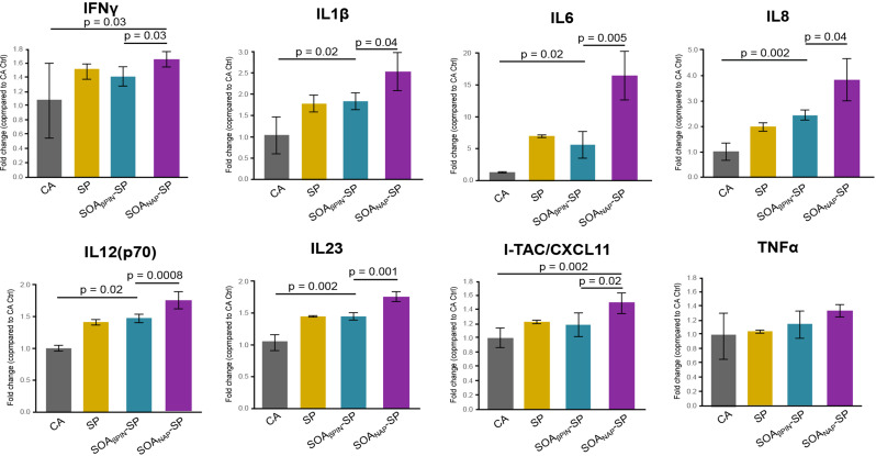
Results: In A549 cells, exposure to naphthalene-derived SOA induced stress-related airway remodeling and an early type I immune response to a greater extent. Transcriptomic analysis of EA.hy926 cells not directly exposed to aerosol and integration with metabolome data indicated generalized systemic effects resulting from the activation of early response genes and the involvement of cardiovascular disease (CVD) -related pathways, such as the intracellular signal transduction pathway (PI3K/AKT) and pathways associated with endothelial dysfunction (iNOS; PDGF). Greater induction following anthropogenic SOA exposure might be causative for the observed secondary genotoxicity.

Conclusion: Our findings revealed that the specific effects of SOA on directly exposed epithelial cells are highly dependent on the chemical identity, whereas non directly exposed endothelial cells exhibit more generalized systemic effects with the activation of early stress response genes and the involvement of CVD-related pathways. However, a greater correlation was made between the exposure to the anthropogenic SOA compared to the biogenic SOA. In summary, our study highlights the importance of chemical aerosol composition and the use of cell systems with cell-to-cell interplay on toxicological outcomes.

Keywords: Airway remodeling; Endothelial dysfunction; Epithelial-endothelial coculture; Inflammation; Secondary organic aerosols (SOA).


Conflict of interest statement

The authors declare no competing interests.

Figures

Fig. 1
Fig. 1
Maximum carbonyl ratio (MCR) and aromaticity of SOANAP-SP and SOAβPIN-SP. Number of oxygen atoms (#O) vs. double bond equivalents (DBE) in SOANAP-SP (top) and SOAβPIN-SP (bottom) with pie charts depicting the percentage of peaks in individual bins of the maximum carbonyl ratio (MCR) and aromaticity index (AI) for ESI in positive mode
Fig. 2
Fig. 2
Transcriptional analysis of aerosol-induced effects in A549 and EA.hy926 cells. (A) Schematic representation of the experimental setup. A coculture model system consisting of A549 lung epithelial cells on one side of the insert membrane and EA.hy926 endothelial cells on the other side were exposed for 4 h at the air-liquid interface (ALI) to two SOA, namely, SOAβPIN-SP and SOANAP-SP and SP. Transcriptional changes in A549 and EA.hy926 cells were also analyzed. (B) Principal component analysis (PCA) of all genes with batch correction and normalization to colors based on exposure to clean air (CA), SOANAP-SP, and SOAβPIN-SP; n = 4 independent experiments and SP of n = 3 experiments of A549 and EA.hy926 cells. (C) Venn diagram of the unique and overlapping genes associated with the up- and downregulated genes (adjusted p-value ≤ 0.05, and log2FC ≥ 0.5 and log2FC ≤ -0.5) in A549 cells after exposure to SP, SOAβPIN-SP or SOANAP-SP compared to the CA control. (D) Gene Ontology (GO) analysis of differentially expressed genes (DEGs) related to biological processes and molecular functions between A549 cells exposed to different aerosols (SP, SOAβPIN-SP, and SOANAP-SP) and the CA Ctrl. (E and F) Same as in (C and D) but based on the analysis of EA.hy926 cells
Fig. 3
Fig. 3
Induction of stress- and inflammatory-related genes in A549 and EA.hy926 cells. (A) Heatmap comparing gene expression values among different aerosol exposures (SP, SOAβPIN-SP and SOANAP-SP) compared to those of the clean air (CA) control originating from GO overrepresentative analysis of A549 cells. (B) Boxplots of the genes depicted from the heatmap in Fig. 3A showing the normalized counts (TMM) in A549 cells after exposure to CA, SP, SOAβPIN-SP or SOANAP-SP. (CD) Same as in (A and B) but based on the analysis of EA.hy926 cells. (E and F) Dot plots showing the level of expression (fold change) of genes activated (filled dots) or suppressed (empty dots) by exposure to SP, SOAβPIN-SP or SOANAP-SP compared to CA exposure in the “mucin-related genes” and “innate immunity” categories for A549 cells I and in the “atherosclerosis” and “endothelial cell biology” categoriesfor EA.hy926 cells (F). * adjusted p-value < 0.05, ** adjusted p-value < 0.01 and *** adjusted p-value < 0.001
Fig. 4
Fig. 4
Circulating cytokine validated the observed toxicogenomic responses. Cytokine validation of IFN-γ, IL1-β, IL6, IL8, IL12(p70), IL23, I-TAC/CXCL11 and TNFα in the cell culture media of the coculture system after exposure to CA, SP, SOAβPIN-SP and SOANAP-SP. The data are presented as the fold change (compared to the CA control) ± SEM from three independent experiments, and the significance of the differences are outlined
Fig. 5
Fig. 5
Integrative analysis of circulating metabolites and gene expression revealed important role of non-directly exposed endothelial cells. (A-C) Volcano plots showing the relationships between differentially abundant metabolite expression (p-value ≤ 0.1 and log2FC ≥ 0.5 and log2FC ≤ -0.5) in the cell culture media after exposure to SP (A), SOAβPIN-SP (B) or SOANAP-SP (C) and the CA control. (D) Venn diagram of the unique and overlapping metabolites identified as up- or downregulated (p-value ≤ 0.1, log2FC ≥ 0.5 and log2FC ≤ -0.5) in the cell culture media after exposure to SP, SOAβPIN-SP or SOANAP-SP compared to the CA control. GC/MS analysis was performed on n = 3 independent experiments
Fig. 6
Fig. 6
Proposed pathomechanisms triggered by aerosol exposure. Aerosols confer most of their effects via damage to the lung (inflammation, oxidative stress, genotoxicity, and airway remodeling toward MUC5AC+ goblet-secretory like cells). This effect seemed to be dependent on the chemical composition of the specific aerosols (organic content, PAHs, functional groups, oxidation state, aromaticity) and was greatest after exposure to SOANAP-SP, followed by SOAβPIN-SP and SP. The primary target organ damaged by aerosols converges at the cardiovascular level by inducing dysfunctions in endothelial cells (secondary genotoxicity), inflammation, metabolic reprogramming, and angiogenesis. These effects seemed to be more independent of the aerosol type and systemic treatment, resulting in the activation of coagulation, atherosclerosis and/or hypertension via iNOS, PDGF and/or PI3/AKT signaling, respectively

References

    1. Manisalidis I, Stavropoulou E, Stavropoulos A, Bezirtzoglou E. Environmental and Health impacts of Air Pollution: a review. Front Public Health. 2020;8:14. - PMC - PubMed
    1. Lelieveld J, Pozzer A, Poschl U, Fnais M, Haines A, Munzel T. Loss of life expectancy from air pollution compared to other risk factors: a worldwide perspective. Cardiovasc Res. 2020;116(11):1910–7. - PMC - PubMed
    1. Pozzer A, Anenberg SC, Dey S, Haines A, Lelieveld J, Chowdhury S. Mortality attributable to Ambient Air Pollution: a review of global estimates. GeoHealth. 2023;7(1):e2022GH000711. - PMC - PubMed
    1. Al-Kindi SG, Brook RD, Biswal S, Rajagopalan S. Environmental determinants of cardiovascular disease: lessons learned from air pollution. Nat Reviews Cardiol. 2020;17(10):656–72. - PMC - PubMed
    1. Brook RD, Rajagopalan S, Pope CA 3rd, Brook JR, Bhatnagar A, Diez-Roux AV, et al. Particulate matter air pollution and cardiovascular disease: an update to the scientific statement from the American Heart Association. Circulation. 2010;121(21):2331–78. - PubMed