. 2024 Sep;13(9):e12506.
doi: 10.1002/jev2.12506.

Plasma-derived extracellular vesicles (EVs) as biomarkers of sepsis in burn patients via label-free Raman spectroscopy

Affiliations

Plasma-derived extracellular vesicles (EVs) as biomarkers of sepsis in burn patients via label-free Raman spectroscopy

Hannah J O'Toole et al. J Extracell Vesicles. 2024 Sep.

Abstract

Sepsis following burn trauma is a global complication with high mortality, with ∼60% of burn patient deaths resulting from infectious complications. Diagnosing sepsis is complicated by confounding clinical manifestations of the burn injury, and current biomarkers lack the sensitivity and specificity required for prompt treatment. There is a strong rationale to assess circulating extracellular vesicles (EVs) from patient liquid biopsy as sepsis biomarkers due to their release by pathogens from bacterial biofilms and roles in the subsequent immune response. This study applies Raman spectroscopy to patient plasma-derived EVs for rapid, sensitive, and specific detection of sepsis in burn patients, achieving 97.5% sensitivity and 90.0% specificity. Furthermore, spectral differences between septic and non-septic burn patient EVs could be traced to specific glycoconjugates of bacterial strains associated with sepsis morbidity. This work illustrates the potential application of EVs as biomarkers in clinical burn trauma care and establishes Raman analysis as a fast, label-free method to specifically identify features of bacterial EVs relevant to infection amongst the host background.

Keywords: bacterial EVs (bEVs); diagnostics; exosomes; systemic inflammatory response syndrome (SIRS).


Conflict of interest statement

The authors declare no conflicts of interest.

Figures
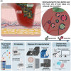
FIGURE 1
FIGURE 1
Extracellular vesicle release during bacterial infection in burn injury; Overview schema of sample preparation and acquisition. (a) The rationale behind our work is that EVs originating from colonizing bacteria are released into circulatory biofluids via damaged endothelium at the site of burn injury. Inflammatory response to injury results in secondary release of EVs from immune and endothelial cells as well. (b) Collection and analysis of EVs in circulating biofluids are a way to determine sepsis in advance of currently available methods.
FIGURE 2
FIGURE 2
Characterization of SEC isolated EVs from septic and non‐septic burn patient plasma. (a–d) CryoEM images of isolated EVs from (a, b) septic and (c, d) non‐septic burn patient samples reveal the expected lipid bilayer structure. Images were acquired at (a, c) 11k and (b, d) 45k magnification. Scale bars are 200 nm. (e) RPS and (f) NTA were used to determine the average size distribution of the EVs isolated from the septic and non‐septic burn patient samples. Shaded regions designate size standard deviations (g). The total EV particle concentration of the samples was measured by RPS and NTA. (h–j) The presence and co‐localization of human EV biomarkers (h) CD63, (i) CD81, and (j) CD9 were determined by an immunofluorescent tetraspanin kit assay using ExoView.
FIGURE 3
FIGURE 3
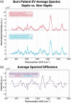
Global averages of septic and non‐septic burn patient EVs. (a) Normalized global average and standard deviation spontaneous Raman spectra for septic burn patient EVs (n = 8) (a), and for control non‐septic burn patient EVs (n = 6). For each patient plasma‐isolated EV sample, a 90s spectrum was acquired at five different spots. (b) The normalized spectral difference between septic and non‐septic burn patients Raman global average spectrum.
FIGURE 4
FIGURE 4
Principal component analysis and confusion matrix results. (a) Normalized spontaneous Raman spectra acquired from patient EV samples (n = 70) underwent principal component analysis and are plotted in PC space along PC1 and PC2. This 2D PC space encompasses 49.2% of the total variance of the dataset. Septic burn patient spectra are shown as pink stars, and non‐septic control burn patients are labelled as blue circles. QDA analysis incorrectly identified four spectra, shown in a yellow overlay on their respective marker. Three spectra were found to be from the same non‐septic burn patient (ARCS barcode number 1879), which were misclassified as septic (false positive, FP). One spectrum from a septic burn‐patient was misclassified as non‐septic (false negative, FN). (b) A confusion matrix visualizes the output of the optimized QDA analysis across the first five PCs (PC1–PC5). Notably, just one of the 70 spectra was classified as a false negative result, yielding a high negative predictive value of 96.4% and a high sensitivity of 97.5%.
FIGURE 5
FIGURE 5
Utilizing bacterial glycoconjugates to elucidate spectral differences between septic and non‐septic burn patient EV Raman fingerprints. (a) Clustering analysis of the four chosen bacterial glycoconjugates’ Raman spectra visualized in three‐dimensional principal component space across the first three PCs, accounting for 90% of the variance. Clear clustering validates spectral differences between the four standards for downstream use as reference analytes. (b) The average spontaneous Raman spectrum of each chosen bacterial glycoconjugate analytical standard following normalization, smoothing, and PLS fitting. Each standard was measured four times then averaged. Shading shows the standard deviation. (c) Comparison of the difference of the average spectrum of non‐septic burn patient EVs from septic burn patient EVs (denoted as difference between population averages, in purple) to the four chosen bacterial glycoconjugates of bacteria commonly associated with sepsis in burn patients. Raman band fits are highlighted in blue. Key in bottom right corner of figure is for panels (b) and (c).

Update of

References

    1. Avni, T. , Levcovich, A. , Ad‐El, D. D. , Leibovici, L. , & Paul, M. (2010). Prophylactic antibiotics for burns patients: Systematic review and meta‐analysis. BMJ, 340, c241. - PMC - PubMed
    1. Bachurski, D. , Schuldner, M. , Nguyen, P. H. , Malz, A. , Reiners, K. S. , Grenzi, P. C. , Babatz, F. , Schauss, A. C. , Hansen, H. P. , Hallek, M. , & Pogge von Strandmann, E. (2019). Extracellular vesicle measurements with nanoparticle tracking analysis—An accuracy and repeatability comparison between NanoSight NS300 and ZetaView. Journal of Extracellular Vesicles, 8(1), 1596016. - PMC - PubMed
    1. Bishop, D. G. , & Work, E. (1965). An extracellular glycolipid produced by Escherichia coli grown under lysine‐limiting conditions. Biochemical Journal, 96, 567–576. - PMC - PubMed
    1. Boehm, D. , & Menke, H. (2021). Sepsis in burns—Lessons learnt from developments in the management of septic shock. Medicina, 58, 26. - PMC - PubMed
    1. Bouza, E. , Onori, R. , Semiglia‐Chong, M. A. , Álvarez‐Uría, A. , Alcalá, L. , & Burillo, A. (2020). Fast track SSTI management program based on a rapid molecular test (GeneXpert® MRSA/SA SSTI) and antimicrobial stewardship. Journal of Microbiology, Immunology, and Infection, 53, 328–335. - PubMed